www.bjournal.com.br
Braz J Med Biol Res, September 2011, Volume 44(9) 920-932
doi: 10.1590/S0100-879X2011007500104
Endothelial dysfunction in cardiovascular and endocrine-metabolic
diseases: an update
A.P. Davel, C.F. Wenceslau, E.H. Akamine, F.E. Xavier, G.K. Couto, H.T. Oliveira and L.V. Rossoni
Institutional Sponsors
The Brazilian Journal of Medical and Biological Research is partially financed by
All the contents of this journal, except where otherwise noted, is licensed under a Creative Commons Attribution License
Faculdade de Medicina de Ribeirão Preto Campus
Ribeirão Preto
Ex plor e H igh - Pe r for m a n ce M S Or bit r a p Te ch n ology I n Pr ot e om ics & M e t a bolom ics
Endothelial dysfunction in cardiovascular and
endocrine-metabolic diseases: an update
A.P. Davel
3, C.F.
Wenceslau
1, E.H.
Akamine
2, F.E.
Xavier
4, G.K. Couto
1,
H.T.
Oliveira
1and L.V. Rossoni
1 1Departamento de Fisiologia e Biofísica, 2Departamento de Farmacologia, Instituto de Ciências Biomédicas, Universidade de São Paulo, São Paulo, SP, Brasil 3Departamento de Anatomia, Biologia Celular e Fisiologia e Biofísica, Instituto de Biologia, Universidade Estadual de Campinas, Campinas, SP, Brasil 4Departamento de Fisiologia e Farmacologia, Centro de Ciências Biológicas, Universidade Federal de Pernambuco, Recife, PE, BrasilAbstract
The endothelium plays a vital role in maintaining circulatory homeostasis by the release of relaxing and contracting factors. Any change in this balance may result in a process known as endothelial dysfunction that leads to impaired control of vascular tone and contributes to the pathogenesis of some cardiovascular and endocrine/metabolic diseases. Reduced endothelium-derived nitric oxide (NO) bioavailability and increased production of thromboxane A2, prostaglandin H2 and superoxide anion in conductance and resistance arteries are commonly associated with endothelial dysfunction in hypertensive, diabetic and obese animals, resulting in reduced endothelium-dependent vasodilatation and in increased vasoconstrictor responses. In addition, recent studies have demonstrated the role of enhanced overactivation of β-adrenergic receptors inducing vascular cytokine production and endothelial NO synthase (eNOS) uncoupling that seem to be the mechanisms underlying endothelial dysfunction in hypertension, heart failure and in endocrine-metabolic disorders. However, some adaptive mechanisms can occur in the initial stages of hypertension, such as increased NO production by eNOS. The present review focuses on the role
of NO bioavailability, eNOS uncoupling, cyclooxygenase-derived products and pro-inflammatory factors on the endothelial dysfunction that occurs in hypertension, sympathetic hyperactivity, diabetes mellitus, and obesity. These are cardiovascular and endocrine-metabolic diseases of high incidence and mortality around the world, especially in developing countries and endothelial dysfunction contributes to triggering, maintenance and worsening of these pathological situations.
Key words: Endothelial dysfunction; Hypertension; Sympathetic hyperactivity; Diabetes; Obesity
Introduction
Correspondence: L.V. Rossoni, Departamento de Fisiologia e Biofísica, ICB, USP, Av. Prof. Lineu Prestes, 1524, Sala 225, 5508-900 São Paulo, SP, Brasil. Fax: +55-11-3091-7285. E-mail: [email protected]
Presented at the XV Simpósio Brasileiro de Fisiologia Cardiovascular, São Paulo, SP, Brazil, February 2-5, 2011.
Received February 14, 2011. Accepted August 1, 2011. Available online August 19, 2011. Published September 16, 2011.
Since the 1980s it has become clear that the endothelium is not only a monolayer that works as a physical barrier between blood and the underlying tissue, but also acts as an endocrine organ able to produce and release different metabolically active substances. Furchgott and Zawadzki (1), in 1980, showed for the first time that endothelial cells have an obligatory role during the relaxation induced by acetylcholine in isolated rabbit aorta. These investigators also demonstrated that acetylcholine activates muscarinic receptors in the endothelial cells and that this activation releases a substance then termed “endothelium-derived relaxing factor - EDRF” (1). Rapoport et al. (2) observed that endothelium-dependent vasodilators induce relaxation
through cyclic GMP-dependent protein phosphorylation, similar to that observed with nitric oxide (NO) donors (e.g., sodium nitroprusside). Later, this EDRF was identified as NO by Ignarro et al. (3) and Palmer et al. (4). Physiologi-cally, NO is synthesized from the amino acid L-arginine by the neuronal (nNOS or NOS I) and endothelial (eNOS or
NOS III) nitric oxide synthase (NOS) enzymes.These
iso-forms are expressed constitutively and the active enzyme is part of a multi-protein complex forming the NOS dimer. The NOS dimer consists of an N-terminal oxygenase domain that contains binding sites for heme, L-arginine
and tetrahydrobiopterin (BH4), and a reductase domain
phosphate (NADPH), flavin mononucleotide (FMN), flavin adenine dinucleotide (FAD), and calmodulin (CaM). Upon activation, the oxidase domain of the NOS isoforms produces
NO from L-arginine using O2, requiring BH4, NADPH, FMN,
and FAD as co-factors that maintain the NOS dimer structure and consequently the NO synthesis (5).
In addition to NO, it is well known that endothelial cells pro-duce and release a series of metabolically active factors. Over the years, it has been shown that endothelium produces several
EDRFs, as well as NO, prostacyclin (PGI2) and
endothelium-derived hyperpolarizing factors (EDHFs). Endothelium also releases contractile factors (EDCFs), as well as angiotensin-II,
endothelin-1, prostaglandins such as thromboxane A2 (TxA2),
prostaglandin H2 (PGH2) and prostaglandin F2α (PGF2α) and
finally reactive oxygen species (ROS). Through a fine balance between the release of EDRFs and EDCFs, the endothelium plays a vital role in maintaining circulatory homeostasis. Any change in this balance may result in endothelial dysfunction.
Endothelial dysfunction is a pathological condition char-acterized by an imbalance between EDRFs and EDCFs. This imbalance is associated with changes in the synthesis, bioavailability and/or action of endothelial factors and results in reduction of endothelium-dependent vasodilatation and/or in an increased response to vasoconstrictor agonists. Thus, endothelial dysfunction impairs vascular function in cardiovas-cular diseases as well as in endocrine-metabolic disorders. Since endothelial cells produce and release many relaxing and contractile factors, extensive studies have analyzed the factors involved in endothelial dysfunction occurring in patho-logical conditions. The present review discusses some recent information concerning the role of endothelial dysfunction in hypertension, sympathetic hyperactivity, diabetes
mellitus, and obesity, which are cardiometabolic diseases of high incidence and mortality around the world, especially in developing countries. We focused on the role of NO bioavailability, eNOS uncoupling, cyclooxygenase (COX)-derived prod-ucts and the role of pro-inflammatory mediators in these cardiometabolic diseases.
Endothelial dysfunction in hypertension
Hypertension is a common disease that affects more than 40% of the adult population in developed countries and about 25% in emerging countries such as Brazil. According to the Brazilian Ministry of Health (6), the proportion of Brazilians diagnosed with hypertension increased from 21.5% in 2006 to 24.4% in 2009. Hypertension is characterized as a medical condition in which blood pressure is chronically elevated and is of multifactorial origin. In this context, physiopathological mechanisms such as endothelial dysfunction present in the blood vessels might contribute to the genesis and/ or maintenance of hypertension.
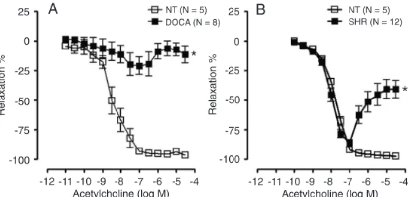
Endothelial dysfunction is a hallmark of hypertension. However, the mechanisms underlying endothelial dysfunction differ among the experimental models of hypertension and the vascular beds studied. Two major functional changes have been frequently observed in the vasculature of hypertensive animals: an increased sensitivity to vasoconstrictor agents and/or a reduced endothelium-dependent relaxation induced by shear stress or agonists such as acetylcholine. These changes have been observed in different experimental hypertension models such as deoxycorticosterone acetate (DOCA)-salt hypertensive rats (volume-dependent hypertension model), spontaneously hypertensive rats (SHR, a genetic model of hypertension that resembles the primary hypertension of humans), ouabain-induced hypertension (an endocrine model of hypertension dependent on the increment of plasma ouabain levels), among others (7-9). As shown in Figure 1, acetylcholine-induced relaxation was impaired in mesenteric resistance arteries from DOCA-salt hypertensive rats and SHR as compared to control normotensive rats. These changes have also been observed in hypertensive patients (10). Konishi and Su (7) first reported that endothelium-dependent acetylcholine-induced relaxation is impaired in aorta from SHR. After that, studies have focused on elucidating the possible pathways involved in this abnormality.
A decline in NO bioavailability in hypertension is one of the most common features of endothelial dysfunction. It can be triggered by reduced eNOS gene and protein expression, impaired eNOS phosphorylation at the serine 1177 residue, decreased availability of eNOS substrate or co-factors, mainly
BH4, altered interaction between eNOS and caveolin-1,
de-creased antioxidant defense, and/or NO inactivation by ROS.
Previous findings have demonstrated an impaired flow-induced vasodilatation in skeletal muscle arterioles from SHR that was due to an impairment of NO-mediated relaxation (11). In addi-tion, aorta from SHR showed impaired endothelium-dependent acetylcholine-induced relaxation that was associated with increased superoxide anion production by NADPH oxidase activity (12).
Rossoni et al. (13) have investigated vascular function in the experimental model of ouabain-induced hypertension in rats. Contrary to that observed in SHR, these investigators demonstrated that treatment with ouabain for 5 weeks was followed by an increase in endothelium-dependent relaxing responses, which reduced the vasoconstrictor response
to α1-adrenergic stimulation and increased the activity of
the Na+,K+-ATPase in conductance vessels. This effect
was mediated by an increase in NO production related to overexpression of eNOS and nNOS (14). These investiga-tors hypothesized that this adjustment was a compensatory mechanism for the high blood pressure observed in
ouabain-treated rats. On the other hand, we also showed that chronic ouabain treatment for 5 weeks increased the expression
of pre-pro-endothelin (endothelin-1 precursor) and ETA
receptor mRNA (15). Furthermore, chronic treatment with
BMS182874, an ETA receptor antagonist, and losartan, an
angiotensin-II type-1 (AT1) receptor antagonist, prevented
ouabain-induced hypertension. Interestingly, only BMS 182874 was able to reduce the protein expression of eNOS in the aorta to control levels (15), suggesting that endothelin-1 underlies both hypertension and nitrergic aortic compensatory adjustment in chronic ouabain-treated rats.
Vascular changes induced by chronic ouabain treatment seems to be time- and vascular bed-dependent (9,16). Thus, 5 and 10 weeks of ouabain treatment did not modify the noradrenaline-induced contraction in mesenteric resistance arteries (9,16) while 20 weeks of treatment with this gly-coside significantly enhanced the contractile response (9). This effect was correlated with a decline in NO modulation (9) (Figure 2). In addition, we also demonstrated that the
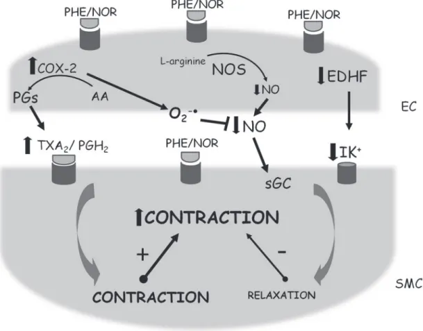
Figure 2. Schematic presentation of the effects elicited by α-adrenoceptor activation in endothelial (EC) and vascular smooth muscle cells (SMC) of small arteries from Wistar rats and spontaneously hypertensive rats (SHR) chronically treated with ouabain. AA = Arachi-donic acid; COX-2 = cyclooxygenase-2; EDHF = endothelium-derived hyperpolarizing factor; NO = nitric oxide; NOR = noradrenaline; NOS = nitric oxide synthase; O2-· = superoxide anion; PGH2 = prostaglandin H2; PGs = prostaglandins; PHE = phenylephrine; sGC = soluble guanylate cyclase; TxA2 = thromboxane A2. ! = increase;
!
treatment of SHR with ouabain for 5 weeks exacerbates the hypertension of SHR associated with a decrease in NO modulation on small arteries (8) (Figure 2).
Previous findings have demonstrated that impaired NO modulation from DOCA-salt, SHR and ouabain-induced hypertensive rats was not only due to its reduced produc-tion. Accelerated degradation by superoxide anion is also observed in these experimental models. In this respect, it has been shown that impaired endothelium-dependent relaxation observed in aorta from DOCA-salt-treated rats was improved by treatment with heparin-binding superoxide dismutase (SOD), a recombinant form of SOD that accu-mulates in the vascular extracellular matrix and potently scavenges superoxide produced by vascular cells (17). Furthermore, removal of the endothelium significantly reduced superoxide levels in DOCA-salt rats but had no effect in normotensive rats, suggesting that high superoxide production in hypertensive vessels is mainly derived from the endothelium (17). Also, in mesenteric resistance arteries from rats treated with ouabain for 20 weeks, the incubation with SOD restored the enhanced noradrenaline-induced contraction (9), showing the involvement of ROS in this model of hypertension.
NADPH oxidase has been suggested to be the most important superoxide anion source in the vascular wall of DOCA-salt and SHR rats (12,18). Additionally, xanthine oxidase and mitochondria also contribute to vascular superoxide anion generation in hypertensive rats (18). Furthermore, recently our group have published that in mesenteric resistance arteries from ouabain-induced hy-pertensive rats, COX-2 is the main source of superoxide anion generation (9) (Figure 2).
There is evidence that persisting oxidative stress would render eNOS uncoupling such that it would no longer produce NO, but superoxide anion (19). This situation characterizes eNOS uncoupling when there is a stoichio-metric mismatch between eNOS protein levels and NO production. Thus, superoxide anion is generated from the dissociation of the heme ferrous-dioxygen complex in the
oxygenase domain of eNOS. The coupling of BH4 to
L-arginine inhibits this production of superoxide anion since
BH4 maintains eNOS dimerization and transfers electrons
during L-arginine oxidation (19). In agreement with this,
in DOCA-salt hypertensive rats, the loss of BH4 induces
eNOS uncoupling, resulting in reduced NO and increased superoxide anion production by this enzyme (20). In this context, NADPH oxidase can further contribute to oxidant stress not only by producing superoxide anion, but also by
initiating BH4 oxidation. Oxidative stress may also impair the
dimethylarginine dimethylaminohydrolase (DDAH) activity, resulting in increasing levels of asymmetric dimethylargi-nine (ADMA), an eNOS endogenous inhibitor. In addition, ADMA contributes to eNOS uncoupling and reduces NO production in hypertension (19).
Endothelial dysfunction has also been associated with
the endothelial release of vasoconstrictor substances. Ac-cordingly, in vascular smooth muscle from hypertensive animals there is an increase in EDCF-mediated signaling, mostly prostanoids, which play an important role in en-dothelial dysfunction. The first study suggesting the role of COX and its products in the endothelial dysfunction of SHR was published years after the discovery of the role of the endothelium in the control of vascular tone (21). This study demonstrated that acetylcholine elicited a biphasic response in aorta from SHR: relaxation at the lowest con-centrations (3 to 100 nM) and a contractile response at the highest concentrations (300 nM to 10 µM) of this agonist. It is important to emphasize that this response is also seen in mesenteric resistance arteries from SHR (Figure 1B). In line with the investigation of the mechanisms involved in this biphasic response, studies demonstrated that the contraction induced by acetylcholine was abolished by
indomethacin, a COX-1/2 inhibitor, and by TxA2/PGH2 (TP)
receptor blockers in both aorta and resistance arteries from SHR (22,23).
Studies have shown that the constitutive COX isoform (COX-1) is the main factor responsible for the acetylcholine-induced contractile response in aorta from SHR (21), since a specific inhibitor of COX-1 (SC-560), but not COX-2 (NS-398), was able to abolish this contraction. However, other studies have demonstrated that COX-2-derived prostanoids play a fundamental role in the endothelial dys-function observed in DOCA-salt and ouabain-hypertensive rats (8,9,24).
Recently, we have shown that both indomethacin (a COX-1/2 inhibitor) and NS-398 (a selective COX-2 inhibitor) reduced the hyperreactivity to noradrenaline in mesenteric resistance arteries from rats with 20-week ouabain-induced hypertension (9). These functional changes were followed by an increase of COX-2 protein expression in mesenteric resistance arteries from ouabain-treated rats (9).
Further-more, incubation with ridogrel (a TxA2 synthase inhibitor and
PGH2/TxA2 receptor antagonist) decreased the contractile
response to noradrenaline in resistance arteries from rats
treated with ouabain for 20 weeks, suggesting that TxA2
and/or PGH2 are the major vasoconstrictor prostanoids
stimulated by noradrenaline in this hypertension model (Figure 2). Consistent with these results, the tail artery from SHR treated with ouabain for 5 weeks showed enhanced phenylephrine-induced contraction that was associated
with TxA2 and/or PGH2 derived from COX-2 metabolism
(8) (Figure 2).
The aforementioned studies suggest the involvement of oxidative stress, COX-derived products, and/or reduced NO bioavailability in endothelial dysfunction in hyperten-sion. Treatment with antioxidant vitamins, NADPH oxidase
inhibitors, SOD mimetics, BH4, or AT1 receptor blockers
(18) underscore the complexity of the interactions among NO, oxidative stress and endothelial dysfunction in hyper-tension. In addition, our group demonstrated that ouabain, besides inducing oxidative stress and COX-2-derived pros-tanoid production, increases the release of proinflammatory cytokines in the arteries (8,9). Thus, it may be possible that treatment with anti-inflammatory drugs and/or inhibitors of ouabain action could become a strategy to reduce the vascular damage in refractory hypertension.
Endothelial dysfunction and sympathetic
hyperactivity
Sympathetic hyperactivity is a common feature of car-diovascular diseases and is a relevant predictor of mortality rate in patients with heart failure (25). In heart failure, the
increased sympathetic outflow contributes to vasocon
-striction and, consequently, to the increment of systemic vascular resistance leading to increased afterload. Elevated afterload further increases cardiac workload and worsens the symptoms of heart failure.
The alterations in vascular tone occurring in heart failure have been linked to endothelial dysfunction. In agreement with this, impairment of endothelium-dependent vasodilatation was demonstrated in heart failure patients, as demonstrated by measurements of forearm blood flow responses to acetylcholine (26). These authors also observed that the blunted vasodilatation in response to acetylcholine was significantly improved by intra-arterial
infusion of phentolamine, an α1-adrenoceptor antagonist
(26). Therefore, an enhanced vascular sympathetic nerve activity could restrain endothelium-mediated vasodilatation in chronic heart failure patients.
Experimentally, coronary artery ligation in rats has been used extensively as a model of chronic left ventricular failure. Using this experimental model, most studies observed an impairment of endothelium-dependent relaxation (27,28). Temporal analysis of endothelial function in aortic rings from myocardial infarcted rats showed that there was no evidence of impaired acetylcholine-induced relaxation after 1 week of coronary artery ligation, with the effect becom-ing evident only at 4 weeks after myocardial infarction and progressively worsening until week 16 (27). This clearly indicates that endothelial dysfunction in heart failure may depend on its stage with time.
An imbalance between NO and superoxide anion production leading to enhanced NO inactivation has been associated with endothelial dysfunction in heart failure and appears to be a common feature of many cardiovascular diseases associated with sympathetic hyperactivity (28).
Heart failure patients and rats after myocardial infarction show elevated plasma norepinephrine levels. However, the myocardial infarction caused by coronary artery ligation, in addition to inducing sympathetic overflow, also induces important humoral changes, including activation of the
renin-angiotensin-aldosterone system (29,30). Activation of plasma and/or tissue renin-angiotensin-aldosterone system plays a key role in vascular dysfunction in heart failure. In this respect, it was demonstrated that
angiotensin-converting enzyme (ACE) inhibitors, AT1 receptor blockade
and mineralocorticoid receptor antagonists could improve the endothelial dysfunction observed in rats after myocardial infarction (29,30). These beneficial effects of blockade of the renin-angiotensin-aldosterone system on endothelial function were accompanied by decreased superoxide anion generation and they are at least part of the mechanism as-sociated with improvement of vasomotor function in heart failure animals (29,30).
Cardiovascular effects of sympathetic overflow can also be mimicked by sustained infusion of low doses of catecholamine, with the activation of adrenoceptors be-ing, in this case, the first signaling event. Moreover, it is known that β-adrenergic stimulation could activate the renin-angiotensin-aldosterone system. The administration of isoproterenol, a non-selective β-adrenoceptor agonist, to laboratory animals for several days is used as a model
of prolonged β-adrenoceptor stimulation. In the vascula
-ture of these animals, we demonstrated that isoproterenol treatment increases the vasoconstrictor response to the
α1-adrenoceptor agonist phenylephrine as well as to the
serotonin (5-HT) agonist in rat (31) and mouse (Figure 3A) aorta, although no changes in acetylcholine-induced relax-ation were observed (31,32). In addition, endothelial removal and L-NAME incubation normalized the hyperreactivity to phenylephrine observed in aortas from isoproterenol-treated rats, indicating that persistent activation of β-adrenoceptors impairs basal endothelial-derived NO (31,32). The reduction of basal NO availability induced by isoproterenol was not related to a reduction in eNOS activity. Indeed, eNOS activity
and protein expression were found to be significantly en
-hanced in aortas from isoproterenol-treated rats (31). How-ever, chronic isoproterenol treatment enhanced superoxide anion levels, measured by hydroethydine fluorescence
in situ in rat (31) and mouse aorta (Figure 3B). Thus, an exacerbated inactivation of basal NO by superoxide anion can be responsible for the increased constrictor reactivity of aortas induced by chronic treatment with isoproterenol. Accordingly, incubation of aortic rings with SOD restored the responsiveness to phenylephrine in rat (31) and mouse aorta from isoproterenol-treated animals (Figure 3A).
The maladaptive effects of chronic activation of β-adrenoceptors were unexpected, since it is known that acute administration of β-adrenoceptor agonists, including isoproterenol, induces vasorelaxation by activation of an adenylyl cyclase-cAMP-PKA pathway, that causes hyperpo-larization and decreases the sensitivity of myofilaments to
Ca2+. In addition, activation of β-adrenoceptors on the en
to oxidative stress through synthesis of superoxide anion (19). In agreement with this hypothesis, we demonstrated that aortas from isoproterenol-treated animals present an increased activity and protein expression of eNOS and nNOS; in addition, L-arginine supplementation was able to normalize the hyperreactivity to phenylephrine (31). Thus, sustained β-adrenoceptor activation could exacerbate eNOS activity and expression, but, in turn, cause eNOS uncoupling that would become a source of superoxide anion production (Figure 4). In addition, aortas from rats with heart failure induced by coronary artery ligation show increased eNOS mRNA and protein expression associated with high superoxide anion levels (28). It is noteworthy that other sources of superoxide anion generation such as NADPH oxidase and xanthine oxidase have been suggested to be activated in blood vessels from rats and patients with heart failure (28,29).
It has been proposed that, in association with oxida-tive stress, an increased production of cytokines and other mediators of inflammation has a pathological role
in generating and/or worsening endothelial dysfunction in many cardiovascular diseases. It is known that cytokines such as interleukin (IL)-1β could impair the endothelium-dependent relaxation in resistance arteries (34). We previ-ously demonstrated that isoproterenol treatment for 7 days significantly enhanced mRNA and protein expression of IL-1β and IL-6 and the activity of the nuclear factor kappa B
(NF-κB) in aortic homogenates (35). Interestingly, suppres
-sion of NF-κB activity via oral treatment with thalidomide improved the hyperreactivity to phenylephrine observed in isoproterenol-treated rats (35). Thus, anti-inflammatory drugs emerge as a therapeutic approach to treat endothe-lial disorders in cardiovascular diseases associated with sympathetic overflow.
Therefore, there is convincing evidence suggesting that reduced NO bioavailability following sympathetic hyperac-tivity and/or increased plasma catecholamine levels is the major contributor to endothelial dysfunction. Generation of superoxide anion by uncoupled eNOS and inflammatory
mediator synthesis induced by chronic β-adrenergic activa
-Figure 3. Effect of 7-day isoproterenol treatment on vascular function and superoxide generation in mouse aorta. A, Contraction response to phenylephrine in aortas with endothelium from control (CT) and isoproterenol (ISO)-treated FVB mice. Superoxide dis-mutase (SOD) incubation (150 U/mL; 30 min) restored the contractile response to phenylephrine of isoproterenol-treated rats. Data are reported as a percentage of the response to 75 mM KCl in each aortic ring. Data are reported as means ± SEM. *P < 0.05 ISO vs
CT; #P < 0.05 ISO vs ISO/SOD (two-way ANOVA). B, Representative fluorescence photomicrographs of slices of aorta from CT and
tion seem to play a central role in the endothelial dysfunction induced by sympathetic overflow. Thus, both mechanisms may be therapeutic targets for cardiovascular diseases associated with endothelial dysfunction and sympathetic hyperactivity.
Endothelial dysfunction in diabetes
mellitus
Diabetes mellitus (DM) is currently considered to be the major epidemic of the century and a public health problem worldwide. In the 1980s it was estimated that 30 million individuals had DM worldwide. This number increased to 135 million in 1995, reaching 173 million in 2002, and 300 million people have been predicted to develop DM by 2030 (36). Approximately two-thirds of these patients live in developing countries, where the epidemic is most
intense, with increasing proportions of affected individuals among younger age groups. DM has been associated with high mortality and a high incidence of cardiovascular complications, which account for about 70% of all deaths in patients with DM. The incidence, prevalence and mortality of cardiovascular disease, such as myocardial infarction, stroke, hypertension, atherosclerosis, and heart failure are approximately 3-5 times higher among diabetic than non-diabetic subjects (36).
Alterations in vascular function are considered to be fac-tors closely related to the development and/or maintenance of cardiovascular complications in patients with DM and may occur even in the early stages of the disease. Although the nature of the pathogenic link between elevated circulating levels of glucose and cardiovascular complications is a source of debate, it is clearly recognized that hyperglycemia is responsible for the pathogenesis of vascular
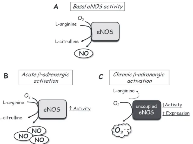
complica-Figure 4. Schematic presentation of the effect of acute and chronic β-adrenergic activation on endothelial nitric oxide synthase (eNOS) activity, expression and uncoupling. A, In normal vessels, basal eNOS activity oxidizes L-arginine, producing L-citrulline and nitric oxide (NO) at physiological rates, contributing to vascular tone maintenance. B, Acute β-adrenergic activation induced by β-adrenoceptor
tions associated with DM. It has been demonstrated that acute or chronic hyperglycemia can cause several changes in vascular function, including a decrease in endothelium-dependent relaxation (37-39), an increased contractile response of vascular smooth muscle (38,40,41) and a pre-disposition to the development of inflammatory, thrombotic and atherosclerotic events (42).
The progression of vascular disease in DM is largely related to endothelial dysfunction mechanisms. Impaired endothelium-dependent vasodilatation has been demon-strated in various vascular beds of different animal models of DM and in human patients with types 1 and 2 DM (37-39). Under these conditions there is vasoconstriction, increased adhesion of platelets and leukocytes, an uncontrolled growth of cells, especially smooth muscle in the vessel wall, an increase in lipids depots in the subendothelium, and an increased tendency to develop vascular thrombosis. The mechanisms associated with these events may be multiple and include changes in synthesis, release, diffusion, or deg-radation of various factors produced by endothelial cells. In DM, endothelial dysfunction is not a homogeneous process in its characteristics and distribution, but it varies depending on the duration of the exposure to hyperglycemia and on the vascular bed evaluated (37,41). Several mechanisms have been proposed to explain the endothelial functional abnormalities associated with the exposure of the vascu-lar tissue to hyperglycemia. The increased activity of the polyol pathway, nonenzymatic glycation of proteins with the production of advanced glycation endproducts (AGEs), oxidative stress, with an increase in ROS production, and alterations in protein kinase C (PKC) are mentioned in the literature as the main mechanisms involved in endothelial dysfunction and atherogenesis, preceding the above-men-tioned vascular complications in patients with DM.
The most notable characteristic of endothelial dysfunc-tion associated with DM is the vascular NO reducdysfunc-tion. Mul-tiple mechanisms are involved in this effect, but increased oxidative stress seems to be the first alteration that triggers several others. The importance of oxidative stress in me-diating the vascular complications of DM is supported by studies showing that antioxidants such as vitamin C and E or SOD correct the endothelium-dependent vasodilator function in humans, in experimental models of DM or in ar-teries exposed to high glucose (42). Hyperglycemia induces oxidative stress by increasing superoxide anion production by the mitochondrial electron transport chain and activation of NADPH-dependent oxidases (42).
In the presence of hyperglycemia, the increased su-peroxide production leads to formation of peroxinitrites
(ONOO-), which may react with the co-factor for NOS, BH
4,
leading to its degradation. In diabetic rats, reduced levels of
BH4 have been reported. According to these results, BH4
supplementation to diabetic rats improves the endothelium-dependent relaxation, suggesting that uncoupled eNOS plays a role in diabetic endothelial dysfunction (39). As
previously described, oxidative stress may also impair DDAH activity, resulting in increasing levels of ADMA. These results therefore suggest that, in addition to its increased degradation, NO synthesis may be decreased by vascular tissue exposure to hyperglycemia.
The sensitivity of vascular smooth muscle may be re-duced even with an adequate supply of NO. Vascular studies in humans and in animal models of DM have commonly used NO donors or nitrovasodilators (e.g., sodium nitroprus-side) as a control for the study of endothelium-dependent vasodilatation. These agents act directly on vascular smooth muscle resulting in an endothelium-independent vasodila-tion. Evidence suggests that the vascular smooth muscle sensitivity to NO is reduced in diabetes and in arteries
exposed to hyperglycemia in vitro, indicating that
hypergly-cemia interferes with the activation of guanylate cyclase by NO (43). On the other hand, other studies have suggested increased production of NO, or increased sensitivity of vascular smooth muscle cells to NO, in diabetes.
As described above, endothelial dysfunction could also be related to the release of vasoconstrictor substances. In vessels of diabetics there is an increase in endothelium-dependent vasoconstrictor mechanisms, mostly mediated by prostanoids, which play an important role in endothelial dysfunction. For example, in mesenteric and coronary arteries from diabetic dogs, exogenous arachidonic acid
induces TxA2-mediated contraction. In contrast, arachidonic
acid leads to PGI2-mediated relaxation in the arteries of
controls (44). Additionally, a reduction in the relaxation induced by acetylcholine in diabetic aortas, mesenteric and femoral arteries is corrected by COX inhibitors (45). An increased participation of vasoconstrictor prostanoids
in the arteries of diabetic rats, such as TxA2 and PGF2α
has also been reported (41,46). Thus, TP receptor antago-nism can restore the impaired relaxation and prevent the endothelium-dependent contraction in arteries exposed to
hyperglycemia (41,46). TxA2 plays a role not only in the
reduction of endothelial function in type 1 DM, but may also be involved in the enhanced contractile response to vasoconstrictor agents (41).
Hyperglycemia selectively increases the expression of COX-2 without affecting COX-1 levels, leading to in-creased release of vasoconstrictor prostanoids (45). The mechanisms by which hyperglycemia leads to COX-2 over-expression are not clear. However, accumulating evidence indicates that ROS activates specific redox-sensitive signal transduction pathways and transcriptional regulatory events that increase COX-2 expression in the vascular wall (45). In support of this view, it has been reported that, under high exposure to glucose, endothelial cells increase the produc-tion of superoxide anion, which was primarily responsible for the enhanced COX-2 expression (47). Hyperglycemia
not only modifies the profile of prostanoids, leading to al
It has been shown that the concentration of diacylglyc-erol (DAG) in hyperglycemia is elevated in vascular cells and tissues. It increases through the acylation of triose intermediates generated by glycolysis, which leads to the synthesis of fatty acids and subsequent acylation of glycerol. DAG is a co-factor, which is rate limiting for PKC (48). PKC and phospholipids generated by calcium-activated
phospho-lipase A2 (cPLA2) regulate several vascular functions such
as cell growth, permeability, contractility, and extracellular matrix protein synthesis. In diabetes, activation of PKC induces an increase in vascular smooth muscle contractility (40) and expression of NADPH oxidase, resulting in ROS
generation (42) and decreased Na+,K+-ATPase activity (49).
However, we have recently reported that diabetic aorta is
also associated with increased Na+,K+-ATPase activity, an
effect, which is also mediated by PKC (50). In these arteries,
TxA2/PGH2 released by COX-2 activates PKC βII, which in
turn increases Na+,K+-ATPase activity (Figure 5). This is
possibly a counter-regulatory mechanism to the vascular dysfunction present in this artery.
Hyperglycemia is also associated with increased pro-duction of AGEs that play a role in endothelial dysfunction associated with DM. Through activation of their receptor, AGEs increase production of ROS via NADPH oxidase (51). Activation of these receptors, when linked to monocytes/ macrophages, also initiates important processes for tis-sue remodeling. The cellular connection of AGE proteins induces cellular responses such as enhanced production of cytokines, growth factors, laminin, fibronectin, and collagen type IV in mesangial cells via PDGF synthesis, as well as increased angiogenesis in endothelial cells. Hyperglycemia can lead to sorbitol accumulation via aldose reductase and
Figure 5. Schematic presentation of the proposed signal transduction pathway involved in altered Na+,K+-ATPase activity induced by
type I diabetes in aorta. The model shows vascular smooth muscle cell (SMC) containing the catalytic α-isoform of Na+,K+-ATPase (NKA) and the abolished effect of endothelium-derived nitric oxide (NO) on this enzyme. Increased protein levels of cyclooxygenase-2 (COX-2) in SMC elevated prostanoid (TxA2 and PGH2) synthesis and their autocrine activity via TxA2/PGH2 receptor enhances the
phosphorylated βII isoform of PKC (p-PKC βII), which increases vascular contractility but in turn activates NKA. EC = Endothelial cell; DAG = diacylglycerol; IP3 = inositol trisphosphate; PIP2 = phosphatidylinositol 4,5-bisphosphate. ! = increase;
!
sorbitol can be converted into fructose by sorbitol dehydro-genase. This increased flow causes elevation of osmotic pressure, decreases the myoinositol concentration and, together with glycolysis, changes the intracellular redox balance.
Therefore, understanding the physiological mechanisms involved in vascular disorders resulting from hyperglyce-mia is essential for the proper use of available therapeutic resources.
Obesity and endothelial dysfunction
The prevalence of obesity is on the rise and has reached epidemic proportions. Obesity is characterized by exces-sive fat accumulation that may impair health. In fact, the excessive accumulation of adipose tissue in the upper body, so-called central or visceral obesity, has a major role in the development of obesity-related metabolic and cardiovascu-lar disorders, such as insulin resistance, impaired glucose tolerance, dyslipidemia, and elevated blood pressure (52). Therefore, risk factors for cardiovascular disease coexist in obese people and the installation of these diseases begins when the risk factors induce endothelial dysfunction.
Despite the fact that obesity-related disorders can individually impair endothelial function, visceral adiposity also affects endothelial function. The severity of endothelial dysfunction correlates with the degree of visceral adiposity (52). Adipocytes and adipose tissue-derived macrophages secrete several factors capable of directly affecting endothe-lial function. Such factors are called adipokines, and com-prise cytokines, chemokines, and hormone-like proteins, such as tumor necrosis factor-alpha (TNF-α), IL-6, leptin, angiotensin-II, plasminogen activator inhibitor-1, resistin, and monocyte chemoattractant protein-1 (52). Uncontrolled secretion of adipokines by obese hypertrophic adipocytes promotes a systemic inflammatory state, which contributes to cardiovascular diseases.
Besides visceral adipose tissue, increased fat depots around blood vessels also contribute to endothelial dys-function. Perivascular adipose tissue has been shown to release vasodilator and vasoconstrictor factors and also a variety of adipokines (53). Although during initial steps of diet-induced obesity perivascular adipose tissue-derived leptin induces an adaptive NO overproduction, contributing to preserving vascular function, obese perivascular adipose
tissue-derived adipokines appear to promote local inflam
-mation (53). This process, together with systemic inflam
-mation, can contribute to alterations of endothelial function. Moreover, we have recently demonstrated an increase in TNF-α protein expression directly in perivascular adipose tissue-cleaned aorta from mice with high-fat diet-induced obesity (54). Whether systemic and perivascular adipose tissue-induced inflammation is involved in the induction of TNF-α expression in vascular cells is not yet known. Whatever the source of inflammatory factors, however, the
endothelium may be negatively affected by proinflamma
-tory cytokines. The exact visceral and perivascular adipose tissue-derived factors implicated in endothelial dysfunction in obesity are not known, but TNF-α and IL-6 have emerged as possible candidates (55).
It has been demonstrated that cytokines impair en-dothelial function, manifested as a reduced NO-dependent relaxation (34). TNF-α-induced endothelial dysfunction can be due to a reduced NO production by inhibition of eNOS gene expression in endothelial cells. Similarly to the TNF-α effect, reduction of eNOS expression was observed in aorta from mice with high-fat diet-induced obesity, which exhibit increased inflammatory markers (54). Obesity modifies post-translational eNOS regulation that results also in reduced NO production. Reduced eNOS phos-phorylation (ser 1177) via phosphatidylinositol 3-kinase/ Akt (56) and uncoupled eNOS (57) are related to impaired endothelium-dependent relaxation in obesity. Moreover, protein expression of caveolin-1, which inhibits eNOS ac-tivity in endothelial cells via protein-protein interactions, is increased in aorta from rats with diet-induced obesity (58). Thus, a reduced NO production is involved in the impaired endothelium-dependent relaxation in obese animals, but how obesity modifies transcriptional and post-translational eNOS regulation is still unclear.
Proinflammatory cytokines not only reduce NO produc
-tion, but also decrease its bioavailability due to increased ROS bioavailability. Several sources of ROS, such as NADPH oxidase, xanthine oxidase, uncoupled eNOS, and mitochondrial respiratory chain, can be stimulated by proinflammatory cytokines in endothelial cells (52). It is well established that oxidative stress is implicated in endothelial dysfunction in the obese condition. Incubation with com-pounds that quench ROS, such as SOD and tiron, restores the reduced endothelium-dependent relaxation in arteries of obese animals (54,57). Although increased vascular cell-produced ROS can be observed in arteries of obese animals (57), obese perivascular adipose tissue-derived ROS can have a paracrine action on the vascular wall and impair the endothelium-dependent relaxation (53). Obesity can also impair the vascular antioxidant capacity. We have demonstrated a decrease in Cu/Zn-SOD protein expres-sion, which could contribute to a decrease in antioxidant capacity and consequently superoxide anion accumulation in the aorta from mice with high-fat-induced obesity (54). Therefore, increase in ROS levels, together with reduced NO production, represent at least part of the mechanism underlying endothelial dysfunction in obesity.
In addition to reducing NO bioavailability, increased ROS levels can also promote inflammation. The NF-κB family members are important redox-sensitive transcriptional
fac-tors that regulate transcription of genes that encode inflam
status by producing ROS and additional cytokines, worsen-ing the pathological processes and leadworsen-ing to endothelial dysfunction. Many of these products of NF-κB activation impact the endothelium-dependent relaxation, such as TNF-α, as described earlier, and the enzyme COX-2 (55). Indeed, an increase in COX-2 protein expression is observed in arteries from obese rats, which contributes to reduced endothelium-dependent vasodilation (57,59). Thus, inhibi-tion of NF-κB may be able to restore the obesity-induced endothelial dysfunction.
The NF-κB pathway appears to be up-regulated in vascular cells from obese individuals. Endothelial cells from obese humans presented elevated NF-κB expression when compared to lean adults (60). Moreover, we have observed a reduction in IκB protein expression in the aorta from obese
mice (54). Although NF-κB is thought to play a role in me
-diating endothelial dysfunction in obesity, there are a few studies showing direct evidence of NF-κB contribution to this condition. Oral administration of salsalate (nonacetylated salicylate) for 4 days inhibits NF-κB in venous endothelial cells and improves flow-mediated endothelium-dependent relaxation in the brachial artery of non-diabetic overweight/ obese middle-aged humans (60). Moreover, inhibition of
NF-κB also reduces adipocyte-induced adhesion mol
-ecule expression in human umbilical venous endothelial cells. Similarly, we have demonstrated a correction of the reduced endothelium-dependent relaxation in obese mice by incubating aortic rings with salicylate (54). This finding suggests the direct participation of the NF-κB pathway in the impaired endothelium-dependent relaxation in obese mice. Taken together, data from these studies demonstrate the direct participation of the up-regulated NF-κB pathway in vascular cells from obese subjects, which could contribute to vascular inflammation and endothelial dysfunction.
The specific mechanism by which inhibition of vascular NF-κB restores endothelial dysfunction in obesity is also not clear. In endothelial cells, NF-κB activation is associated with an increase in NADPH oxidase-derived superoxide levels.
Since both inhibition of NF-κB activity and antioxidant com
-pounds showed similar effects on endothelium-dependent vasodilation (54,60), inhibition of NF-κB might improve endothelial function by reducing ROS levels. However, the sequence of events leading to endothelial dysfunction still needs to be elucidated, including the possibilities that NF-κB could increase ROS availability or that ROS could activate NF-κB.
It is clear that systemic and local inflammation associated with oxidative stress is implicated in endothelial dysfunc-tion in obesity. Many pharmacotherapies (e.g., glitazones, fibrates, metformin, and statins) and even weight loss, which
have a cardioprotective effect, reduce systemic inflamma
-tion, at least in part by inhibiting inflammatory signaling in adipocytes. A direct effect of vascular NF-κB activation appears to make an important contribution to this condition, which may be partially mediated by oxidative stress. Addi-tional studies aimed at elucidating specific mechanisms of endothelial function impairment by NF-κB would most likely support vascular NF-κB as a crucial target in a therapeutic strategy to treat obesity-induced cardiovascular disease.
Conclusion
In conclusion, endothelial dysfunction is a hallmark of most cardiovascular and endocrine/metabolic diseases. It is induced by complex changes in the balance between EDRF and EDCF production, bioavailability and action in smooth muscle cells. Reduction of NO bioavailability due to impaired NO synthesis by eNOS, increased oxidative stress and eNOS uncoupling are common mechanisms underly-ing endothelial dysfunction in many cardiovascular and endocrine diseases. In addition, COX-2-derived prostanoids and pro-inflammatory cytokines could be overproduced in hypertension, diabetes and obesity. Although much is known about the importance of endothelial dysfunction in the pathophysiology of cardiovascular diseases, these diseases still have a high mortality worldwide. Thus, further research of the vascular pathophysiology of cardiovascular diseases is needed in order to determine exact tools for a proper treatment in each case.
Finally, the present review reinforces the importance of anti-inflammatory drugs (e.g., NF-κB and COX-2 inhibitors)
and eNOS co-factors (e.g., BH4) as an effective
pharma-cotherapy strategy to improve endothelial dysfunction present in classical cardiovascular diseases associated with sympathetic hyperactivity such as heart failure and hypertension, and also in endocrine/metabolic disorders including type I and II diabetes and obesity. In addition, drugs that chronically inhibit the vascular action of oua-bain represent a strategy for reducing vascular damage in refractory hypertension. The potential beneficial effects of these drugs on endothelial function could be effective in improving the local control of vascular tone in worldwide cardiovascular and metabolic diseases.
Acknowledgments
1. Furchgott RF, Zawadzki JV. The obligatory role of endothelial cells in the relaxation of arterial smooth muscle by acetyl-choline. Nature 1980; 288: 373-376.
2. Rapoport RM, Draznin MB, Murad F. Endothelium-depen-dent relaxation in rat aorta may be mediated through cyclic GMP-dependent protein phosphorylation. Nature 1983; 306: 174-176.
3. Ignarro LJ, Buga GM, Wood KS, Byrns RE, Chaudhuri G. Endothelium-derived relaxing factor produced and released from artery and vein is nitric oxide. Proc Natl Acad Sci U S A 1987; 84: 9265-9269.
4. Palmer RM, Ashton DS, Moncada S. Vascular endothelial cells synthesize nitric oxide from L-arginine. Nature 1988; 333: 664-666.
5. Alp NJ, Channon KM. Regulation of endothelial nitric oxide synthase by tetrahydrobiopterin in vascular disease. Arterio-scler Thromb Vasc Biol 2004; 24: 413-420.
6. Ministério da Saúde. Portal da Saúde. http://portal.saude.gov.br/ portal/aplicacoes/noticias/default.cfm?pg=dspDetalheNoticia&id_ area=124&CO_NOTICIA=11290. Accessed April 1, 2011. 7. Konishi M, Su C. Role of endothelium in dilator responses of
spontaneously hypertensive rat arteries. Hypertension 1983; 5: 881-886.
8. Xavier FE, Davel AP, Fukuda LE, Rossoni LV. Chronic ouabain treatment exacerbates blood pressure elevation in spontaneously hypertensive rats: the role of vascular mechanisms. J Hypertens 2009; 27: 1233-1242.
9. Wenceslau CF, Davel AP, Xavier FE, Rossoni LV. Long-term ouabain treatment impairs vascular function in resistance arteries. J Vasc Res 2011; 48: 316-326.
10. Taddei S, Virdis A, Mattei P, Salvetti A. Vasodilation to acetylcholine in primary and secondary forms of human hypertension. Hypertension 1993; 21: 929-933.
11. Koller A, Huang A. Impaired nitric oxide-mediated flow-induced dilation in arterioles of spontaneously hypertensive rats. Circ Res 1994; 74: 416-421.
12. Zalba G, Beaumont FJ, San Jose G, Fortuno A, Fortuno MA, Etayo JC, et al. Vascular NADH/NADPH oxidase is involved in enhanced superoxide production in spontaneously hyper-tensive rats. Hypertension 2000; 35: 1055-1061.
13. Rossoni LV, Salaices M, Marin J, Vassallo DV, Alonso MJ. Alterations in phenylephrine-induced contractions and the vascular expression of Na+,K+-ATPase in ouabain-induced hypertension. Br J Pharmacol 2002; 135: 771-781. 14. Rossoni LV, Salaices M, Miguel M, Briones AM, Barker LA,
Vassallo DV, et al. Ouabain-induced hypertension is accom-panied by increases in endothelial vasodilator factors. Am J Physiol Heart Circ Physiol 2002; 283: H2110-H2118. 15. Xavier FE, Yogi A, Callera GE, Tostes RC, Alvarez Y, Salaices
M, et al. Contribution of the endothelin and renin-angiotensin systems to the vascular changes in rats chronically treated with ouabain. Br J Pharmacol 2004; 143: 794-802. 16. Xavier FE, Rossoni LV, Alonso MJ, Balfagon G, Vassallo DV,
Salaices M. Ouabain-induced hypertension alters the partici-pation of endothelial factors in alpha-adrenergic responses differently in rat resistance and conductance mesenteric arteries. Br J Pharmacol 2004; 143: 215-225.
17. Somers MJ, Mavromatis K, Galis ZS, Harrison DG. Vascular superoxide production and vasomotor function in
hyperten-sion induced by deoxycorticosterone acetate-salt. Circula-tion 2000; 101: 1722-1728.
18. Touyz RM. Reactive oxygen species, vascular oxidative stress, and redox signaling in hypertension: what is the clini-cal significance? Hypertension 2004; 44: 248-252.
19. Forstermann U. Nitric oxide and oxidative stress in vascular disease. Pflugers Arch 2010; 459: 923-939.
20. Landmesser U, Dikalov S, Price SR, McCann L, Fukai T, Holland SM, et al. Oxidation of tetrahydrobiopterin leads to uncoupling of endothelial cell nitric oxide synthase in hyper-tension. J Clin Invest 2003; 111: 1201-1209.
21. Luscher TF, Vanhoutte PM. Endothelium-dependent con-tractions to acetylcholine in the aorta of the spontaneously hypertensive rat. Hypertension 1986; 8: 344-348.
22. Gluais P, Paysant J, Badier-Commander C, Verbeuren T, Vanhoutte PM, Feletou M. In SHR aorta, calcium ionophore A-23187 releases prostacyclin and thromboxane A2 as endothelium-derived contracting factors. Am J Physiol Heart Circ Physiol 2006; 291: H2255-H2264.
23. Lang MG, Noll G, Luscher TF. Effect of aging and hyperten-sion on contractility of resistance arteries: modulation by endothelial factors. Am J Physiol 1995; 269: H837-H844. 24. Adeagbo AS, Zhang X, Patel D, Joshua IG, Wang Y, Sun X,
et al. Cyclo-oxygenase-2, endothelium and aortic reactivity during deoxycorticosterone acetate salt-induced hyperten-sion. J Hypertens 2005; 23: 1025-1036.
25. Barretto AC, Santos AC, Munhoz R, Rondon MU, Franco FG, Trombetta IC, et al. Increased muscle sympathetic nerve activity predicts mortality in heart failure patients. Int J Cardiol 2009; 135: 302-307.
26. Santos AC, Alves MJ, Rondon MU, Barretto AC, Middlekauff HR, Negrao CE. Sympathetic activation restrains endotheli-um-mediated muscle vasodilatation in heart failure patients.
Am J Physiol Heart Circ Physiol 2005; 289: H593-H599. 27. Teerlink JR, Clozel M, Fischli W, Clozel JP. Temporal
evolu-tion of endothelial dysfuncevolu-tion in a rat model of chronic heart failure. J Am Coll Cardiol 1993; 22: 615-620.
28. Bauersachs J, Bouloumie A, Fraccarollo D, Hu K, Busse R, Ertl G. Endothelial dysfunction in chronic myocardial infarction despite increased vascular endothelial nitric oxide synthase and soluble guanylate cyclase expression: role of enhanced vascular superoxide production. Circulation 1999; 100: 292-298.
29. Sartorio CL, Fraccarollo D, Galuppo P, Leutke M, Ertl G, Ste-fanon I, et al. Mineralocorticoid receptor blockade improves vasomotor dysfunction and vascular oxidative stress early after myocardial infarction. Hypertension 2007; 50: 919-925.
30. Schafer A, Fraccarollo D, Tas P, Schmidt I, Ertl G, Bauer-sachs J. Endothelial dysfunction in congestive heart failure: ACE inhibition vs angiotensin II antagonism. Eur J Heart Fail
2004; 6: 151-159.
31. Davel AP, Kawamoto EM, Scavone C, Vassallo DV, Rossoni LV. Changes in vascular reactivity following administration of isoproterenol for 1 week: a role for endothelial modulation.
Br J Pharmacol 2006; 148: 629-639.
32. Fukuda LE, Davel AP, Verissimo-Filho S, Lopes LR, Ca-chofeiro V, Lahera V, et al. Fenofibrate and pioglitazone do not ameliorate the altered vascular reactivity in aorta of
isoproterenol-treated rats. J Cardiovasc Pharmacol 2008; 52: 413-421.
33. Ferro A, Queen LR, Priest RM, Xu B, Ritter JM, Poston L, et al. Activation of nitric oxide synthase by beta 2-adreno-ceptors in human umbilical vein endothelium in vitro. Br J Pharmacol 1999; 126: 1872-1880.
34. Jimenez-Altayo F, Briones AM, Giraldo J, Planas AM, Salaices M, Vila E. Increased superoxide anion production by interleukin-1beta impairs nitric oxide-mediated relaxation in resistance arteries. J Pharmacol Exp Ther 2006; 316: 42-52.
35. Davel AP, Fukuda LE, De Sa LL, Munhoz CD, Scavone C, Sanz-Rosa D, et al. Effects of isoproterenol treatment for 7 days on inflammatory mediators in the rat aorta. Am J Physiol Heart Circ Physiol 2008; 295: H211-H219.
36. Murray CJ, Lopez AD. Mortality by cause for eight regions of the world: Global Burden of Disease Study. Lancet 1997; 349: 1269-1276.
37. Pieper GM. Enhanced, unaltered and impaired nitric oxide-mediated endothelium-dependent relaxation in experimental diabetes mellitus: importance of disease duration. Diabeto-logia 1999; 42: 204-213.
38. Davel AP, Rossoni LV, Vassallo DV. Effects of ouabain on the pressor response to phenylephrine and on the sodium pump activity in diabetic rats. Eur J Pharmacol 2000; 406: 419-427.
39. Akamine EH, Kawamoto EM, Scavone C, Nigro D, Carvalho MH, de Cassia AT, et al. Correction of endothelial dysfunc-tion in diabetic female rats by tetrahydrobiopterin and chronic insulin. J Vasc Res 2006; 43: 309-320.
40. Abebe W, MacLeod KM. Protein kinase C-mediated contrac-tile responses of arteries from diabetic rats. Br J Pharmacol
1990; 101: 465-471.
41. Xavier FE, Davel AP, Rossoni LV, Vassallo DV. Time-depen-dent hyperreactivity to phenylephrine in aorta from untreated diabetic rats: role of prostanoids and calcium mobilization.
Vascul Pharmacol 2003; 40: 67-76.
42. Cohen RA, Tong X. Vascular oxidative stress: the common link in hypertensive and diabetic vascular disease. J Cardio-vasc Pharmacol 2010; 55: 308-316.
43. Chan NN, Vallance P, Colhoun HM. Endothelium-dependent and -independent vascular dysfunction in type 1 diabetes: role of conventional risk factors, sex, and glycemic control.
Arterioscler Thromb Vasc Biol 2003; 23: 1048-1054. 44. Gimeno AL, Sterin-Borda L, Borda ES, del Castillo EJ,
Gimeno MF. Arachidonate evokes constriction of coronary and mesenteric arteries isolated from diabetic dogs. Adv Prostaglandin Thromboxane Leukot Res 1983; 12: 235-240.
45. Bagi Z, Erdei N, Papp Z, Edes I, Koller A. Up-regulation of vascular cyclooxygenase-2 in diabetes mellitus. Pharmacol Rep 2006; 58 (Suppl): 52-56.
46. Tesfamariam B, Brown ML, Deykin D, Cohen RA. Elevated glucose promotes generation of endothelium-derived vaso-constrictor prostanoids in rabbit aorta. J Clin Invest 1990; 85: 929-932.
47. Cosentino F, Eto M, De Paolis P, van der Loo B, Bachschmid M, Ullrich V, et al. High glucose causes upregulation of cyclooxygenase-2 and alters prostanoid profile in human
endothelial cells: role of protein kinase C and reactive oxy-gen species. Circulation 2003; 107: 1017-1023.
48. Wild S, Roglic G, Green A, Sicree R, King H. Global preva-lence of diabetes: estimates for the year 2000 and projec-tions for 2030. Diabetes Care 2004; 27: 1047-1053. 49. Xia P, Kramer RM, King GL. Identification of the mechanism
for the inhibition of Na+,K+-adenosine triphosphatase by hyperglycemia involving activation of protein kinase C and cytosolic phospholipase A2. J Clin Invest 1995; 96: 733-740.
50. Gallo LC, Davel AP, Xavier FE, Rossoni LV. Time-dependent increases in ouabain-sensitive Na+,K+-ATPase activity in aortas from diabetic rats: The role of prostanoids and protein kinase C. Life Sci 2010; 87: 302-308.
51. Wautier MP, Chappey O, Corda S, Stern DM, Schmidt AM, Wautier JL. Activation of NADPH oxidase by AGE links oxidant stress to altered gene expression via RAGE. Am J Physiol Endocrinol Metab 2001; 280: E685-E694.
52. Sturm W, Sandhofer A, Engl J, Laimer M, Molnar C, Kaser S, et al. Influence of visceral obesity and liver fat on vascular structure and function in obese subjects. Obesity 2009; 17: 1783-1788.
53. Eringa EC, Bakker W, Smulders YM, Serne EH, Yudkin JS, Stehouwer CD. Regulation of vascular function and insulin sensitivity by adipose tissue: focus on perivascular adipose tissue. Microcirculation 2007; 14: 389-402.
54. Kobayasi R, Akamine EH, Davel AP, Rodrigues MA, Car-valho CR, Rossoni LV. Oxidative stress and inflammatory mediators contribute to endothelial dysfunction in high-fat diet-induced obesity in mice. J Hypertens 2010; 28: 2111-2119.
55. Kralisch S, Sommer G, Stangl V, Kohler U, Kratzsch J, Stepan H, et al. Secretory products from human adipocytes impair endothelial function via nuclear factor kappaB. Ath-erosclerosis 2008; 196: 523-531.
56. Zecchin HG, Priviero FB, Souza CT, Zecchin KG, Prada PO, Carvalheira JB, et al. Defective insulin and acetylcholine induction of endothelial cell-nitric oxide synthase through insulin receptor substrate/Akt signaling pathway in aorta of obese rats. Diabetes 2007; 56: 1014-1024.
57. Lobato NS, Filgueira FP, Akamine EH, Davel AP, Rossoni LV, Tostes RC, et al. Obesity induced by neonatal treatment with monosodium glutamate impairs microvascular reactiv-ity in adult rats: Role of NO and prostanoids. Nutr Metab Cardiovasc Dis 2010 [doi: 10.1016/j.numecd.2010.02.006]. 58. Yang N, Ying C, Xu M, Zuo X, Ye X, Liu L, et al. High-fat diet
up-regulates caveolin-1 expression in aorta of diet-induced obese but not in diet-resistant rats. Cardiovasc Res 2007; 76: 167-174.
59. Sanchez A, Contreras C, Martinez P, Villalba N, Benedito S, Garcia-Sacristan A, et al. Enhanced cyclooxygenase 2-mediated vasorelaxation in coronary arteries from insulin-resistant obese Zucker rats. Atherosclerosis 2010; 213: 392-399.