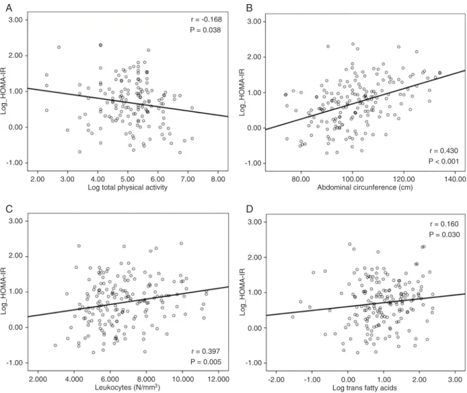
Trans fatty acid intake is associated with insulin sensitivity but independently of inflammation
Texto
Imagem
Documentos relacionados
Objective: To investigate the role of Acylation Stimulating Protein (ASP) receptor C5L2 in skeletal muscle fatty acid accumulation and metabolism as well as insulin sensitivity in
activation, associated with persistent low-grade systemic inflammation in patients with lower CD4 counts at ART initiation, probably plays a role in their lower insulin sensitivity,
Earlier Menarche Is Associated with Lower Insulin Sensitivity and Increased Adiposity in Young Adult Women..
Furthermore, Pudgy mutants do not ingest more than control animals (Figure S3A and S3A 9 ) and have reduced expression of key lipogenic genes such as Acetyl-CoA Carboxylase (ACC)
Fatty acid induced insulin resistance in rat-1 fibroblasts overexpressing human insulin receptors: Impaired insulin-stimulated mitogen-activated protein kinase activity. Effect
Increased adipose tissue PC-1 protein content, but not tumour necrosis factor-alpha gene expression, is associated with a reduc- tion of both whole body insulin sensitivity and
Effects of supplementation with omega-3 on insulin sensitivity and non-esteriied free fatty acid (NEFA) in type 2 diabetic patients.. Efeito da suplementação com ômega-3 sobre
Conclusions: The increase in serum uric acid showed a positive statistical correlation with insulin resistance and it is associated with and increased risk of insulin resistance
