Inte stinal pe rm e ability in
stro ngylo idiasis
1Laboratório de Investigação Médica (LIM 07),
Departamento de Gastroenterologia, Hospital das Clínicas, and Departamentos de 2Radiologia and 3Patologia, Faculdade de Medicina,
Universidade de São Paulo, São Paulo, SP, Brasil A.L. Werneck-Silva1,
A.M. Sipahi1,
A.O .M.C. Damião1,
C.A. Buchpiguel2,
K. Iriya3 and
A.A. Laudanna1
Abstract
The objective of the present study was to assess intestinal permeability in patients with infection caused by Strongyloides stercoralis. Twenty-six patients (16 women and 10 men), mean age 45.9, with a diagnosis of strongyloidiasis were evaluated. For comparison, 25 healthy volun-teers (18 women and 7 men), mean age 44.9, without digestive disorders or intestinal parasites served as normal controls. Intestinal permeability was measured on the basis of urinary radioactivity levels during the 24 h following oral administration of chromium-labeled ethylenediaminetetraacetic acid (51Cr-EDTA) expressed as
percent-age of the ingested dose. The urinary excretion of 51Cr-EDTA was
significantly reduced in patients with strongyloidiasis compared to controls (1.60 ± 0.74 and 3.10 ± 1.40, respectively, P = 0.0001). Intestinal permeability is diminished in strongyloidiasis. Abnormali-ties in mucus secretion and intestinal motility and loss of macromol-ecules could explain the impaired intestinal permeability.
Co rre spo nde nce
A.M. Sipahi
Rua Joaquim Floriano, 72 Cj 72 04534-000 São Paulo, SP Brasil
Fax: + 55-11-820-7457 E-mail: aytan@ usp.br
Publication supported by FAPESP.
Received May 15, 2000 Accepted January 9, 2001
Ke y wo rds
·Intestinal permeability
·51Cr-EDTA
·Strongyloidiasis
·Small bowel
Intro ductio n
The intestinal epithelium plays a major role in the digestion and absorption of fluids and nutrients and also represents an efficient barrier that protects the host from environ-mental pathogens and antigens (1). Impaired barrier function intensifies antigen access, which in turn leads to an exaggerated im-mune stimulation that initiates or perpetu-ates inflammation (2). Increased intestinal permeability has been described in inflam-matory bowel disease (3-11), atopic eczema (12), celiac disease and dermatitis
herpeti-formis (13), cystic fibrosis (14,15), alcohol consumption (16), use of nonsteroidal anti-inflammatory drugs (17-20), and acute in-fectious diarrhea (12,21). On the other hand, decreased intestinal permeability has been demonstrated in Blastocystis hominis infec-tion (22).
layer of the intestine, although the whole intestinal wall may be involved in severe cases (23-26). Also, our group has described small bowel overgrowth (27) which could explain in part the high frequency of Gram-negative sepsis associated with the severe cases with widespread dissemination of lar-vae (28). Intestinal permeability has not been evaluated in strongyloidiasis. Thus, in the present study, intestinal permeability was investigated by means of chromium-labeled ethylenediaminetetraacetic acid (51
Cr-EDTA) in patients infected with Strongy-loides stercoralis.
Mate rial and Me tho ds
Twenty-six patients (16 women and 10 men) with strongyloidiasis were prospec-tively studied. Their ages ranged from 23 to 70 years (mean = 45.9). The diagnosis of strongyloidiasis was confirmed by identifi-cation of the worm in at least two stool examinations by the method of Rugai et al. (29) (three samples). For comparison, 25 asymptomatic healthy volunteers (18 women and 7 men) aged 24 to 68 years (mean = 44.9) without digestive disorders or intesti-nal parasites and with normal upper gas-trointestinal endoscopy served as normal controls. Exclusion criteria included: a) other parasitoses detected by the methods of Faust (30) and Hoffmann et al. (31) (three samples); b) nonsteroidal anti-inflammatory drugs or alcohol consumption; c) acute or chronic renal insufficiency, diabetes mellitus and systemic arterial hypertension; d) celiac dis-ease, inflammatory bowel disease or gastro-enteritis; e) previous gastric or intestinal sur-gery. Our Hospital Ethics Committee ap-proved the research protocol and informed consent was obtained from all subjects.
Blood samples were taken from patients and controls for laboratory data (blood count, glucose, urea/creatinine ratio, and sodium and potassium levels). Enteroscopy (Entero-scope SIF 10, Olympus) was performed in
the patients with strongyloidiasis and four duodenal and four jejunal biopsies were taken. All specimens were examined by the same pathologist (K.I.). Inflammation was scored as mild, moderate and severe accord-ing to the classification of De Paola et al. (23). The control group was submitted to digestive endoscopy (GIF PQ 10, Olympus) to rule out inflamed or ulcerated lesions. Intestinal permeability was measured by the method described by Bjarnason et al. (32) using 51Cr-EDTA. Briefly, after an 8-h
pe-riod of fasting, the patients ingested a solu-tion with 0.6 µmol of 51Cr-EDTA containing
about 100 µCi of radioactivity. The patients then drank 300 ml of water and after 2 h were permitted to eat normally. A 24-h urine sample was then analyzed for radioactivity. Permeability data were expressed as per-centage of excreted radioactivity in relation to the total administered dose.
Results are reported as means ± SD. The chi-square and Student t-tests were used to determine differences between the two groups. The probability value accepted as significant was P<0.05.
Re sults
The two groups were similar regarding age, gender, distribution and all laboratory data, except for eosinophilia (>400 eosino-phils/mm3) which was detected in 56% of
mild in 15, moderate in 5, and severe in 1. In these 21 patients, an increased number of goblet cells was observed. Villous atrophy was only detected in one case (severe). Strongyloides stercoralis larvae were identi-fied histologically in 4 cases, with infesta-tion scored as mild in 1, moderate in 2, and severe in 1. Four patients presented larvae in both duodenal and histological material.
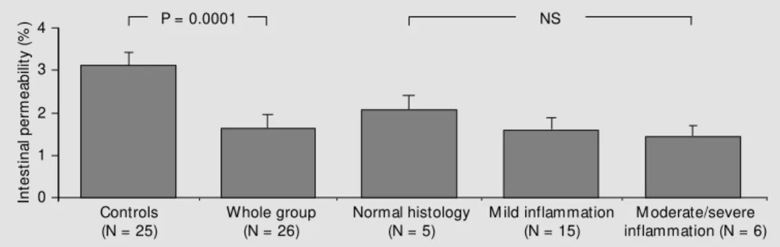
Figure 1 shows the intestinal permeabil-ity data for patients with strongyloidiasis and controls. The urinary excretion of 51
Cr-EDTA was significantly reduced in patients with strongyloidiasis compared to controls (1.60 ± 0.74 and 3.10 ± 1.40, respectively, P = 0.0001). Patients with histologically mod-erate or severe inflammation had lower val-ues than those with mild inflammation (1.38 ± 0.71 versus 1.54 ± 0.75) and normal histol-ogy (2.08 ± 1.68), but the difference was not significant (P = 0.19).
D iscussio n
In the present investigation, we have stud-ied intestinal permeability in patients in-fected with Strongyloides stercoralis (mild/ moderate disease), a nematode particularly found in tropical and subtropical areas. Uri-nary radioactivity levels during the 24 h following oral administration of 51Cr-EDTA,
expressed as percentage of the ingested dose, were significantly reduced in patients with strongyloidiasis (Figure 1). Similarly, Zucker-man et al. (22) also demonstrated decreased intestinal permeability to 51Cr-EDTA in
pa-tients infected with Blastocystis hominis. It is important to point out that our results for intestinal permeability to 51Cr-EDTA in
healthy controls (3.10 ± 1.40) are higher than those obtained in England (1.90%) (13), Canada (1.34%) (8), Belgium (2.51%) (12), Norway (2.45%) (33) and Italy (2.25%) (34). However, they are similar to the data from Israel (4.05%) (35) and from another group in Brazil (3.14%) (36). Methodological dif-ferences are probably not the reason for these differences since we used the same method for measuring intestinal permeabil-ity as used by these investigators. Differ-ences in diet, socioeconomic and nutritional conditions, frequency of bacterial infections and parasitosis may account for these dis-crepant results (8,34,37,38).
The mechanisms underlying intestinal permeability to molecular markers like EDTA in this parasitosis are still unknown. The following possible mechanisms have been suggested. First, the intestinal inflammatory response in strongyloidiasis is accompanied by enhanced mucus secretion (39). In fact, in our series, most of the patients showed an increased number of goblet cells on histol-ogy. The increase in the surface mucus layer in strongyloidiasis could render the intesti-nal barrier less permeable to EDTA. Also, the intestinal cell turnover is highly increased in strongyloidiasis (40) and could impair the paracellular route of intestinal permeability markers like EDTA. Second, most of our patients (62%) had diarrhea. Accordingly, accelerated intestinal transit has frequently
In
te
s
ti
n
a
l
p
e
rm
e
a
b
ili
ty
(
%
) 4
Controls (N = 25)
Whole group (N = 26)
Normal histology (N = 5)
M ild inflammation (N = 15)
M oderate/severe inflammation (N = 6) 3
2
1
0
P = 0.0001 NS
been described in strongyloidiasis (41) and it is possible that the little time for contact between EDTA and the intestinal mucosa affected permeation by the marker. How-ever, this assumption must be evaluated with caution in view of a recent report stating that small intestinal transit has no influence on the excretion of 51Cr-EDTA, at least in
healthy individuals (42). Finally, protein-losing enteropathy has been demonstrated in strongyloidiasis (43) and one may speculate that EDTA is lost by the same intestinal pathway as protein.
Whatever the mechanisms involved, our first description of diminished intestinal per-meability in strongyloidiasis is relevant in
view of the high prevalence of this parasito-sis in some developing countries and its physiopathologic implications. Whether very severe cases of strongyloidiasis with mul-tiple small bowel ulcers or hyperinfection will disclose the same intestinal permeabil-ity pattern we have seen in our patients with mild and moderate disease remains to be elucidated.
Ackno wle dgm e nts
The authors gratefully acknowledge the expert editorial assistance of Maria Laura Lacava Lordello.
Re fe re nce s
1. Kraehenbuhl J-P, Pringault E & Neutra M R (1997). Intestinal epithelia and barrier functions. Alimentary Pharmacology and Therapeutics, 11 (Suppl 3): 3-9.
2. Sartor RB (1995). Current concepts of the etiology and pathogenesis of ulcerative colitis and Crohn’s disease. Gastroenter-ology Clinics of North America, 3: 475-507.
3. Ainsw orth M , Eriksen J, Waever Rasmus-sen J & Schaffalitzk de M uckadell OB (1989). Intestinal permeability of 51 Cr-la-belled ethylenediaminetetraacetic acid in patients w ith Crohn’s disease and their healthy relatives. Scandinavian Journal of Gastroenterology, 24: 993-998.
4. Bjarnason I, O’M orain C, Levi AJ & Peters TJ (1983). Absorption of 51 chromium-la-beled ethylenediaminetetraacetate in in-flammatory bow el disease. Gastroenter-ology, 85: 318-322.
5. Hollander D, Vadheim CM , Brettholz E, Petersen GM , Delahunty T & Rotter JI (1986). Increased intestinal permeability in patients w ith Crohn’s disease and their relatives. Annals of Internal M edicine, 105: 883-885.
6. Hollander D (1988). Crohn’s disease - a permeability disorder of the tight junc-tion? Gut, 29: 1621-1624.
7. Hollander D (1992). The intestinal perme-ability barrier: a hypothesis as to its regu-lation and involvement in Crohn’s disease (Review ). Scandinavian Journal of
Gastro-enterology, 27: 721-726.
8. Jenkins RT, Jones DB, Goodacre RL, Collins SM , Coates G, Hunt RH & Bienen-stock JT (1987). The reversibility of in-creased intestinal permeability to 51 Cr-EDTA in patients w ith gastrointestinal in-flammatory diseases. American Journal of Gastroenterology, 82: 1159-1164. 9. Jenkins RT, Ramage JK, Jones DB, Collins
SM , Goodacre RL & Hunt RH (1988). Small bow el and colonic permeability to 51Cr-EDTA in patients w ith active
inflam-matory bow el disease. Clinical and Inves-tigative M edicine, 11: 151-155.
10. O’M orain CA, Abelow AC, Chervu LR, Fleischner GM & Das KM (1986). Chro-mium51-ethylenediaminetetraacetate test: a useful test in the assessment of inflam-matory bow el disease. Journal of Labora-tory and Clinical M edicine, 108: 430-435. 11. Zuckerman M J & Watts M T (1993). Intes-tinal permeability to 51 Cr-ethylenedia-minetetraacetate in patients w ith ulcer-ative colitis. American Journal of Gastro-enterology, 88: 1978-1979.
12. Forget P, Sodoyez-Goffaux F & Zappitelli A (1985). Permeability of small intestine to [51Cr] EDTA in children w ith acute gas-troenteritis or eczema. Journal of Pediat-ric Gastroenterology and Nutrition, 4: 393-396.
13. Bjarnason I, M arsh M N, Price A, Levi AJ & Peters TJ (1985). Intestinal permeability in patients w ith coelic disease and
derma-titis herpetiformis. Gut, 26: 1214-1219. 14. Escobar H, Perdomo M , Vasconez F,
Camarero C, Del Olmo M T & Suárez L (1992). Intestinal permeability to 51 Cr-EDTA and orocecal transit time in cystic fibrosis. Journal of Pediatric Gastroenter-ology and Nutrition, 14: 204-207. 15. Leclerq-Foucart J, Forget P,
Sodoyez-Goffaux F & Zappitelli A (1986). Intestinal permeability to [51Cr]-EDTA in children w ith cystic fibrosis. Journal of Pediatric Gastroenterology and Nutrition, 5: 384-387.
16. Bjarnason I, Wark K & Peters TJ (1984). The leaky gut of alcoholism: possible route of entry for toxic compounds. Lan-cet, 1: 179-182.
17. Aabakken L & Osnes M (1990). 51 Cr-eth-ylenediaminetetraacetic acid absorption test. Effects of naproxen, a non-steroidal, ant i-inf lam m at ory drug. Scandinavian Journal of Gastroenterology, 25: 917-924. 18. Bjarnason I, So A, Levi AJ, Peters TJ, Williams P, Zanelli GD, Gumpel JM & Ansell B (1984). Intestinal permeability and inflammation in rheumatoid arthritis: effects of non-steroidal anti-inflammatory drugs. Lancet, 2: 1171-1173.
20. Jenkins AP, Trew DR, Crump BJ, Nukajam WS, Foley JA, M enzies IS & Creamer B (1991). Do non-steroidal anti-inflammatory drugs increase colonic permeability? Gut, 32: 66-69.
21. Zuckerman M J, Watts M T, Bhatt BD & Ho H (1993). Intestinal permeability to [51Cr] EDTA in infectious diarrhea. Diges-tive Diseases and Sciences, 38: 1651-1657.
22. Zuckerm an M J, W at t s M T, Ho H & M eriano FV (1994). Blastocystis hominis infection and intestinal injury. American Journal of the M edical Sciences, 308: 96-101.
23. De Paola D, Braga-Dias L & Silva JR (1962). Enteritis due to Strongyloides stercoralis. American Journal of Digestive Diseases, 7: 1086-1098.
24. Britt DP, Khan HA & Florentino AO (1989). Strongyloides infection in an immunosup-pressed patient. M edical Principles and Practice, 1: 63-64.
25. Cook GC (1987). Strongyloides stercoralis hyperinfection syndrome: how often is it missed? Quarterly Journal of M edicine, 244: 625-629.
26. Purtilo DT, M eyers WM & Connor DH (1974). Fatal strongyloidiasis in immuno-suppressed patients. American Journal of M edicine, 56: 488-493.
27. Sipahi AM , Damião AOM C, Simionato CS, Bonini N, Santos M AA, M oraes-Filho JPP, Laudanna AA & Betarello A (1991). Small bow el bacterial overgrow th in strongy-loidiasis. Digestion, 49: 120-124. 28. Igra-Siegman Y, Kapila R, Sen P, Kaminski
Z & Louria DB (1981). Syndrome of
hyper-infection w ith Strongyloides stercoralis. Review s of Infectious Diseases, 3: 397-407.
29. Rugai E, M attos T & Brisola AP (1954). Nova técnica para isolar larvas de nema-tóides das fezes. M odificação do método de Baerm ann. M em órias do Instituto Adolfo Lutz, 14: 5-8.
30. Faust EC (1939). Human Helminthology. 2nd edn. Lea & Febiger, Philadelphia. 31. Hof f m ann W A, Pons JA & Janer JL
(1934). The sedimentation-concentration m et hod in Schist osom iasis m ansoni. Puerto Rico Journal of Health and Tropical M edicine, 9: 283-294.
32. Bjarnason I, Peters TJ & Veall N (1983). A persistent defect in intestinal permeabil-ity in coeliac disease demonstrated by 51Cr-labelled EDTA absorption test.
Lan-cet, 1: 323-325.
33. Aabakken L (1989). 51 Cr-ethylenediamine-tetraacetic acid absorption test. M ethod-ologic aspects. Scandinavian Journal of Gastroenterology, 24: 351-358.
34. Pironi L, M iglioni M , Ruggeri E, Levorato M , Dallasta M A, Corbelli C, Nibaldi M G & Barbara L (1991). Relation betw een intes-tinal permeability to [51Cr] EDTA and in-flammatory activity in asymptomatic pa-tients w ith Crohn’s disease. Digestive Dis-eases and Sciences, 35: 582-588. 35. Peled Y, Watz C & Gilat T (1985). M
eas-urement of intestinal permeability using 51Cr-EDTA. American Journal of
Gastro-enterology, 80: 770-773.
36. Troncon LEA, Pires CR, Kraus OA & Iazigi N (1996). Estudo da permeabilidade intes-tinal pelo teste do 51Cr-EDTA. Arquivos
de Gastroenterologia, 33: 66-73. 37. Ukabam SO, Homeida M M A & Cooper
BT (1986). Small intestinal permeability in normal Sudanese subjects: evidence of tropical enteropathy. Transactions of the Royal Society of Tropical M edicine and Hygiene, 80: 204-207.
38. M enzies IS, Zuckerman M J, Nukajam WS, Somasundaram SG, M urphy B, Jenkins AP, Crane RS & Gregory GG (1999). Ge-ography of intestinal permeability and ab-sorption. Gut, 44: 483-489.
39. Faust EC (1935). The pathology of Stron-gyloides infection. Archives of Pathology, 19: 769-806.
40. Da Costa LR (1971). Small intestinal cell turnover in patients w ith parasitic infec-tions. British M edical Journal, 3: 281-283. 41. Garcia FT, Sessions JT, St rum W B, Schw eistris E, Tripathy K, Bolaños O, Lotero H, Duque E, Ramelli D & M ayoral LG (1977). Intestinal function and mor-phology in strongyloidiasis. Am erican Journal of Tropical M edicine and Hygiene, 26: 859-865.
42. M adsen JL, Scharf f O, Rabøl A & Krogsgaard OW (1996). Relationship be-tw een small intestinal transit rate and in-testinal absorption of 14C-labelled manni-tol and 51Cr-labelled ethylenediaminetet-raacetic acid in healthy subjects. Scandi-navian Journal of Gastroenterology, 31: 254-259.