RESUmo
Estudo experimental laboratorial que com-parou a ação de cinco métodos de
desin-fecção na remoção de bioilme em endos
-cópios gastrintesinais. Foram uilizados
como corpos de prova tubos novos
trans-parentes de politetraluoreileno (Telon®) simulando os canais lexíveis dos endos
-cópios. Após limpeza prévia os tubos fo -ram contaminados intencionalmente com Pseudomonas aeruginosa para formação
de bioilme e submeidos à desinfecção.
Como resultado, nenhum deles removeu
100% dos bioilmes. O que mais removeu isicamente o bioilme foi o glutaraldeído 2% em processadora automáica, prova
-velmente jusiicado pela dupla limpeza, já que o equipamento conta com essa fase no
início do seu ciclo. O método que se mos
-trou menos eiciente para remoção de bio
-ilme e outros resíduos foi água eletrolíica ácida. Esses resultados sugerem que a lim
-peza é mais impactante na remoção de bio
-ilmes do que a desinfecção consecuiva, uma vez que o glutaraldeído, desinfetante da máquina que se mostrou mais eiciente, é um ixador de resíduos orgânicos.
dEScRitoRES Endoscópios
Bioilmes
Desinfecção
Almoxarifado Central
Hospitalar Enfermagem
Removing biofilm from a endoscopic: evaluation
of disinfection methods currently used
*O
riginal
a
r
ticle
AbStRAct
Laboratory experimental study that
com-pared the efeciveness of ive methods of disinfecion for the removal of bioilm in gastrointesinal endoscopes. New trans
-parent tubes of polytetraluoroethylene (Telon®) were used as specimens to simu
-late the channels of lexible endoscopes. Ater pre-cleaning the tubes were inten
-ionally contaminated with Pseudomonas aeruginosa and subjected to disinfecion methods. As a result, none removed 100% of these bioilms. What else physically re
-moved bioilm was 2% glutaraldehyde in an automaic processor, probably jusiied
by the double clean, since the equipment has this phase at the beginning of your
cycle. The method less efecive for re
-moving plaque and other debris was the acidic electrolyic water. These results su -ggest that the cleaning is most striking in
the removal of bioilms that disinfecion of consecuive since glutaraldehyde disin
-fectant by machine is more eicient, it is a fastener organic waste.
dEScRiPtoRS Endoscopes
Bioilms Disinfecion
Central Supply Hospital
Nursing
RESUmEn
Un estudio experimental en el laboratorio en el que se comparó la acción de los cinco métodos de desinfección en la eliminación
de bioilm en los endoscopios gastrointes
-inales. Fueron uilizados como muestras tubos nuevos transparentes de politetralu
-oroeileno (Telon®) simulando los canales de los endoscopios lexibles. Después de pre-limpieza los tubos fueron contamina -das intencionadamente con Pseudomonas
aeruginosa y se someió a métodos de de
-sinfección. Como resultado, ningún método hay removido 100% de las biopelículas. El método que más hay removido ísicamente fue 20% glutaraldehído en un procesador automáico, probablemente jusiicado por
la doble limpio, ya que el equipo iene esta fase en el comienzo de su ciclo. El método es menos eicaz para eliminar la placa y la otra ruina era el agua ácida electrolíica. Estos resultados sugieren que la limpieza es más notable en la eliminación de las biopelículas que la desinfección de forma consecuiva desde desinfectante glutaraldehído de la máquina es más eiciente, es un cierre de los residuos orgánicos.
dEScRiPtoRES Endoscopios
Bioilmes
Desinfección
Central de Suministros Hospital
Enfermería
Ana Cristina Balsamo1, Kazuko Uchikawa Graziano2, René Peter Schneider3,
Manoel Antunes Junior4, Rúbia Aparecida Lacerda5
Remoção de biofilme em canais de endoscópios: avaliação de métodos de desinfecção atualmente utilizados
Remoción de biofilm de canales de los endoscopios: evaluación de los métodos de desinfección Que se utilizan actualmente
*Taken from the dissertation “Evaluation of high-level cleaning and disinfection effectiveness for the removal of bioilm in endoscopic channels”, Graduate Program in Adult Health, University of São Paulo School of Nursing, 2009. 1RN. Ph.D. in Nursing, Graduate Program in Adult Health, University of São Paulo
School of Nursing. Nurse, Healthcare Related Infection Control and Prevention Service, University Hospital, University of São Paulo. São Paulo, SP, Brazil. [email protected] 2RN. Full Professor, Medical-Surgical Nursing Department, University of São Paulo School of Nursing, Brazil. [email protected]
3Ph.D., Professor, Environmental Microbiology Department, Institute of Biomedical Sciences, University of São Paulo. São Paulo, SP, Brazil. schneide@icb.
intRodUction
Endoscope equipment is used in specialized services with a high demand for exams. Because of their high cost, their inventory tends to be restricted. Reuse of the equip
-ment is approved, despite its complex structure, with long channels internally covered with polytetraluorethylene and small luminal diameter, favoring the atachment of
organic material and microorganisms and, consequently,
the formaion of bioilm.
Bioilms knowingly impede eicient processing and represent a challenge for material reuse. They consist of muliple layers of bacterial cells or fungi, grouped and in -volved in amorphous extracellular material composed of
bacterial exopolysaccharides (EPS), whose funcion is to closely unite the cells to and between the biomaterial sur
-faces, consituing an extracellular matrix fundamentally
composed of carbohydrates and proteins,
with the presence of extracellular DNA and
dead cell debris(1-2).
According to the material cleaning dif
-iculty evaluaion criteria proposed in one
study(3), gastrointesinal endoscopes rep
-resent a high risk score as, besides their
complex coniguraion, they are neither dismountable nor transparent, which ham
-pers their internal visualizaion and can thus compromise the evaluaion of their cleaning process. As their internal structure permits organic material accumulaion and bioilm formaion and direct fricion with a brush is not always possible, diiculies may arise to perform the cleaning required.
In this context, endoscopes are classiied
as material that represents a great challenge
for processing. On the other hand, they per
-mit the entry and exit of water, as well as the use of internal cleaning arifacts, and can be immersed in detergent soluion that facilitates the dissoluion of dirt.
Although diferent specialized socieies have well es
-tablished gastrointesinal endoscope cleaning and disin
-fecion recommendaions, various studies discuss that the transmission of microorganisms or adverse efects in paients submited to gastrointesinal endoscopes may be due to the formaion and permanence of bioilms, mak -ing them responsible for cross-transmission of bacteria
and viruses. Therefore, their authors propose the need for studies to evaluate adherence to cleaning and disinfecion protocols, the elaboraion of methods that permit moni
-toring the processing and tests to check its eiciency(1,4-8). As bioilm formaion is unavoidable in structures like endoscope channels and a causal link exists between the current causes of exogenous infecions related to lexible
endoscopes and bad processing quality(9-10), the aims of
this study were to evaluate the efeciveness of high-level disinfecion ater previous brushing for bioilm removal in sample specimens that simulate lexible endoscope chan -nels, besides comparing the methods available at health
services. This research contributes by unveiling the ex
-tent to which bioilms can be eliminated from endoscope
channels, using currently available resources for cleaning
and high-level disinfecion.
mEtHod
In this comparaive experimental laboratory research, the eiciency of ive high-level disinfecion methods for bioilm removal was tested.
In all tests, new transparent lexible tubes were used, with a length of 1m20 and an internal diameter of 2.8 mm, covered with polytetraluorethylene (Tef
-lon®), the same material that covers origi
-nal endoscope channels. These tubes were submited to chemical composiion analysis
through scanning electron microscopy,
con-irming their similarity with the originals. To airm bioilm removal diferences among the ive disinfecion methods, prob
-ability was set at 99.98%. Ater professional staisic advice, a total number of 70 tubes was determined, equally distributed among the methods. From each of the 14 tubes,
in turn, three surface segments of
approxi-mately 3 mm2 were taken, represening their (previously ideniied) start, middle and end, totaling 210 sample segments, 42 for each processing method. The opion to
remove three segments increased the
prob-ability of detecing the presence of bioilm. To obtain bioilm in the sample speci
-mens, challenge contaminaion with Pseu-domonas aeruginosa (ATCC 27853) was used, a microorganism capable of producing bioilm. Originally obtained from the culture inventory of the Mi -crobiology Laboratory at the University of São Paulo
Uni-versity Hospital, this microorganism was inoculated on a MacConkey Agar plate on the day before preparing the suspension. On the date of the experiment, a suspension was prepared with 1x10⁶ colony forming units per milli-liter (CFU/mL) of this microorganism in 10% TSC culture medium, using the colorimeter. To complete each lumen of the tubes, the quanity of suspension introduced was established by calculaing the internal volume in rela
-ion to the tube length, totaling about eight milliliters per tube. Before contaminaion by this suspension, the 70 new tubes were previously submited to manual cleaning, using water and neutral detergent, drying and sterilizaion in a steam autoclave.
although different
specialized societies
have well established gastrointestinal
endoscope cleaning
and disinfection
recommendations,
various studies discuss that the transmission
of microorganisms or adverse effects in
patients submitted
to gastrointestinal
endoscopes may be
due to the formation
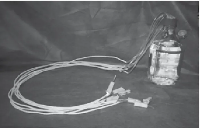
For bioilm formaion, a system (Figure 1) was set up with the following materials: two-liter glass lasks with lid, polyvinyl chloride (PVC) and silicon extensions, clamps and an 0.2 micrometer ilter to ilter air from the system and prevent contaminaion of the culture medium. In the lid of the lasks, eight perforaions were made to introduce eight PVC extensions, ixed with Araldite® rapide epoxy
glue. This set was sterilized in a steam autoclave at 134° C during ive minutes. The soluion with the TSB culture me
-dium was prepared in the glass lasks and then autoclaved at 121° C during 15 minutes. On the day of the experi
-ment, about eight milliliters of Pseudomonas aeruginosa
inoculum – 106 CFU/mL – were injected in each of the
tubes of the sample specimens and their extremiies were atached in circular shape and incubated at 37° Celsius for one hour. This procedure allowed the microorganism to interact with the surface of the sample specimens(11).
Figure 1 – Experimental model for bioilm formation on sample
specimens
At the end of this period, the lid with the PVC exten
-sions was screwed onto the mouths of the lask containing the culture medium, their extremiies were atached to the tubes previously contaminated with the Pseudomo-nas aeruginosa suspension and the culture medium ran
into the distal end of the system, permiing a slow and constant low, controlled by the clamps, for a random six-hour period to promote bioilm formaion. The choice of this period was established to simulate the endoscope us
-age ime in a hospital context, which funcions in six-hour shits, although the bioilm was obtained ater one hour in the laboratory result. Then, the clamps were closed, the Telon® tube removed from the system, the clamp on the extremity it was connected to was ideniied with the let
-ter F to indicate the end (inal in Portuguese) of the tube and the specimens were forwarded to the Endoscopy Ser
-vice for the cleaning and disinfecion phases. Posiive and negaive controls were performed for each disinfecion method. In the posiive control, contaminated sample specimens were not submited to cleaning and disinfec
-ion to prove that uniform bioilm forma-ion occurred across the extent of the lumens. In the negaive control, a new and clean tube was submited to the steam autoclave sterilizaion process. Both controls took place at the same ime as the experiments.
All contaminated sample specimens were previously submited to manual cleaning ater immersion in Rioquími
-ca® enzymaic detergent (containing proteases, amylases
and lipases) and then distributed among the ive diferent disinfecion methods: 1) basic 2% glutaraldehyde soluion (Cidex® – Johnson&Johnson) in manual method; 2) basic 2% glutaraldehyde soluion (Cidex® – Johnson&Johnson) in automated method, using the Lifemed® Endolav® endo
-scope cleaner/disinfector; 3) 0.09%-0.15% acive peraceic acid (Anios® Anioxyde 1000) in manual method; 4) 35% peraceic acid in automated method, using the Steris® Sys-tem sterilizer, whose acive principle is 35% peraceic acid itself (sterilizing concentrate STERIS® 20); 5) acidic elec
-trolyic water produced in situ in the Cleantop® processor. The cleaning and disinfecion methods followed the recom
-mendaions in the Endoscope Processing Manual of the Brazilian Gastrointesinal Endoscopy Nursing Society(12).
In the manual disinfecion methods, the tubes were immersed in the test-disinfectant ater cleaning and their lumens were illed with the help of a 10-ml sterilized sy
-ringe. Contact imes with the disinfectant were estab -lished according to the product manufacturers’
recom-mendaions. Ater removing the soluion, the tubes were rinsed under running tap water using a water pistol, the lumens were dried with a compressed air pistol and the external surface was dried with a clean cloth.
For the automated methods, prototypes (Figure 1) were created which permited the adequate iing of the
test tubes into the machine connectors, guaranteeing
contact between the disinfectant and the internal and ex
-ternal surfaces of each tube.
At the end of the processing phase (cleaning and disinfection), the three segments were removed from each of the 70 tubes, which were prepared with plat -inum in high vacuum and stored in a desiccator until
they were subject to scanning electron microscopy (Brand FEI, model Quanta 600 – FEG) analysis to detect
the presence or not of residual biofilms. The likelihood
ratio test(8)was used to compare the segments from the
start, middle and end of the tubes for each
disinfec-tion method. Significance was set at 5% and statistical power at 95%.
RESULTS
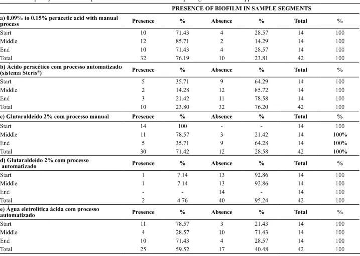
Table 1 demonstrates the results for the presence of bioilm atached to the sample segments ater each tested disinfecion method.
Table 1 – Frequency distribution of presence of bioilm in different sample segments after the application of the tested disinfection methods PRESENCE OF BIOFILM IN SAMPLE SEGMENTS
a) 0.09% to 0.15% peracetic acid with manual
process Presence % Absence % Total %
Start 10 71.43 4 28.57 14 100
Middle 12 85.71 2 14.29 14 100
End 10 71.43 4 28.57 14 100
Total 32 76.19 10 23.81 42 100
b) Ácido peracético com processo automatizado
(sistema Steris®) Presence % Absence % Total %
Start 5 35.71 9 64.29 14 100
Middle 2 14.28 12 85.72 14 100
End 3 21.42 11 78.58 14 100
Total 10 23.80 32 76.20 42 100
c) Glutaraldeído 2% com processo manual Presence % Absence % Total %
Start 14 100 - - 14 100
Middle 11 78.57 3 21.42 14 100%
End 5 35.71 9 64.28 14 100%
Total 30 71.42 12 28.58 42 100%
d) Glutaraldeído 2% com processo
automatizado Presence % Absence % Total %
Start 1 7.14 13 92.86 14 100
Middle 1 7.14 13 92.86 14 100
End - - 14 - 14 100
Total 2 4.76 40 95.24 42 100
e) Água eletrolítica ácida com processo
automatizado Presence % Absence % Total %
Start 11 78.57 3 21.43 14 100
Middle 4 28.57 10 71.43 14 100
End 10 71.43 4 28.57 14 100
Total 25 59.52 17 40.48 42 100
Based on Table 1, it is veriied that none of the process
-ing methods was able to completely remove the bioilms. The result of the automated method using 2% glutaral
-dehyde can be considered saisfactory though, removing almost all inoculum, followed by the automated process using peraceic acid (Steris® system), totaling ten sample segments contaminated with bioilm. In the other meth
-ods, the bioilm that remained was pracically equivalent, totaling between 25 and 32 sample segments.
As for the presence of bioilm in the segments (start x middle x end), a staisically signiicant diference was found among them in the manual disinfecion method us -ing 2% glutaraldehyde and in the automated method
us-ing acidic electrolyic water, indicaus-ing that results were not uniform in the same sampling unit.
In the manual method with glutaraldehyde, this difer
-ence was obtained between the iniial and inal porions (p < 0.001), showing that the bioilm remained atached in 35.71% of the sample segment surfaces. On the other hand, no staisically signiicant diference (p = 0.067) was found between the iniial and middle porions but, when comparing the middle and inal segments, signiicant dif
-In the automated method with acidic electrolyic wa
-ter, a signiicant diference was found between the iniial and middle segments (p = 0.039). The other pairs showed no signiicant mutual diferences. Using electron micros
-copy, however, it was veriied that segments without bio
-ilm were not clean. Some segments contained countless isolated or grouped bacteria, while others only revealed the EPS layer without the presence of bacteria (Table 2). The presence of bioilm or isolated bacteria beyond the
bioilm, or EPS without the presence of bacteria, observed
through electron microscopy, indicated ineicient clean
-ing of the channels.
In the automated method using 2% glutaraldehyde, no
signiicant diference was observed when comparing all segments: start x middle (p = 0.999; start x end, p = 0.466; middle x end, p = 0.466). Although this method removed the bioilm from most sample segment surfaces, only 19.04% (8/42) of these contained no debris – bacteria or EPS (Table 2).
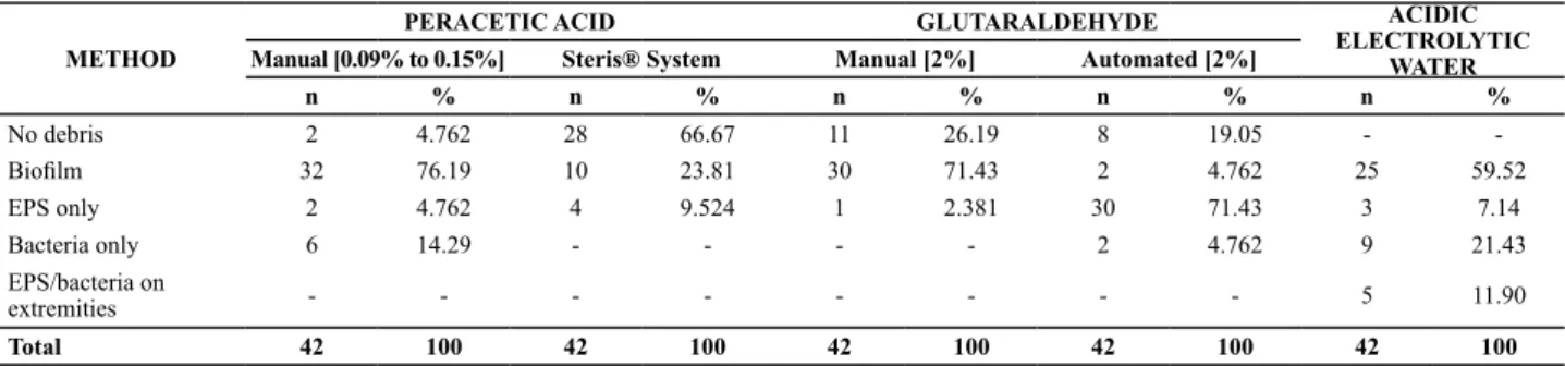
Table 2 – Frequency distribution of presence of bioilm and other debris after manual cleaning and disinfection with different methods METHOD
PERACETIC ACID GLUTARALDEHYDE ACIDIC
ELECTROLYTIC WATER
Manual [0.09% to 0.15%] Steris® System Manual [2%] Automated [2%]
n % n % n % n % n %
No debris 2 4.762 28 66.67 11 26.19 8 19.05 -
-Bioilm 32 76.19 10 23.81 30 71.43 2 4.762 25 59.52
EPS only 2 4.762 4 9.524 1 2.381 30 71.43 3 7.14
Bacteria only 6 14.29 - - - - 2 4.762 9 21.43
EPS/bacteria on
extremities - - - 5 11.90
Total 42 100 42 100 42 100 42 100 42 100
As for the peraceic acid, neither the manual nor auto
-mated methods presented signiicant diferences between the segment posiions (respecively, 0.15% start x middle, p = 0.802; start x end, p = 0.999; middle x end, p = 0.802; and start x middle, p = 0.487; start x end, p = 0.222; middle x end, p = 0.487). In the automated method, the presence of atached bacteria without the exopolysaccharide (EPS) layer or of EPS without the bacteria was considered as debris instead of bio
-ilm. When using this processor, a proporion of 28 clean seg
-ments in 42 sample units (66.66%) was obtained (Table 2).
Table 2 summarizes the results obtained for the
presence of biofilm and debris in the different sample
specimen segments. In many samples, the biofilm re
-mained attached when using 0.09% to 0.15% peracetic acid, and in less samples when using automated disin
-fection with 2% glutaraldehyde. The presence of the
EPS layer at one end of the sampling unit but only
bac-terial cells at the other end was observed in the same
segment only in the method using acidic electrolytic
water.
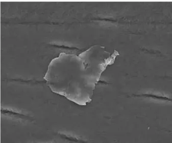
Figure 4 - Bioilm after disinfection using 0.09% to 0.15% pe
-racetic acid
Figure 5 - Bioilm after automated disinfection using 2% gluta
-raldehyde
Figure 6 - Bioilm after manual disinfection using 2% glutaral
-dehyde
Figure 7 - EPS fragment after manual disinfection using 2% glu
-taraldehyde
Figure 9 – EPS fragment after disinfection using acidic elec
-trolytic water
Figure 10 – Bacterial cells after disinfection using acidic elec
-trolytic water
diScUSSion
This study showed that processing through the meth
-ods commonly used in Brazil was unable to remove all bio
-ilm atached to the lumen surfaces of the test specimens that simulated the lexible endoscopic channels. The results
enhanced the understanding that the processing of this
care equipment sill represents a challenge to insituions, researchers and oicial health eniies. The phases of this
process need to be reconsidered and discussed, including
the use of manual cleaning arifacts, eicacy of cleaning agents, microbial acivity of disinfectants and, mainly, the ability of this whole apparatus to remove bioilm.
A research showed that the number of imes the endo
in gastroscopes as well as colonoscopes(4). The authors conclude that these indings relect the microorganism’s capacity to survive the cleaning and disinfecion process.
At two Brazilian hospital, it was also demonstrated
that Pseudomonas aeruginosa was the microorganism
most isolated from ready-to-use endoscope samples and highlighted the biofilm formation ability of this
microorganism(14).
Authors consider that, although diicult, the removal of bioilm can be achieved through mechanic cleaning and
brushing, but some disinfectants are capable of
remov-ing the bioilm while others are not(1,15). In our research, diferences were found in the disinfectants’ efect on the bioilms. While some removed the bioilm from most of
the sample segments, others only removed the
exopoly-saccharide (EPS) layer, while bacterial cells remained, and vice-versa.
Cleaning agents are more efecive to remove bioilm
than disinfectants, as the forms are able to detach the
bio-ilm from the surface(15). In this research, the best process -ing result involved two clean-ing phases us-ing enzymaic detergent.
In one study, the authors elaborated a system to
produce bioilm in 16 hours, using Escherichia coli
bac-teria, in Telon® tubes, used for endoscope channels(16). The goal of these authors was to check the eicacy of
detergents recommended for endoscope cleaning to
remove bioilm, three of which contained enzymes and one did not. The cleaning procedures did not use a brush
to scrub the surfaces, but merely immersion during eight
minutes. Diferences among the products were found, as the non-enzymaic detergent removed more bioilm.
In the topic Leters to the editor, it was published that
the non-enzymaic detergent, called Matrix®, tested in
that research, contains a signiicant quanity of quater
-nary ammonium compound (not cited by the research author), resuling in the removal of the bioilm from the Telon® tubes. The same author airms that the bioilm was reduced because the detergent was associated with
the disinfectant(17).
In our research, although the internal surfaces of the
sample tubes were scrubbed with a brush, the results showed that the cleaning process was insuicient to re
-move the bioilm. It should be reminded that the presence of dry organic material makes cleaning more diicult and enhances the creaion of bioilms. These, in turn, make it diicult for the chemical agent to penetrate and, thus, microbial death does not occur. In places without a nurs
-ing team to perform equipment clean-ing and disinfecion immediately ater use, like at night or during emergency care, the endoscope awaits processing by the regular team, which only happens on the next workday. Hence,
the endoscope surface is exposed to organic material for a
long ime, suicient for bioilm to form.
Endoscope processors ofer advantages in comparison with manual processing: they automaize and standard
-ize important processing phases, reduce the probability of omiing a phase and minimize the team’s exposure to chemical products. The processor reduces the possibil -ity of human errors and generally involves devices to be
connected to water ilter systems, which avoid the equip
-ment’s contaminaion by opportunisic microorganisms found in reservoir water(18). The same author describes that previous manual cleaning with a brush can be more efecive than automated cleaning and that adding this phase would increase total processing ime by 40 to 60 minutes. Also, it is reported that automaic processors neither use 70% alcohol for the inal lush nor monitor the concentraion of the chemical product automaically, and
that machine compartments should be frequently
evalu-ated to avoid any accumulevalu-ated dirt and bioilm formaion in its internal circuits.
In our study, the best bioilm removal result was ob -tained in the automated processing method that includes
another cleaning phase with enzymaic detergent. These indings allow us to infer that the diference obtained is
more related to the accomplishment of another cleaning phase than to the chemical product used, in this case 2%
glutaraldehyde, which is an organic waste fastener. One
important issue is the fact that endoscope processors
come with their own devices that it into the holes of the endoscope, guaranteeing contact between the cleaning
agent and chemical product in the internal equipment
channels through a direct, pressurized low without re
-turn. The automaic processor with 2% glutaraldehyde most removed bioilm, but maintained a high percentage of segments with EPS layers (more than 60%). Manual processing with 0.09% to 0.15% peraceic acid and 2% glu
-taraldehyde were the methods that most retained bioilm. Peraceic acid has been recommended for alternaive high-level disinfecion instead of aldehyde derivaives be
-cause of its low toxicity and biodegradability, although its animicrobial efeciveness is similar(19). In a previous pub -licaion, however, the same author demonstrated, with other collaborators, that some peraceic acidic formulae ixed bioilm while other did not(20). They found that stabi -lized, i.e. ready-to-use peraceic acid did not ix the bioilm and concluded that, when choosing a disinfectant product for high-level disinfecion, not only the germicide’s bacte
-rial acivity should be considered, but also its ability not to ix bioilm.
CONCLUSION
In conclusion, none of the disinfecion methods tested totally removed the bioilm; the most eicient was the use
of 2% glutaraldehyde in automated equipment, and the
-method, and also due to the fact that this product fastens debris, the present study results suggest that cleaning is
more efecive to remove bioilm than consecuive disin
-fecion; this is jusiied by the fact that the automaic pro
-cessor with this product includes a cleaning phase at the start of its cycle. This research alerts to the capacity of mi
-croorganisms to form bioilms within one hour ater con
-taminaion, reinforcing the need to clean the endoscope soon ater its use, so as to avoid environments that favor their development. As the microorganisms present in rins
-ing water are capable of form-ing bioilm, we suggest us
-ing bacterial ilters for the endoscopes’ rins-ing water, as well as tesing other disinfectants available in the market in disinfecing washers.
REFEREncES
1. Pajkos A, Vickery K, Cossart Y. Is bioilm accumulaion on en
-doscope tubing a contributor to the failure of cleaning and decontaminaion? J Hosp Infect. 2004; 58(3):224-9.
2. Lindsay D, Von Holy A. Bacterial bioilms within the clinical seing: what healthcare professionals should know. J Hosp Infect. 2006; 64(4):313-25.
3. Graziano KU, Balsamo AC, Lopes CL, Zotelli MF, Couto AT, Pas
-choal ML. Criteria for evaluaing diiculies in cleaning single-use items. Rev Laino Am Enferm. 2006;14(1):70-6.
4. Bisset L, Cossart Y, Selby W, West R, Caterson D, O’Hara K, et
al. A prospecive study of the eicacy of rouine decontamina
-ion for gastrointesinal endoscopes and the risk factors for failure. Am J Infect Control. 2006;34(5):274-80.
5. Obee PC, Griith CJ, Cooper RA, Cooke RP, Bennion NE, Lewis M. Real-ime monitoring in managing the decontaminaion of lexible gastrointesinal endoscopes. Am J Infect Control. 2005;33(4):202-6. 6. BloB R, Kampf G. Test models to determine cleaning eicacy
with diferent types of bioburden and its clinical correlaion. J Hosp Infect. 2004;56 Suppl 2: 544-8.
7. Alfa MJ, Memes R. Inadequacy of manual cleaning for repro-cessing single-use, triple-lumen sphinctertomes: simulated-use tesing comparing manual with automated cleaning methods. Am J Infect Control. 2003;31(4):193-207.
8. Zuhlsdorf B, Emmrich M, Floss H, Mariny H. Cleaning eicacy of nine diferent cleaners in a washer-disinfector designed for lexible endoscopes. J Hosp Infect. 2002; 52(3):206-11. 9. Graziano KU, Lacerda RA, Turrini RT, Bruna CQM, Silva CPR,
Schmit C, et al. Indicators for evaluaion of processing den
-tal-medical-hospital supplies: elaboraion and validaion. Rev Esc Enferm USP [Internet]. 2009 [cited 2012 abr. 19];43(n. spe 2):1174-80. Available from: htp://www.scielo.br/pdf/ reeusp/v43nspe2/en_a05v43s2.pdf
10. Costa EAM, Costa EA, Graziano KU, Padoveze MC. Medical device reprocessing: a regulatory model proposal for Bra-zilian hospitals. Rev Esc Enferm USP [Internet]. 2011 [cited 2012 Jan 17];45(6):1459-65. Available from: htp://www. scielo.br/pdf/reeusp/v45n6/en_v45n6a26.pdf
11. Murga R, Miller JM, Donlan RM. Bioilm formaion by gram-negaive bactéria on central venous catheter connerctors: efect of condiioning ilms in a laboratory model. J Clin Mi-crobiol. 2001;39(6):2294-97.
12. Muller S, Graziano KU, Hoefel HK; Agência Nacional de Vigilância Sanitária; Sociedade Brasileira de Enfermagem em Endoscopia e Gastrointesinal. Manual de limpeza e desin-fecção de aparelhos endoscópicos [Internet]. Brasília; 2012 [citado 2012 abr. 12]. Disponível em: htp://www.anvisa. gov.br/servicosaude/manuais/sobeeg_manual.pdf
13. Abramson JH. Winpedi (PEPI – for windows): computer programs for epidemiologists. Epidemiol Perspect Innov. 2006;1(1):6.
14. Machado PA, Pimenta ATM, Gonijo PP, Geocze S, Fischman O. Microbiologic proile of lexible endoscope disinfecion in two brazilian hospitals. Arq Gastroenterol. 2006;43(4):255-8. 15. Marion K, Freney J, James G, Bergeron E, Renaud FN,
Coster-ton Jw. Using an eicient bioilm detaching agent: an essen-ial step for the improvement of endoscope reprocessing protocols. J Hosp Infect. 2006;64(2):136-42.
16. Vichery K, Pajkos A, Cossart Y. Removal of bioilm endo-scopes: evaluaion of detergent eiciency. Am J Infect Con-trol. 2004;32(3):170-6.
17. Sava A. Bioilm digeston: more confusion than answers [let-ter]. Am J Infect Control. 2005;33 (10):614.
18. Muscarella LF. Advantages and limitaions of automaic lexible endoscope reprocessors. Am J Infect Control. 1996;24(4):304-9.
19. Loukili NH, Granbasien B, Faure K, Guery B, Beaucaire G. Ef-fect of diferent stabilized preparaions of peraceic acid on bioilm. J Hosp Infect. 2006;63(1):70-2.