Article
0103 - 5053 $6.00+0.00*e-mail: [email protected]
Liposome Encapsulation of Lipophilic
N
-Alkyl-propanediamine Platinum Complexes:
Impact on their Cytotoxic Activity and Inluence of the Carbon Chain Length
Heveline Silva,a Ana Paula S. Fontes,a Miriam Teresa P. Lopesband Frédéric Frézard*,c
aDepartamento de Química, Universidade Federal de Juiz de Fora,
36036-900 Juiz de Fora-MG, Brazil
bDepartamento de Farmacologia, Universidade Federal de Minas Gerais,
31270-901 Belo Horizonte-MG, Brazil
cDepartamento de Fisiologia e Biofísica, Universidade Federal de Minas Gerais,
31270-901 Belo Horizonte-MG, Brazil
Complexos de platina(II) derivados de N-alquil-propanodiamina com cadeia carbônica
variável (C8, C10, C12 ou C14) foram incorporados em lipossomas e a atividade citotóxica dessas
formulações foi avaliada em linhagens tumorais (A549, MDA-MB-231, B16-F1 and B16-F10) e
não-tumorais (BHK-21 and CHO). Suspensões de lipossomas estáveis e monodispersas incorporando os complexos de platina foram obtidas com uma composição lipídica de
diestearoil-sn-glicero-3-fosfocolina, colesterol, e 1,2-diestearoil-sn-glicero-3-fosfoetanolamina-N
-(metoxi(polietileno-glicol)-2000) na razão molar 5:3:0,3. A eiciência da incorporação dos complexos de platina em lipossomas aumentou com o tamanho da cadeia carbônica e foi maior que 80% com os derivados C12 e C14. O efeito da encapsulação em lipossomas na atividade citotóxica dos complexos mostrou-se dependente do tamanho da cadeia carbônica. Os dados indicam que a biodisponibilidade da platina a partir das formulações de lipossomas foi maior para o complexo apresentando uma cadeia carbônica (C12) e uma partição entre a membrana e a fase aquosa intermediárias.
Antitumor platinum(II) complexes derived from N-alkyl-propanediamine differing in the
length of their carbon chain (C8, C10, C12 and C14) were incorporated in liposomes and the
cytotoxic activity of these formulations was evaluated against tumor (A549, MDA-MB-231, B16-F1
and B16-F10) and non-tumor (BHK-21 and CHO) cell lines. Stable and monodisperse liposome suspensions incorporating the platinum complexes were obtained from the lipid composition consisting of distearoyl-sn-glycero-3-phosphocholine, cholesterol and
1,2-distearoyl-sn-glycero-3-phophoethanolamine-N-(methoxy(polyethylene glycol)-2000) at 5:3:0.3 molar ratio. The
entrapment eficiency (EE%) of the platinum complexes in liposomes increased with the carbon chain length. EE% was higher than 80% in C12- and C14-derivatives. The effect of liposome encapsulation on the cytotoxic activity of the complexes was found to depend on the carbon chain length. These data indicate that the highest drug bioavailability from liposome formulations was achieved with the complex showing intermediate carbon chain length and partition between the liposome membrane and aqueous phase.
Keywords: liposomes, platinum complexes, cytotoxic activity
Introduction
Cancer is one of the leading causes of death in the world, with chemotherapy being a valuable tool for the treatment of the disease. The development of novel compounds which are more effective and less toxic and which could become
eventual anti-tumor drugs constitutes a research area of great relevance.
Platinum complexes are well established anti-tumor agents in clinical use, with cisplatin being the most
widely used compound of this class.1-3 However, their use
has been compromised by its propensity to cause several toxicities including nephrotoxicity, neurotoxicity and
specially caused by the reduction of intracellular platinum
accumulation.7,8 Lipophilic derivatives of platinum(II) have
been investigated for their ability to enhance the cellular uptake of platinum in both cisplatin-sensitive and resistant
cell lines.9,10
Nanocarriers have been a fundamental tool in cancer research. Currently, natural and synthetic polymers and lipids are typically used as components of drug delivery nanovectors. The family of nanocarriers includes polymer conjugates, polymeric nanoparticles, lipid-based carriers such as liposomes and micelles, dendrimers, carbon nanotubes, and gold nanoparticles, including nanoshells and nanocages. Rationally designed nanocarriers can offer several therapeutic advantages over free drugs. Liposomes have attractive biological properties, including general biocompatibility, biodegradability, isolation of drugs from the surrounding environment, and the ability to entrap both hydrophilic and hydrophobic drugs and today are approved by regulatory agencies to carry a range of
chemotherapeutics.11
The encapsulation of lipophilic derivatives of cisplatin in liposomes has been explored to allow for the safe
parenteral administration of these complexes.12-14 In
addition to their solubilization properties, liposomes have been extensively studied for the passive and active
targeting of drug to tumors.15 For instance, the use of
polyethylene glycol as PEG-coated liposomes was found to prolong the circulation time of liposomes in blood, to passively enhance their tumor localization, and improve the therapeutic eficacy of platinum complexes, exploring
the EPR (enhanced permeability and retention) effect.16
As another potential beneit, liposome encapsulation may also circumvent drug resistance of tumor cells, as reported
previously for cisplatin.17,18
Although several lipophilic drugs have been prepared and evaluated under the encapsulated form in liposomes, the physicochemical factors that determine the bioavailability of a lipophilic drug from a liposome carrier are still poorly understood. In particular, the inluence of the carbon chain length/partition coeficient has not been systematically investigated.
This work describes the effective and stable
encapsulation in PEGylated liposomes of four N
-alkyl-propanediamine platinum complexes differing in the length of their carbon chain. The cytotoxic activity of these liposome formulations was assessed against tumor and normal cell lines and compared to that of the free platinum compounds, cisplatin and carboplatin. The influence of hydrophilic/hydrophobic balance of the platinum complex is discussed.
Experimental
Materials
Distearoyl-sn-glycero-3-phosphocholine (DSPC),
1,2-distearoyl-sn-glycero-3-phophoethanolamine-N
-(methoxy(polyethylene glycol)-2000 (DSPE-PEG) and soybean phosphatidylcholine (SPC) were obtained from Lipoid AG. 1-(4,5-Dimethylthiazol-2-yl)-3,5-diphenylformazan (MTT) , cholesterol (CH) and dicetylphosphate (DCP) were obtained from Sigma Chemical Co (St. Louis, MO, USA). All other chemicals were reagent-grade and were used without further puriication.
Synthesis of platinum complexes
Complexes 1, 2, 3 and 4 (Figure 1) were synthesized as
previously described.19 Briely, the platinum(II) complexes
were obtained by reaction of the corresponding ligands with potassium tetrachloroplatinate(II) in a water/methanol mixture at room temperature for 24 h, and were isolated by simple iltration. The results of the characterization of these complexes by elemental analyses, infrared
spectroscopy and 1H, 13C and 195Pt NMR spectroscopy have
been presented in our previous work.19 The purity of the
compounds was conirmed by elemental analyses.
Preparation of the liposome formulation of platinum complex
Multilamellar vesicles (MLV) containing the platinum complexes were prepared according to the thin film hydration method, followed by the extrusion process for
liposome size calibration.20,21
Briefly, lipids and each platinum complex were mixed and dissolved in chloroform. The following lipid compositions were evaluated: CH/complex at 3:7 molar ratio, DCP/CH/complex at 2:3:5 molar ratio, SPC/CH/ complex at 5:3:1 molar ratio, SPC/CH/Complex/DSPE-PEG2000 at 5:3:1:0.3 molar ratio, DSPC/CH/complex at
5:3:1 molar ratio and DSPC/CH/Complex/DSPE-PEG2000 at 5:3:1:0.3 molar ratio.
The chloroform was removed under vacuum for 12 h leading to a thin lipid ilm. The MLV were formed by
hydrating the lipid ilm with 0.15 mol L-1 NaCl aqueous
solution at 55 °C (inal lipid concentration of 60 mmol L-1)
and then shaking vigorously and vortexing the suspension for few minutes. The resulting liposome suspension was then submitted to repeated extrusions (ive cycles) through two stacked polycarbonate membranes with pore diameter of 200 nm at 60 °C and under high pressure of argon (500 psi).
Characterization of liposomal platinum complex and stability
In order to evaluate the stability of the drug encapsulation in liposomes, 1 mL of liposome suspension was submitted to dialysis across regenerated cellulose membrane (Spectra/
Por®Biotech, MWCO:15000 Da) and against 1 L of
0.15 mol L-1 NaCl under stirring at 25 °C for 48 h, changing
the external solution each 12 h.
The liposome samples obtained either before or after dialysis, were freeze-dried overnight.
The freeze-dried process was performed overnight using a Freeze dryer equipment (Labconco, Freezone 4.5) with frozen samples in liquid nitrogen. The dried samples were digested at 400 °C for 2 h and then solubilized in HCl/
HNO3 (3:1, v/v) acid solution at 60 °C.
The drug quantiication was based on the platinum determination by lame atomic absorption (AA) using nitrous oxide-acetylene lame at 3000 °C (Hitachi 8200 AA spectrometer). The method used was selective with optimal
linear range up to 80 µg mL-1. Under the optimum condition
the detection limit was found as 0.8 µg mL-1 for platinum
in HCl/HNO3 (3:1, v/v).
The encapsulation eficiency (EE%) was calculated as: EE% = 100×(amount of Pt in the sample by AA)/(initial amount of added Pt).
Particle size distribution was analyzed by photon correlation spectroscopy at 25º and 90º C scattering angle using a channel correlation (Malvern Instruments, type 3000HS) in conjunction with a He/Ne laser (wavelength 633 nm). The samples were prepared by dilution of the
liposome suspension in 0.15 mol L-1 NaCl aqueous solution.
The mean hydrodynamic diameter and polydispersity index were determined.
Cytotoxicity assays
The cytotoxic activity was investigated against
tumor cell lines such as A549 -human non-small cell lung
carcinoma (Cell Bank of the Federal University of Rio de Janeiro - Brazil), B16-F1 - mouse non-metastatic skin melanoma, B16-F10 - mouse metastatic skin melanoma, MDA-MB-231 - human breast adenocarcinoma (Ludwig Institute of Cancer Research - São Paulo - Brazil) and normal cell lines such as BHK-21 - Baby Hamster Kidney and CHO - Chinese Hamster Ovary (Centro Panamericano de Febre Aftosa – Rio de Janeiro – Brazil). All the cell lines were propagated in culture medium RPMI 1640 (Roswell Park Memorial Institute) pH 7.4, supplemented with 10% heat-inactivated fetal bovine serum (FBS), hepes (4.0 mmol
L-1), NaHCO
3 (14.0 mmol L
-1), ampicillin (0.27 mmol L-1),
and streptomycin (0.06 mmol L-1).
Cells were harvested by trypsinization and seeded in 96-well tissue culture plates (100 µL/well) at different
densities according to the cell line (0.5×10³ to 2×10³
viable cells/well) and incubated at 37 °C in a humidiied
atmosphere containing 5% CO2 for 24 h. Stock solutions
of the test substances in DMSO were serially diluted in cell culture medium ( < 1% DMSO). After drug exposure for
120 h at 37 °C and 5% CO2, cells were incubated with MTT
(0.01 mol L-1 in water solution - 10 µL/well) for 4 h at 37 °C
and 5% CO2.22 MTT is metabolized by viable cells resulting
in a violet complex product that, after solubilization in 100 µL of DMSO, can be quantiied through colorimetric assay using ELISA reader (absorbance at 570 nm).
The negative control (100% value of viability) was obtained with cells exposure by RPMI 1640 medium supplemented with 10% FBS. Positive controls were also used against the cell lines, such as cisplatin and carboplatin.
The raw data were normalized to the untreated control cells and set into relation to the metabolic activity of the
viable treated cells. IC50 values were calculated by four
parametric nonlinear regression using GraphPad Prism 5.0
software (San Diego, USA).23
Results and Discussion
Preparation and characterization of the liposome formulations
In a irst set of experiments, liposome formulations of
complex 3 were prepared using different lipid compositions,
the sides of the round bottom lask following mechanical agitation) or non-effective iltration across polycarbonate membrane (the membrane has a tendency to foul with the liposomes suspension). On the other hand, the formulation made from DSPC/CH/complex (5:3:1 molar ratio) showed both effective hydration and extrusion steps, however it formed precipitates and, therefore, did not show adequate colloidal stability. When PEG-lipid was incorporated in the latter formulation (DSPC/CH/complex/DSPE-PEG2000 at 5:3:1:0.3 molar ratio), a stable colloidal suspension was achieved. Such improvement of the colloidal stability of the liposome suspension was indeed expected from the previous
works performed with PEGylated liposomes.24
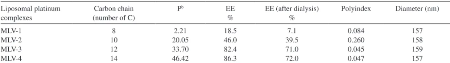
In a second set of experiments, the four different platinum complexes were incorporated in liposomes made from the selected lipid composition. Table 1 shows the characteristics of the resulting liposome preparations with respect to platinum encapsulation eficiency (EE%) and particle size distribution (mean hydrodynamic diameter and polydispersity index).
EE% was found to depend on the carbon chain length. complex 1 with the shortest alkyl chain (C8) showed the lowest value of EE%, whereas the other complex with a longer chain exhibited higher values. Such an increase in EE% as a function of the chain length of the complex most probably arises from the enhanced binding of the complex to the liposome membrane as a result of its higher lipophilicity. Indeed, as shown in Table 1, determination of the lipophilicity of the different complexes through
measurement of the octanol/water partition coeficient (P)25
established a clear correlation between the lipophilicity of the complex and its alkyl chain length.
P appears as a crucial parameter for the design of new
platinum-based anticancer drugs, as it markedly inluences important biological processes such as absorption, transmembrane transport, drug-receptor interactions and
toxicity of molecules.26 In the present work, P is shown
to inluence directly the EE% in multilamellar vesicles. The correlation between the EE% and the lipophilicity of the complex also supports the model that the most
lipophilic complexes are predominantly located at the water/lipid interface of the lipid vesicles. Nevertheless, more specific studied are still needed to confirm this localization.
According to the polydispersity index (PI) of the
liposome suspensions, MLV-1, MLV-3 and MLV-4, with
PI < 0.2, could be considered as monodisperse. The mean
hydrodynamic diameter of the liposome suspensions was in the range of 150-160 nm. Following storage of the formulations at 4 ºC for 30 days, no change in the particle size distribution was observed. This is a strong indication of colloidal stability, even though the period of time is short when compared to the 6 months period of evaluation required by ANVISA, Agência Nacional de Vigilância Sanitária (http://www.anvisa.gov.br), for pharmaceutical formulations. The ability of the formulation to retain the encapsulated platinum complex was assessed by submitting the liposome suspension to dialysis for 48 h at 25 ºC against saline
at physiological concentration (0.15 mol L-1 NaCl). As
shown in Table 1, only limited release of platinum was observed, indicating high retention of the drug from these formulations.
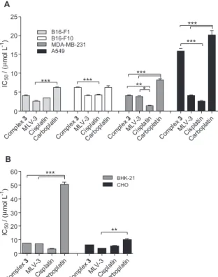
Cytotoxic activity
The cytotoxic activities of the complexes 1, 2, 3 and
4 and MLV-2, MLV-3 and MLV4 were assessed against
different tumor and normal cell lines. Table 2 displays the
results of the IC50 for these different drugs. The liposomal
formulation containing complex 1 (MLV-1) was not
evaluated because of very low EE%. Liposomes prepared without the platinum complex (empty liposomes) did not show signiicant cytotoxicity (data not shown) conirming that the cytoxicicity is only caused by the complex.
Interestingly, the impact of liposome encapsulation on the cytotoxic activity of the complex was found to depend on the carbon chain length.
As illustrated in Figure 2, encapsulation of the
C12-derivative (complex 3) in liposomes (MLV-3) tended
to increase its cytotoxicity in all cell lines. The most
Table 1. Characteristics of the liposome formulationa of the different N-alkyl-propanediamine platinum complexes. Encapsulation eficiency (EE%) before
and after dialysis, mean hydrodynamic diameter, polydispersity index (polyindex)
Liposomal platinum complexes
Carbon chain (number of C)
Pb EE
%
EE (after dialysis) %
Polyindex Diameter (nm)
MLV-1 MLV-2 MLV-3 MLV-4
8 10 12 14
2.21 20.05 33.70 46.42
18.5 46.0 82.4 86.3
7.1 39.5 71.0 72.0
0.084 0.260 0.045 0.047
157 158 159 157
aLiposome formulation: DSPC/CH/Complex/DSPE-PEG2000 at 5:3:1:0.3 molar ratio. bPartition Coeficient (P) of platinum complexes, values from
pronounced effect was observed in the A549 cell line, with a 3- to 4-fold increase. The MLV-3 liposome formulation was more effective than carboplatin in all cell lines and showed results as good as those of cisplatin in melanoma cell lines (B16-F1 and B16-F10). MLV-3 appeared to be less toxic than cisplatin in normal kidney cells (p < 0.001,
t-student test). This suggests a lower nephrotoxicity during
the therapy, one of the most severe side effects caused by cisplatin.
On the other hand, the encapsulation of the
C14-derivative (complex 4) in liposomes (MLV-4) showed
an opposite tendency. The complex showed a higher cytotoxicity in the free form, presumably because of lower bioavailability and tendency to transfer from the liposome to the cell membrane.
Finally, no general tendency was observed in the case
of the C10-derivative (complex 2) in liposomes (MLV-2).
These data suggest that the highest drug bioavailability from liposome formulations is achieved with the complex showing an intermediate carbon chain length and partition between the liposome membrane and aqueous phase.
In previous work, these complexes were investigated, in the non-encapsulated form, to evaluate the inluence of the carbon chain length on their cellular uptake. It was reported that the increase of the carbon chain of the complex facilitated its uptake by cells and enhanced its cytotoxicity against GLC4 (lung cancer cell line) and
K562 (leukemia myelogenic cell line).19 The same trend of
cytotoxicity was observed in the case of non-encapsulated
complexes: complex 2 < complex 3 ≈ complex 4.
However, a different cytotoxicity proile is now reported in the case of liposome-encapsulated complexes:
complex 2 < complex 3 > complex 4, supporting the idea
that liposome encapsulation modulates the drug activity.
Figure 2. Effect of several N-alkyl-propanediamine platinum complexes on the viability of (A) tumor cells (B16-F1, B16-F10, MDA-MB-231 and A549) and (B) normal cells (BHK-21 and CHO); incubated for 120 h in complete RPMI-1640 medium at 37 °C in a 5% CO2. The cytotoxicity was assessed employing tetrazolium bromide reduction method (MTT). Values represent the average of two independent experiments performed in triplicate. The differences among the experimental groups were compared using the variance analysis (ANOVA) followed by Bonferroni, * (p < 0.05), ** (p < 0.01) and *** (p < 0.001).
Table 2. Cytotoxicity of lipophilic platinum complexes and their respective liposomes formulationsa
Complexes IC50 (µmol L-1)b
Tumor cell lines Non-tumor cell lines
B16-F1 B16-F10 MDA-MB-231 A549 BHK-21 CHO
1 20.8 10.9 7.2 19.1 4.2 1.8
2 16.5 15.8 16.5 16.5 12.5 9.5
MLV-2 14.4 6.3 25.1 14.4 22.9 3.6
3 4.1 6.3 4.2 15.8 7.6 6.3
MLV-3 2.7 4.2 3.9 4.2 7.2 3.9
4 5.1 8.3 8.5 4.6 5.5 5.2
MLV-4 14.5 9.8 15.9 19.1 19.0 6.2
Cisplatin 3.5 4.2 1.4 2.7 3.6 5.5
Carboplatin 6.3 6.3 8.3 20.1 50.6 9.8
aLiposome formulation: DSPC/CH/Complex/DSPE-PEG2000 at 5:3:1:0.3 molar ratio. bInhibitory concentration of 50% of cell growth are presented as
Conclusions
The present work brings new insights into the inluence of the carbon chain length/partition coeficient of lipophilic platinum drugs on their bioavailability from a liposome carrier. Importantly, our data indicates that the highest drug bioavailability from the liposome formulation was achieved with the complex having intermediate carbon chain length and partition between the liposome membrane and aqueous phase. Thus, it is suggested that lipophilic drugs with high partition into the liposome membrane may not be advantageous for liposome-mediated delivery to tumor cells. The liposome formulation containing the C12-derivative platinum complex was more effective against tumor cell lines than the free drug and appears as
the most promising formulation for future in vivo antitumor
activity evaluations.
Acknowledgments
To CNPq and FAPEMIG for inancial support and fellowship.
References
1. Pratt, W. B.; Ruddon, R. W.; Ensminger, W. D.; Maybaum, J.;
The Anticancer Drugs, Oxford University Press: New York; 1994, pp. 133-139.
2. Hambley, T. W.; Coord. Chem. Rev.1997, 166, 181. 3. Natile, G.; Coluccia, M.; Coord. Chem. Rev.2001, 216-217,
383.
4. Sykes, A. G.; Platinum Metals Rev.1988, 32, 170.
5. Pasini, A.; Zunino, F.; Angew Chem., Int. Ed. Engl.1987, 26, 615.
6. Farrell, N.; Qu, Y.; Hacker, M. P.; J. Med. Chem.1990, 33, 2179. 7. Siddik, Z. H.; Oncogene2003, 22, 7265.
8. Johnson, S. W.; Ferry, K. V.; Hamilton, T. C.; Drug Res. Updates
1998, 1, 243.
9. Ye, Q. S.; Lou, L. G.; Liu, W. P.; Yu, Y.; Chen, X. Z.; Hou, S. Q.; Gao, W. Q.; Liu, Y.; Bioorg. Med. Chem. Lett.2007,17, 2144.
10. Halámidová, A.; Heringová, P.; Kaspárková, J.; Intini, F. P.; Natile, G.; Nemirovski, A.; Gibson, D.; Brabec, V.; J. Inorg. Biochem.2008, 102, 1077.
11. Peer, D.; Karp, J. M.; Hong, S.; Farokhzad, O. C.; Margalit, R.; Langer, R.; Nature Nanotech.2007, 2, 751.
12. Perez-Soler, R.; Khokhar, A. R.; Cancer Res.1992, 52, 6341. 13. Han, I.; Jun, M. S.; Kim, M. K.; Kim, J. C.; Sohn, Jpn., Y. S.;
J. Cancer Res.2002, 93, 1244.
14. Fahr, A.; Hoogevest, P.; May, S.; Bergstrand, N.; Leigh, M. L. S.; Eur. J. Pharmacol. Sci.200526, 251.
15. Harrington, K. J.; Rowlinson-Busza, G.; Syrigos, K. N.; Uster, P. S.; Vile, R. G.; Stewart, J. S. W.; Clin. Cancer Res.2000, 6,
2528.
16. Verrecchia, T.; Spenlehauer, G.; Bazile, D. V.; Murry-Brelier, A.; Archimbau, Y.; Veillard M.; J. Control. Releas.1995, 36, 49. 17. Carvalho Júnior, A. D.; Vieira, F. P.; De Melo, V. J.; Lopes, M.
T. P.; Silveira, J. N.; Ramaldes, G. A.; Garnier-Suillerot, A.; Pereira-Maia, E. C.; De Oliveira, M. C.; Braz. J. Med. Biol. Res.2007, 40, 1149.
18. Mayer, L. D.; Shabbits, J. A.; Cancer Metastasis Rev. 2001, 20, 87.
19. Silva, H.; Barra, C. V.; da Costa, C. F.; de Almeida, M. V.; César, E. T.; Silveira, J. N.; Garnier-Suillerot, A.; de Paula, F. C. S.; Pereira-Maia, E. C.; Fontes. A. P. S.; J. Inorg. Biochem.2008, 102, 767.
20. Frezard, F.; Schettini, D. A.; Rocha, O. G. F.; Demicheli, C.;
Quim. Nova 2005, 28, 511.
21. Nayar, R.; Hope, M. J.; Cullis, P. R.; Biochim. Biophys. Acta
1989, 986, 200.
22. Silva, H.; Barra, C.; Rocha, F.; de Almeida, M. V.; Cesar, E. T.; Siqueira, L. M. S.; Lopes, M. T. P.; Fontes, A. P. S.; Chem. Biol. Drug Des.2010, 75, 407.
23. Garmann, D.; Warnecke, A.; Kalayda, G. V.; Kratz, F.; Jaehde, U.; J. Control. Release2008, 131, 100.
24. Woodle, M. C.; Lasic, D. D.; Biochim. Biophys. Acta1992,
1113, 171.
25. Guerra, W.; Silva, I. R.; Azevedo, E. A.; Monteiro, A. R. S.; Bucciar- elli-Rodriguez, M.; Chartone-Souza, E.; Silveira, J. N.; Fontes, A. P .S.; Pereira-Maia, E. C.; J. Braz. Chem. Soc.
2006, 17, 1627.
26. Tetko, I. V.; Jaroszewicz, I.; Platts, J. A.; Kuduk-Jaworska, J.;
J. Inorg. Biochem. 2008, 102, 1424.
Submitted: January 30, 2010