Article
0103 - 5053 $6.00+0.00*e-mail: [email protected]
Synthesis, X-ray Crystal Structure and Theoretical Calculations of Antileishmanial
Neolignan Analogues
Josenaide P. do Nascimento,a Lourivaldo S. Santos,a Regina Helena A. Santos,b
Érica Tozzo,b Janaina G. Ferreira,b,c Maria Carolina L. do Carmo,a
Davi S. B. Brasila and Cláudio N. Alves*,a
aInstituto de Ciências Exatas e Naturais, Universidade Federal do Pará, Rua Augusto Corrêa 01, 66075-110 Belém-PA, Brazil
bInstituto de Química de São Carlos, Universidade de São Paulo, Av. do Trabalhador São-carlense 400, CP 780, 13560-970 São Carlos-SP, Brazil
cInstituto de Química de Araraquara, Universidade Estadual Paulista, Rua Francisco Degni s/n, 14800-900 Araraquara-SP, Brazil
A síntese e a estrutura cristalina por difração de raios-X de dois análogos de neolignanas, 2-(4-clorofenil)-1-feniletanona (20) e 2-[tio(4-clorofenil)]-1-(3,4-dimetoxifenil)propan-1-ona
(12) são descritas. O composto 12 apresenta atividade intracelular contra Leishmania donovani e Leishmania amazonensis de amastigotas que causam a leishmaniose tegumentar e visceral. Além
disso, a teoria do funcional de densidade (DFT) com o funcional híbrido B3LYP foi empregado para calcular um conjunto de descritores moleculares para dezenove análogos sintéticos de neolignanas com atividades antileishmaniose. Posteriormente, a análise discriminante stepwise foi realizada
para investigar possíveis relações entre a estrutura molecular e atividades biológicas. Por meio dessa análise os compostos foram classiicados em dois grupos ativos e inativos de acordo com seu grau de atividade biológica, e as propriedades mais importantes foram as cargas de alguns átomos, a ainidade eletrônica e o ClogP.
The synthesis and X-ray crystal diffraction structure of two analogues of neolignans, 2-(4-chlorophenyl)-1-phenylethanone (20) and 2-[(4-chlorophenyl)thio]-1-(3,4-dimethoxyphenyl)
propan-1-one (12) is described. The compound 12 presents activity against intracellular Leishmania donovani and Leishmania amazonensis amastigotes that cause cutaneous and visceral leishmaniasis.
In addition, the density functional theory (DFT) with the B3LYP hybrid functional was employed to calculate a set of molecular descriptors for nineteen synthetic analogues of neolignans with antileishmanial activities. Afterwards, the stepwise discriminant analysis was performed to investigate possible relationship between the molecular descriptors and biological activities. Through this analysis the compounds were classiied into two groups active and inactive according to their degree of biological activities, and the more important properties were charges on some key atoms, electronic afinity and ClogP.
Keywords: neolignans, antileishmanial, B3LYP, X-ray crystal structure
Introduction
The leishmaniases are parasitic diseases caused by protozoa of the genus Leishmania and remain a severe
public health problem, particularly in many tropical and subtropical regions. The infection is transmitted by bite from female sandlies, which are of the genus Phlebotomus
or Lutzomyin. This infection has several diverse clinical
manifestations: cutaneous, mucosal and most notably visceral leishmaniasis, and is caused by Leishmania amazonensis, Leishmania braziliensis, and Leishmania donovani species.1-4 An estimated 1.5 to 2 million new
vaccine are considered to be two stumbling blocks in the combat of this disease.10
Treatment approaches and responses to chemotherapy vary by regions. Antimony remains the therapeutic cornerstone in all regions; however, antimony is expensive, toxic and requires administration of high doses resulting in serious side effects.11-13 Therefore, there remains an urgent need for development of less toxic drugs that are effective against all forms of leishmaniases. The search of new drugs against leishmaniasis, potentially compounds and their derivatives have been described in the literature.14-16
Neolignans are groups of compounds that show a wide range of biological effects including antifungal,17-19 anti-schistosomal,20-23 antiplasmodial,24 trypanocidal,25-27 antibacterial,28 anti-PAF,29,30 antipsychotic,31 antioxidant,32,33 activities, and biological activity against Escherichia coli,34,35
Paracoccidioides brasiliensis.36 Usually, neolignans are
organic dimmers derived from oxidative coupling of allyl and propenyl phenols.16,22 Previous studies have evaluated the antileishmanials activities of twenty-two sulfur and oxygen synthetic analogues of neolignans against parasite species that cause cutaneous and visceral Leishmaniasis.14,15 These compounds were synthesized and their activities against both intracellular amastigotes of L. donovani and L. amazonensis were compared.
In the work reported here, two neolignan analogues were synthesized, the 2-[(4-chlorophenyl)thio]-1-(3,4-dimethoxyphenyl)propan-1-one (12) and the
2-(4-chlorophenyl)-1-phenylethanone (20), and their
molecular structures were obtained by X-ray diffraction (Figure 1 and 2). The synthesis of 2-[(4-chlorophenyl) thio]-1-(3,4-dimethoxyphenyl)propan-1-one (12) was
described in previous studies14,15 and their biological activity was evaluated by Aveniente et al.14 However, the X-ray experimental results for this molecule together with synthesis and crystallographic of 2-(4-chlorophenyl)-1-phenylethanone (20) are described for the irst time here.
In addition, the DFT method was employed in order to calculate forty-one physicochemical descriptors, including electronic, steric and hydrophobic of nineteen synthetic analogues of neolignans reported by Aveniente et al. 14
(see Figure 3 and Table 1). Recently, we successfully used molecular descriptors in structure-activity relationship (SAR) studies of neolignan compounds with anti-schitosomal activity,20,21 antifungal activity,37 and synthetic neolignans with biological activity against Escherichia coli34 and Paracoccidiodes brasiliensis.36These descriptors
are successful and well accepted.38-45
Our purpose is to investigate, in a qualitative way, the structure activity relationship of neolignan compounds using quantum chemical descriptors. Stepwise discriminant
analysis (SDA) was employed to analyze the data set, in order to build models for predicting the relationship between these descriptors and the antileishmanial activities of the compounds.
Experimental
Instruments
1H NMR (100 MHz) spectrum was recorded on a Varian
XL-100 spectrometer. Chemical shifts were reported in ppm from tetramethylsilane on the d scale and coupling constants J are expressed in Hz. IR spectra was recorded in
KBr ilm and measured with a Bomen model MB series II spectrophotometer. Electrothermal melting point apparatus are uncorrected.
General
Within the great structural variety of neolignans the β-ketoether, β-ketosulfide, β-ketosulfoxide and Figure 1. Structural skeleton numbering (a) and ORTEP view of molecule 12 (b), with the atom labelling scheme and thermal ellipsoids vibration with 50% of probability.
β-ketosulfone derivatives are closely related to natural 8,4’-oxyneolignans, which are of interest because of their moderate antileishmaniasis activity against both intracellular amastigotes of L. donovani and L. amazonensis.14 Insight into the biological and physicochemical functions of complex neolignans at the molecular level requires a precise understanding of their three-dimensional structures. Figure 3 shows the basic skeletons of the twenty compounds studied and Table 1 lists their classes and biological response. Two of these neolignan derivatives, β-ketosulide
12 and β-ketoether 20 were synthesized from condensation
reactions among α-bromoketone and thiophenol or phenol
derivatives, respectively, with their structures elucidated by X-ray crystal diffraction. The synthesis of 12 was
mentioned previously,15 but the crystal data and structural features were not published. Here, we report the synthesis and the X-ray crystallographic studies of 12 and 20 with a
comparison of their three-dimensional structures.
Synthesis
In the present study, 2-[(4-chlorophenyl)thio]-1-(3,4-dimethoxyphenyl)propan-1-one (12) was prepared by
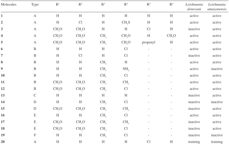
reaction of α-bromo-3,4-dimethoxypropiophenone and Table 1. Biological response for the neolignans studied. See Figure 1 for the positions of substituents ‘R’ listed in the irst line of the table
Molecules Type R1 R2 R3 R4 R5 R6 Leishmania
donovani
Leishmania amazonensis
1 A H H H H H H active active
2 A H Cl H CH3S H H active active
3 A CH3O CH3O H H Cl H inactive active
4 A CH3O CH3O CH3 CH3O H CH3O active active
5 A CH3O CH3O CH3 CH3O propenyl H active active
6 B H H H Cl - - active active
7 B H Cl H Cl - - inactive active
8 B H H CH3 H - - active active
9 B H H CH3 NH2 - - active inactive
10 B H H CH3 Cl - - active active
11 B CH3O CH3O CH3 CH3 - - active active
12 B CH3O CH3O CH3 Cl - - active active
13 C H H H H - - inactive active
14 D H H CH3 Cl - - inactive inactive
15 D CH3O CH3O CH3 CH3 - - inactive active
16 E H H CH3 Cl - - active active
17 E CH3O CH3O CH3 CH3 - - inactive active
18 E CH3O CH3O CH3 Cl - - inactive active
19 F H H CH3 Cl - - inactive inactive
20 A H H H H Cl H training training
Figure 3. Strucutal skeleton and numbering of twenty neolignan derivatives.
O O
R1
R2 R
3
R4
R6 R5
S O
R1
R2 R
3
R4
S O
R1
R2 R
3
R4
O
S O
R1
R2
R3
R4
O O
S R1
R2 R
3
R4
OH
S
R3
Cl OAc
A B C
D E F
1
2 3 4 5
6
7 8 4' 5' 6'
3'
4-chlorothiophenol in anhydrous ethyl methyl ketone, in accordance with the method described in the literature.14,15 The β-ketoether 2-(4-chlorophenyl)-1-phenylethanone (20) was prepared by reacting equimolecular amounts of α-bromoacetophenone and 4-chlorophenol, in basic solution. The synthetic route of compounds is outlined in Figure 4. The structures of compounds were conirmed through analytical and spectral data (1H NMR, IR, and MS).
2-[(4-Chlorophenyl)thio]-1-(3,4-dimethoxyphenyl)propan-1-one (12)
The 2-[(4-chlorophenyl)thio]-1-(3,4-dimethoxyphenyl) propan-1-one (12) was obtained from a solution of 1.36 g (9.43 mmol) of 4-chlorothiophenol and 2.27 g (16.48 mmol) of anhydrous K2CO3 in 45 mL of anhydrous ethyl methyl ketone was stirred for 10 min at room temperature. After this period, a solution of 2.50 g (9.16 mmol) of the α-bromo-3,4-dimethoxypropiophenone in 20 mL of anhydrous ethyl methyl ketone was added dropwise and the mixture was stirred and reluxed for 8 h. The solution was cooled at room temperature, iltered, and the residue washed with CH2Cl2. The solution was concentrated in vacuum (to eliminate the ethyl methyl ketone), diluted with H2O, and extracted thoroughly with CH2Cl2 (3×). Organic extracts were combined, washed with water, 5% NaHCO3 solution, NaCl saturated solution, dried over Na2SO4 and then iltered and concentrated in vacuum. The residue was puriied by crystallization with MeOH giving 12 (2.52 g) as colorless crystals, mp 92-93 °C in 81.7% yield. The spectrometric data is in concordance with the literature.14, 15
2-(4-Chlorophenyl)-1-phenylethanone(20)
The 2-(4-chlorophenyl)-1-phenylethanone (20)
was obtained from a solution of 0.66 g (5.12 mmol) of 4-chlorophenol and 1.25 g (9.04 mmol) of anhydrous K2CO3 in 20 mL of anhydrous ethyl methyl ketone was stirred for 10 min at room temperature. After this period, a solution of 1.0 g (5.02 mmol) of the α-bromo-acetophenone in 7.5 mL of anhydrous ethyl methyl ketone was added dropwise and the
mixture was stirred and reluxed for 1.5 h. The solution was cooled at room temperature, iltered, and the residue washed with CH2Cl2. The solution was concentrated in vacuum (to eliminate the ethyl methyl ketone), diluted with H2O, and extracted thoroughly with CH2Cl2 (3×). Organic extracts were combined, washed with water, 5% NaHCO3 solution, NaCl saturated solution, dried over Na2SO4 and then iltered and concentrated in vacuum. The residue was puriied by crystallization with MeOH giving 20 (1.18 g) as colorless crystals, mp 99-100 °C in 95% yield. IR νmax/cm-1 2910, 1700 (C=O), 1600, 1580, 1490, 1440, 1230 (KBr); 1H NMR (100 MHz , CDCl3) d 5.22 (2H, s, 2 H-8), 6.90 (2H, d, J 7.0 Hz,
H-3´/H-5´), 7.20 (2H, d, J 7.0 Hz, H-2´/H-6´), 7.35-7.60 (3H,
m, H-3, H-4 and H-5), 7.95 (2H, dd, J 6.8 Hz and J 1.5 Hz,
H-2/H-6); EIMS (probe) 70 eV, m/z (rel. int.) 246 [M]+ (40),
139 [C7H5O]+ (100).
Crystallographic data
Suitable colorless single crystals of the neolignan derivatives 12 and 20, with approximate dimensions 0.05×0.05×0.10 and 0.05×0.10×0.10 mm, respectively, were selected and the diffraction data were collected using an Enraf-Nonius CAD4 diffractometer with graphite monochromated KαMo radiation (λ = 0.71073 Å), in the ω-2θ scan mode, at room temperature. The unit cell parameters were determined using 25 automatically centered relections. Intensities of the relections were corrected by absorption factors [µ (MoKα) = 0.308 and 0.367, for compounds 12 and 20, respectively] using the PSISCAN method.46 Information concerning to the crystallographic data collection and reinement of the structures are given in Table 2. The structures were solved by SIR-9247 and reined by full matrix least squares and difference Fourier synthesis by SHELXL-97,48 using the WinGX software package.49 All non-hydrogen atoms were reined anisotropically. The hydrogen atoms were located in their ideal positions and not reined. The structural analysis was performed by PLATON system.50 The graphic representations of the molecules were made using ORTEP3 for Windows.51 The crystal data are deposited at Cambridge Crystallographic Data Centre, CCDC 703864 and703867, for compound 12 and 20, respectively.
Computational methods
The molecular geometries obtained from X-ray diffraction data of 12 and 20 were used here as a starting point for our calculations of the remaining molecules, just changing the substituent. All compounds were optimized using the B3LYP hybrid functional,52,53 together with the
6-311++G(d,p) basis sets in the Gaussian 03 molecular
package.54 Vibrational analysis was carried out for the complete equilibrium geometry obtained by the procedure in the Gaussian 03 package at the DFT level with the B3LYP/6-311++G(d,p) level in the gas phase, ensuring
that each gradient optimization located was indeed a true minimum energy structure (no imaginary frequencies). In addition, the conformational analysis were carried out to conirm the minimum energy structure to molecules 12 and 20, by carrying out a series of partial optimizations constraining the concerned dihedral angle step by step within the appropriate range, with a step size of 5°, these calculations were carried out using the B3LYP/6-31G* basis set, the dihedral angles analyzed were C1–C7–C8–O and C4’–O–C8–C7 for molecule 12 and C1–C7–C8–S and C4’–S–C8–C7 for molecule 20. Previous studies about conformational preferences in solid state (crystal) and in solution has been published55-57 for a large number of compounds analogues of the compounds 12 and 20, studied in the present paper. The geometrical structures of the radicals studied were optimized independently from the neutral molecules prior to the calculations of energies, treated as open shell systems DFT/UB3LYP. Thus, the more relevance electronic descriptors to antileishmaniasis activities were calculated such as: eletrostatic potential atomic charges (QN - net atomic charge on atom N), occupied and unoccupied molecular orbital energies (εHOMO and εLUMO), electronegativity (χ), hardness (η), softness
(1/η), chemist potential (µ), electrophilic index (ω) and electronic afinity (EA) were calculated with the DFT/ B3LYP level, together with the 6-311++G(d,p) basis sets.
The χ was calculated as the sum mean between the energies of HOMO and LUMO (χ = –(εHOMO + εLUMO)/2).58 The η is simply the energy difference between LUMO and HOMO energies, while the 1/η is the inverse of the hardness.59 The µ is simply Koopmans’ approximation.60 From equation ω = µ2/2η we obtained the electrophilic index.58 The IP was calculated as the energy differences between a radical cation (Ec) and the respective neutral molecule (En); IP = Ec – En.61 The volume (Vol), molecular refractivity (MR), polarizability (Pol), partition coeficient (ClogP) and hydration energy (HE) were obtained by using the Hyperchem 7.5 molecular package.62 The transport of a compound through membranes can be modeled by molecular hydrophobicity, which can be described by octanol/water partition coeficients (ClogP). The value of this property was obtained by using the Chem-3D molecular package.63 Table 3 shows almost molecular descriptors.
The atomic charges were obtained by employing the electrostatic potential method, which was used because the charges derived from the electrostatic potential method are physically more satisfactory than Mulliken’s charges, especially when related to biological activity. The choice of the best descriptors to correlate with the biological activities was performed using stepwise discriminant analysis (SDA) built in the Minitab 14 statistical software.64
Table 2. Crystallographic data for compounds 12 and 20
Formula C17H17 O3ClS (12) C14H11ClO2 (20)
Formula weight 336.83 246.68
Crystal system monoclinic monoclinic
Space group P21 P21/c
a (Å) 8.765(1) 5.840(1)
b (Å) 7.975(1) 7.374(2)
c (Å) 11.8155(1) 27.492(8)
β (°) 93.663(7) 90.26(2)
Z 2 4
V (Å3) 824.3(2) 1184.5(5)
Crystal size (mm) 0.05 × 0.05 × 0.10 0.05 × 0.10 × 0.10
Crystal color colorless colorless
µ (MoKα) (mm−1) 0.367 0.308
Relections: total; unique; Rint 1880; 1791; 0.035 2654; 2411; 0.027
Observed Relections (I > 2σ(I)) 1598 1422
R; wR2 0.0322; 0.0920 0.0552; 0.1796
GOF; Npar 1.05; 202 1.03; 154
Min. and max. resd. dens. [e/Å3] −0.20, 0.17 −0.22, 0.39
The molecular electrostatic potential (MEP) surface was generated using the geometry optimized in B3LYP/6-311++G(d,p) and an isodensity surface of 0.002 a.u. On
the MEP surface, regions indicating the excess of negative potential correspond to excess negative charges, i.e.,
attraction of the positively charged probe. The MEP surface was calculated and analyzed visually using the PC Spartan PRO molecular package.65
Results and Discussion
Synthesis
The molecules 12 and 20 (Figure 4) were synthesized
through procedures described in the literature14,15 using the condensation reaction of α-bromoketones with a phenol or thiophenol derivatives, in basic medium. Compound 12 was
synthesized from the condensation reaction of α -bromo-3,4-dimethoxypropiophenone and 4-chlorothiophenol in anhydrous ethyl methyl ketone, in 81.7% yield. The spectroscopic data of compound 12 is in concordance with
the literature.14,15 Compound 20 was synthesized in the same conditions, from reaction of α-bromoacetophenone and 4-chlorophenol, in 95% yield. Spectroscopic data and physical properties of compound 20 are described for the
irst time in this paper. The compounds were puriied by PTLC and purity done by GC-MS. Compound 20 was
isolated as a colorless crystals and molecular formula was determined by elemental analysis and MS M+ C
14H11O2Cl [246 (40%)]. Its 1H NMR spectrum showed typical signals of an aromatic compound. The singlet signal at d 5.20 ppm was assigned to the two H-8 protons. The two doublets at d
6.90 and 7.20 ppm with orto coupling (J7.0 Hz) belong to
the pairs H-3´/H-5´ and H-2´/H-6´ protons of the aromatic ring-B, respectively. Multiplet signals at d 7.35-7.60 ppm are due to H-3, H-4 and H-5 protons of the aromatic ring-A and the signal at d 7.95 ppm (dd, J 6.8 Hz and J 1.5 Hz)
was assigned to the H-2 and H-6 protons.
X-ray crystallography
The ORTEP representations of the molecular structure of 2-[(4-chlorophenyl)thio]-1-(3,4-dimethoxyphenyl)propan-1-one (12) and 2-(4-chlorophenyl)-1-phenylethanone (20)
with the atom labelling scheme are depicted in Figures 1 and 2, respectively, using thermal vibration factors with 50% of probability.
As shown in the crystal representation (see Figures 1 and 2), both compounds have a Cl atom in position 4′ (ring B), whereas compound 12 has also an –OCH3 group in positions Table 3. The eight most important descriptors that classiied the twenty neolignans used in the SDA study
Derivatives EA (eV) ClogP Q1 Q2 Q6 (O or S) Q1’ Q5’
1 0.38 3.02 −0.02 −0.16 −0.01 −0.54 0.10 −0.27
2 0.78 4.00 −0.02 −0.18 −0.05 −0.44 0.12 0.07
3 0.35 3.77 −0.14 −0.17 −0.08 −0.52 −0.16 −0.31
4 0.26 2.61 −0.12 −0.14 −0.09 −0.39 0.11 0.40
5 0.25 4.22 −0.18 −0.13 −0.14 −0.50 −0.27 0.34
6 0.62 4.43 −0.04 −0.12 −0.02 −0.28 −0.16 −0.18
7 0.86 5.22 −0.06 −0.15 −0.02 −0.28 −0.16 −0.21
8 0.48 3.97 −0.04 −0.10 −0.03 −0.31 0.09 −0.04
9 0.45 2.74 −0.05 −0.19 0.01 −0.34 −0.81 −0.09
10 0.61 4.74 −0.08 −0.07 0.01 −0.31 −0.15 −0.07
11 0.28 4.36 −0.15 −0.11 −0.08 −0.31 −0.17 −0.03
12 0.45 4.63 −0.17 −0.09 −0.07 −0.31 −0.14 −0.07
13 0.87 3.10 −0.08 −0.16 −0.04 −0.02 −0.12 −0.07
14 1.01 3.05 −0.04 −0.10 −0.03 0.99 −0.12 −0.04
15 0.62 2.61 −0.12 −0.16 −0.16 0.97 −0.23 0.00
16 −0.01 4.37 0.08 −0.20 −0.05 −0.41 −0.14 −0.16
17 −0.19 3.76 0.04 −0.18 −0.23 −0.37 −0.22 −0.10
18 0.01 4.03 −0.10 −0.20 −0.08 −0.44 −0.14 −0.16
19 −0.09 5.23 0.05 0.07 −0.21 −0.25 −0.14 −0.22
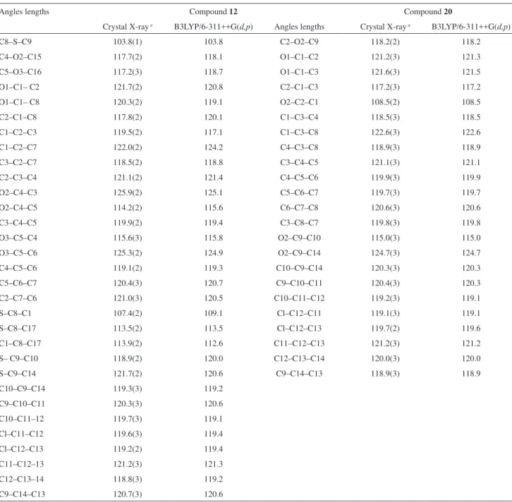
3 and 4 (ring A). The bond distances for the single bonds C–O are 1.418(4) and 1.369(4) Å for compound 20, and the C–S, as expected, are 1.838(3) and 1.776(3) Å for 12. No signiicant differences were found in the bond distances and angles of the two molecules, the others bond lengths C=O, C–Cl and C–C are in the expected ranges. The distances and angles parameters are listed in Table 4 and 5, respectively.
The most relevant structural difference is that the structure of 20 is almost planar, with the dihedral angle between the two benzene planes of 2.8(1)°, while the structure of 12 is itself twisted out of the plane of the aromatic rings, as can be seen from the C1–C2–S–C9 torsion angle of 123.3(5)°. The dihedral angle between the rings A and B is 34.4(1)°, which presumably relects some lexibility of the molecule, enabling it to rotate and deform in order to minimize any unfavorable intramolecular interactions due to the presence of the methyl group. Also, in compound 12 the keto atom is more coplanar in relation to the ring A. The keto atom O1 is 0.254(3) Å out of the least-squares plane of the ring
A. In compound 20 the same keto atom is 0.464(2) Å out of the plane A. The three-dimensional structure of the compound 12 and 20 and the non-covalent interactions formed in their crystal structure along the b-axis are shown in Figure 5 and 6.
In the crystal packing of 12, the oxygen from the methoxy group participates in two non conventional C–H…O hydrogen bonds. The molecules of 12 are joined
via intermolecular hydrogen bonds (C10…O3 3.491(4) Å,
H10–O3 2.57 Å, 170.0° and C16…O2 3.323(5) Å, H16C–O2 2.60 Å, 132.0°) giving rise to the formation of the zig-zag supramolecular chain, as shown in the Figure 5. The crystal packing of 20 also shows the existence of an intermolecular C-H…O hydrogen bond (C2…O1 3.424(5) Å, H…O1 2.56 Å, C2-H2B…O1 148°). As can be observed in Figure 6, this hydrogen bond are responsible for the self-assembly into one-dimensional chain, formed by the interaction at the end of the neighboring molecules while in the 12 it was formed in the middle of adjacent molecules.
Table 4. Crystal X-ray and B3LYP/6-311++G(d,p) selected bond lengths (Å) for compounds 12 and 20
Bond distances Compound 12 Compound 20
Crystal X-ray a B3LYP/6-311++G(d,p) Bond distances Crystal X-ray a B3LYP/6-311++G(d,p)
S–C8 1.838(3) 1.875 O2–C2 1.418(4) 1.418
S–C9 1.776(3) 1.795 O2–C9 1.369(4) 1.369
O1–C1 1.217(3) 1.221 O1–C1 1.212(4) 1.212
O2–C4 1.361(3) 1.358 C1–C2 1.506(4) 1.506
O3–C5 1.352(3) 1.352 C1–C3 1.481(4) 1.481
O2–C15 1.410(4) 1.423 C3–C4 1.393(4) 1.393
O3–C16 1.435(4) 1.423 C3–C8 1.394(4) 1.394
C1–C2 1.484(3) 1.492 C4–C5 1.377(5) 1.376
C1–C8 1.518(4) 1.530 C5–C6 1.382(5) 1.383
C2–C3 1.402(4) 1.410 C6–C7 1.387(5) 1.387
C2–C7 1.386(4) 1.393 C7–C8 1.384(5) 1.384
C3–C4 1.366(4) 1.383 C9–C10 1.383(4) 1.382
C4–C5 1.419(4) 1.421 C9–C14 1.395(4) 1.395
C5–C6 1.378(4) 1.393 C10–C11 1.376(5) 1.376
C6–C7 1.386(4) 1.396 C11–C12 1.379(4) 1.380
C8–C17 1.511(4) 1.523 C12–C13 1.374(5) 1.374
C9–C10 1.390(4) 1.399 C13–C14 1.385(5) 1.385
C9–C14 1.388(4) 1.399 Cl–C12 1.742(3) 1.743
C10–C11 1.375(5) 1.393
C11–C12 1.374(5) 1.391
C12–C13 1.384(5) 1.391
C13–C14 1.379(5) 1.392
Cl–C12 1.739(3) 1.758
Theoretical calculations
The distance lengths and angles for both X-ray and B3LYP/6-311G++(d,p) optimized structures of 12 and 20 presented in Tables 4 and 5 are normal for this type of compound. The values of C–S and C–O bond distances calculated for compounds 12 and 20 are 1.875and 1.795 Å, and 1.418 and 1.369 Å, respectively. The C1–C2–S–C9 torsion angle experimentally observed are 123.3(5)º and 34.4(1)º, while the values calculated were of 123.3o and 35.7o for compounds 12 and 20, respectively. These
results are in agreement with the experimental values. No signiicant differences were found between these structures (12 and 20) and theremaining molecules. The harmonic frequency showed that the molecular geometrics obtained by DFT calculations correspond to a local minimum energy structure, no imaginary frequencies. In addition, Table S1 (supplementary material) presents the results to conformational analysis carried out to molecules 12 and 20. These results indicate that the structures optimized with the 6-311++G(d,p) basis sets using X-ray diffraction data
as starting points have the lowest energy, indicating global Table 5. Crystal X-ray and B3LYP/6-311++G(d,p) selected angles lengths (°) for compounds 12 and 20
Angles lengths Compound 12 Compound 20
Crystal X-ray a B3LYP/6-311++G(d,p) Angles lengths Crystal X-ray a B3LYP/6-311++G(d,p)
C8–S–C9 103.8(1) 103.8 C2–O2–C9 118.2(2) 118.2
C4–O2–C15 117.7(2) 118.1 O1–C1–C2 121.2(3) 121.3
C5–O3–C16 117.2(3) 118.7 O1–C1–C3 121.6(3) 121.5
O1–C1– C2 121.7(2) 120.8 C2–C1–C3 117.2(3) 117.2
O1–C1– C8 120.3(2) 119.1 O2–C2–C1 108.5(2) 108.5
C2–C1–C8 117.8(2) 120.1 C1–C3–C4 118.5(3) 118.5
C1–C2–C3 119.5(2) 117.1 C1–C3–C8 122.6(3) 122.6
C1–C2–C7 122.0(2) 124.2 C4–C3–C8 118.9(3) 118.9
C3–C2–C7 118.5(2) 118.8 C3–C4–C5 121.1(3) 121.1
C2–C3–C4 121.1(2) 121.4 C4–C5–C6 119.9(3) 119.9
O2–C4–C3 125.9(2) 125.1 C5–C6–C7 119.7(3) 119.7
O2–C4–C5 114.2(2) 115.6 C6–C7–C8 120.6(3) 120.6
C3–C4–C5 119.9(2) 119.4 C3–C8–C7 119.8(3) 119.8
O3–C5–C4 115.6(3) 115.8 O2–C9–C10 115.0(3) 115.0
O3–C5–C6 125.3(2) 124.9 O2–C9–C14 124.7(3) 124.7
C4–C5–C6 119.1(2) 119.3 C10–C9–C14 120.3(3) 120.3
C5–C6–C7 120.4(3) 120.7 C9–C10–C11 120.4(3) 120.3
C2–C7–C6 121.0(3) 120.5 C10–C11–C12 119.2(3) 119.1
S–C8–C1 107.4(2) 109.1 Cl–C12–C11 119.1(3) 119.1
S–C8–C17 113.5(2) 113.5 Cl–C12–C13 119.7(2) 119.6
C1–C8–C17 113.9(2) 112.6 C11–C12–C13 121.2(3) 121.2
S– C9–C10 118.9(2) 120.0 C12–C13–C14 120.0(3) 120.0
S–C9–C14 121.7(2) 120.6 C9–C14–C13 118.9(3) 118.9
C10–C9–C14 119.3(3) 119.2
C9–C10–C11 120.3(3) 120.6
C10–C11–12 119.7(3) 119.1
Cl–C11–C12 119.6(3) 119.4
Cl–C12–C13 119.2(2) 119.4
C11–C12–13 121.2(3) 121.3
C12–C13–14 118.8(3) 119.2
C9–C14–C13 120.7(3) 120.6
minimum energy structures. So the geometries obtained by B3LYP/6-311G++(d,p) level were used to calculate
forty-one physicochemical descriptors.
Before applying the SAR analysis to the nineteen compounds under study, each calculated property (variables or descriptors) was auto scaled. In the auto scaling method, each variable is scaled to a mean of zero and a variance of unity. This method is very important because each variable is weighted equally and this provides a measure of the ability of a descriptor to discriminate classes of compounds. With this method, we can compare all variables at the same level although presenting different units.
SAR analysis has been carried out using molecular descriptors that were selected by stepwise discriminant analysis (SDA). The main objective of SDA is to determine discriminant functions using the measured variables that
separate the groups as distinctly as possible. In this study, we considered two groups: active and inactive molecules against L. donovani and L. amazonensis activities. The SDA
linear function is based on the Fisher’s test (Ftest) for the signiicance of the variables. In each step one variable is selected based on its signiicance and after several steps, the more signiicant variables are extracted from the whole data set under investigation.
From the twenty-two synthetic analogues of neolignans reported by Aveniente et al.14 three of them were not included in the analysis, because they not have experimental values (values reported as “nd” not determined). Therefore, the equations presented in this study were constructed with nineteen molecules – compounds 1-19 – that present antileishmaniasis activities (values expressed as percent inhibition of parasite growth at 80 µg mL–1 concentration). The SDA results will be used for molecule 20 to verify if this new molecule would be active or inactive. The allocation rule derived from the SDA results, when the antileishmanials activity of a new neolignan compound is investigated, is: (i) initially one
calculates, for the new neolignan compound, the values for the more important descriptors obtained with the SDA; (ii) substitute these values in the two discriminant
functions obtained in this study; (iii) check out which
discriminant function (active or inactive compounds) presents the higher value. The new neolignan compound is active if the higher value is related to the active discriminant function and vice versa.
SDA for the Leishmania donovani activity
In this analysis, we considered two groups: active molecules (1, 2, 4-6, 8-12 and 16) and inactive molecules (3, 7, 13-15, 17-19) against L. donovani. The SDA indicated
that the descriptors: electronic afinity (AE), charges on C6, O or S and C5’ atoms were the most important in order to get the separation of active and inactive compounds. The discriminant functions for L. donovani activity obtained
with nineteen compounds are given in equation 1 and 2:
Inactive compounds = – 1.01 + 1.17 (EA) – 2.15 Q6 + 0.60
(O or S) – 1.46 Q5’ (1)
Active compounds = – 0.54 – 0.85 (EA) + 1.56 Q6 – 0.44
(O or S) + 1.06 Q5’ (2)
Through the discriminant functions above (equation 1 and 2) and the values of each variable for the compounds studied (Table 3); we obtain the classiication matrix by using all compounds in the analysis (Table 6).
Figure 5. View along the crystallographic b-axis of supramolecular
arrangement supported by the intermolecular hydrogen bonds between the adjacent molecules in compound 12, indicated as dashed lines.
The SDA allowed correct classiication scores of 100% (active compounds), 87.5% (inactive compounds) and 94.7% (total), resulting in a better performance in the separation of the two groups (Table 6). Derivative 18 was incorrectly classiied in the group of active compounds. Probably this occurred because this compound showed a sulfur bond in position 8 that increases activity if compared to compounds bearing oxygen bond, like previous results shown by Aveniente et al.14 Furthermore, the molecule 18 shows a meta chlorine substituents in the
B ring, a methyl substituents in position 7, and a meta
methoxyl substituents in the A ring, similar to molecule 13 which is active.
In accordance with equation 1 and Table 3, in general, active neolignans against L. donovani have more positive
charges on atoms 6 and 5’ and less positive charges on heteroatom. The charge is electronic descriptor; therefore, we can conclude that electronic effects have a very important role when one is trying to understand the activity of neolignan derivatives. The charges on the atoms S or O in compounds 14 and 15 have more positive values, because the inductive effect on the B ring, which have a Cl substituent in the molecule 14 and a methyl substituent in the molecule 15, in general, molecules with more positive values for S or O charge are inactives. In Figure 7 is shown the box plot for Q6 (see supplementary material Figure S2 for the Q5’ charges).
In order to verify if a new molecule would be active or inactive against L. donovani, we had to apply the results
obtained with the discriminant functions for the compound 20. From the results obtained, we can see that this molecule was classiied as active, and from it we can conclude that the model obtained with SDA can be applied to new neolignan compounds whose biological activity is unknown.
Recently, we successfully used three-dimensional MEP surfaces to deine the most probable sites of protonation of dipyridamole66, aparisthman,67 cordatin, 8-epicordatin;68 on the study of the molecular mechanisms the Diels-Alder reaction69 and to get some clues about the transition state of the catalyzed reaction.40 The MEP surfaces of compound 1 (active) and compound 14 (inactive) in terms of total electron density show that the lowest electronic potential is in the proximity of oxygen atoms of the carbonyl (O1), and oxygen heteroatom (Figure 8). The large negative potential of oxygen atoms may be regarded to a nucleophilic suction pump, acting as a possible magnet for electrophilic attack of a biological receptor. The surfaces of 1 and 14 are different and compound 1 (active) provides a much more intense region of negative electrostatic potential than compound 14 (inactive).
SDA for the Leishmania amazonensis activity
For this activity the molecules were also classiied in two groups: active molecules (1-8, 10-13, 15-18) and inactive molecules (9, 14 and 19). The most signiicant descriptors selected by SDA were obtained with nineteen compoundsand is given in equation 3 and equation 4:
Inactive compounds = – 6.21 – 3.40 ClogP + 2.87 Q1 +
5.25 Q2 – 4.20 Q1’ (3)
Active compounds = – 0.22 + 0.64 ClogP – 0.54 Q1 – 0.98
Q2 + 0.79 Q1’ (4)
Figure 8. MEP surfaces of (a) compound 1 (active) and (b) compound
17 (inactive). The increase of negative charges goes from positive (dark gray) to negative.
Figure 7. Box plot of L. donovani activity for Q6 considering nineteen neolignan compounds.
Table 6. Classiication matrix obtained using the SDA method for L. donovani activity
Classiied group True group
% Inactivity Activity
Inactivity 87.5 7 1
Activity 100 0 11
Total 94.7 7 12
In such case, the four descriptors (ClogP and charges on 1, 2 and 1’substituent in the carbon atoms) represent the strength of a molecular association by electronic interaction. By using the quantities given in the discriminant functions above, we can obtain the classiication summary showed in Table 7. The classiication error rate was zero, resulting in a satisfactory separation of the two groups.
In accordance with equations 3 and 4 and Table 3, we can observe that, in general, molecules with more negative charge on C1 and C2 atoms are actives, while more positive charge on C1’ atom are inactives. In addition, Figure 9 shows that, in general, molecules with more positive values for Q1’ charge are actives, and the molecules with more negative values for Q1’ charge are inactives. The ClogP is a measure of hydrophobicity; molecules with large value of ClogP have higher hydrophobicity and consequently better transport through cell membranes. In general, we can observe that molecules with high ClogP values are active, while the molecules with low values of ClogP are inactive (see supplementary material Figure S3). In other words, the hydrophobic character of molecules improves to L. amazonensis activity, what can indicate that the
active compounds must interact with a target system such as an enzyme or receptor where the binding site is usually hydrophobic.
In order to verify if a new molecule would be active or inactive against L. amazonensis, we need to apply
the results obtained with discriminant functions for the
molecule 20. From the results obtained in this work we can conclude that the models attained here with SDA can be applied to new neolignans compounds whose biological activity is unknown.
In Figure 10 we can observe that compound 2 (active) provides a much more intense region of negative electrostatic potential than compound 9 (inactive), thus compound 2 has a more attractive cation-binding site. In general, we observed that active compounds have more intense region of negative electrostatic potential than inactive ones.
Conclusions
We have carried out a synthesis and X-ray crystallographic investigation of the molecular structures of compounds 12 and 20. No significant differences
were found in the bond distances and angles of the two molecules. The most relevant structural difference is that the structure of 20 is almost planar, while the structure of 12 is itself twisted out of the plane of the aromatic rings.The
results show that the agreement between theory and X-ray diffraction data is excellent. In fact the structure geometrics used in theoretical studies are more stable conformers; suggest that calculated structures for molecules 12 and 20
are the global minimums. In addition, the SDA method is quite eficient to classify the nineteen neolignans studied here in two groups, actives and inactives, according to their antileishmanials activities, and only some descriptors as atomic charges in positions 1 (Q1), 2 (Q2), 6 (Q6), O or S, 1’ (Q1’) and 5’ (Q5’) atoms, the electronic afinity (EA) and ClogP are responsible for the separation between the active and inactive compounds. Four different sets of descriptors Table 7. Classiication matrix obtained using the SDA method for
L. amazonensis activity
Classiied group True group
% Inactivity Activity
Inactivity 100 3 0
Activity 100 0 16
Total 100 3 16
Figure 9. Box plot of L. amazonensis activity for Q1’ considering nineteen neolignan compounds.
Figure 10. MEP surfaces of (a) compound 2 (active) and (b) compound 9 (inactive). The increase of negative charges goes from positive (dark gray) to negative.
were found to correlate with the two different biological activities, which may indicate that the interaction between the receptor and the binding site and/or mode of action must depend on the type of biological activity.
Supplementary Information
CCDC 703864 and CCDC 703867 contain the supplementary crystallographic data for this paper. These data can be obtained free of charge at www.ccdc. can.ac.uk/conts/retrieving.html or from the Cambridge Crystallographic Data Center (CCDC), 12 Union Road, Cambridge CB2 1EZ, UK; fax: +44(1233)336-033; e-mail: [email protected].
Other informations are available free of charge at http://jbcs.sbq.org.br, as pdf ile.
Acknowledgments
The authors would like to thank CAPES, CNPq and FINEP (Brazilian agencies) for the inancial support in this work.
References
1. Desjeux, P.; Clin. Dermatol. 1996, 14, 417.
2. Desjeux, P.; Comp. Immunol. Microbiol. Infect. Dis. 2004, 27, 305.
3. Boelaert, M.; Rijal, S.; Regmi, S.; Singh, R.; Karki, B.; Jacquet, D.; Chappuis, F.; Campino, L.; Desjeux, P.; Le Ray, D.; Koirala, S.; Van Der Stuyft, P.; Am. J. Trop. Med. Hyg. 2004, 70, 72. 4. Herwaldt, B. L.; Lancet 1999, 354, 1191.
5. Murray, H. W.; Berman, J. D.; Davies, C. R.; Saravia, N. G.; Lancet 2005, 366, 1561.
6. Reithinger, R.; Coleman, P. G.; Bmc Infect. Dis. 2007, 7, 3. 7. Reithinger, R.; Dujardin, J. C.; J. Clin. Microbiol. 2007, 45, 21. 8. Reithinger, R.; Dujardin, J. C.; Louzir, H.; Pirmez, C.;
Alexander, B.; Brooker, S.; Lancet Infect. Dis. 2007, 7, 581. 9. Rijal, S.; Chappuis, F.; Singh, R.; Bovier, P. A.; Acharya, P.;
Karki, B. M. S.; Das, M. L.; Desjeux, P.; Loutan, L.; Koirala, S.; Trans. Royal Soc. Trop. Med. Hyg. 2003, 97, 350. 10. Coler, R. N.; Reed, S. G.; Trends Parasitol. 2005, 21, 244. 11. Cook, G. C.; J. Antimicrob. Chemother. 1993, 31, 327. 12. Croft, S. L.; Coombs, G. H.; Trends in Parasitol. 2003, 19, 502. 13. Olliaro, P. L.; Bryceson, A. D. M.; Parasitol. Today 1993, 9, 323. 14. Aveniente, M.; Pinto, E. F.; Santos, L. S.; Rossi-Bergmann, B.;
Barata, L. E. S.; Bioorg. Med. Chem. 2007, 15, 7337. 15. Barata, L. E. S.; Santos, L. S.; Ferri, P. H.; Phillipson, J. D.;
Paine, A.; Croft, S. L.; Phytochemistry 2000, 55, 589. 16. Gottlieb, O. R.; Mem. Inst. Oswaldo Cruz 1991, 86, 25. 17. Zacchino, S.; Rodriguez, G.; Pezzenati, G.; Orellana, G.; Enriz,
R.; Sierra, M. G.; J. Nat. Prod. 1997, 60, 659.
18. Zacchino, S.; Rodriguez, G.; Santecchia, C.; Pezzenati, G.; Giannini, F.; Enriz, R.; J. Ethnopharmacol. 1998, 62, 35. 19. Zacchino, S. A.; Lopez, S. N.; Pezzenati, G. D.; Furlan, R. L.;
Santecchia, C. B.; Munoz, L.; Giannini, F. A.; Rodriguez, A. M.; Enriz, R. D.; J. Nat. Prod. 1999, 62, 1353.
20. Alves, C. N.; Barroso, L. P.; Santos, L. S.; Jardim, I. N.; J. Braz. Chem. Soc. 1998, 9, 577.
21. Alves, C. N.; de Macedo, L. G. M.; Honorio, K. M.; Camargo, A. J.; Santos, L. S.; Jardim, I. N.; Barata, L. E. S.; da Silva, A. B. F.; J. Braz. Chem. Soc. 2002, 13, 300.
22. Braga, A. C. H.; Zacchino, S.; Badano, H.; Sierra, M. G.; Ruveda, E. A.; Phytochemistry 1984, 23, 2025.
23. Isogai, A.; Suzuki, A.; Tamura, S.; Murakosh, S.; Agric. Biol. Chem. 1973, 37, 1479.
24. Kraft, C.; Jenett-Siems, K.; Kohler, I.; Tofern-Reblin, B.; Siems, K.; Bienzle, U.; Eich, E.; Phytochemistry 2002, 60, 167. 25. Abe, F.; Nagafuji, S.; Yamauchi, T.; Okabe, H.; Maki, J.; Higo,
H.; Akahane, H.; Aguilar, A.; Jimenez-Estrada, M.; Reyes-Chilpa, R.; Biol. Pharm. Bull. 2002, 25, 1188.
26. Luize, P. S.; Ueda-Nakamura, T.; Dias, B. P.; Cortez, D. A. G.; Nakamura, C. V.; Biol. Pharm. Bull. 2006, 29, 2126.
27. Nocito, I.; Castelli, M. V.; Zacchino, S. A.; Serra, E.; Parasitol. Res. 2007, 101, 1453.
28. Lima, O. A.; Magalhães, M. T.; Gottlieb, O. R.; Phytochemistry 1972, 11, 2031.
29. Barata, L. E. S.; Santos, L. S.; Fernandes, A. M. A. P.; Ferri, P. H.; Queiroz, M.; Neal, R.; Jourdan, M. C.; Abstracts of II Brazilianisch-Deustsches Symposium für Naturstoffchemie, Hannover, Germany, 1991.
30. Sartorelli, P.; Benevides, P. J. C.; Ellensohn, R. M.; Rocha, M. V. A. F.; Moreno, P. R. H.; Kato, M. J.; Plant Sci. 2001, 161, 1083.
31. Son, Y. K.; Lee, M. H.; Han, Y. N.; Arch. Pharmacal Res. 2005, 28, 34.
32. Konya, K.; Varga, Z.; Antus, S.; Phytomedicine 2001, 8, 454. 33. Lee, W. S.; Baek, Y. I.; Kim, J. R.; Cho, K. H.; Sok, D. E.; Jeong,
T. S.; Bioorg. Med. Chem. Lett. 2004, 14, 5623.
34. Camargo, A. J.; Mercadante, R.; Honorio, K. M.; Alves, C. N.; da Silva, A. B. F.; J. Mol. Struct. 2002, 583, 105.
35. Ferri, P. H.; Barata, L. E. S.; Phytochemistry 1992, 31, 1375. 36. Camargo, A. J.; Honorio, K. M.; Mercadante, R.; Molfetta, F.
A.; Alves, C. N.; da Silva, A. B. F.; J. Braz. Chem. Soc. 2003, 14, 809.
37. Pinheiro, A. A. C.; Borges, R. S.; Santos, L. S.; Alves, C. N.; J. Mol. Struct. 2004, 672, 215.
38. Alves, C. N.; Pinheiro, J. C.; Camargo, A. J.; de Souza, A. J.; Carvalho, R. B.; da Silva, A. B. F.; J. Mol. Struct. 1999, 491, 123. 39. Dias, J. C.; Rebelo, M. M.; Alves, C. N.; J. Mol. Struct. 2004,
676, 83.
41. Lameira, J.; Alves, C. N.; Moliner, V.; Silla, E.; Eur. J. Med. Chem.2006, 41, 616.
42. Reis, M.; Lobato, B.; Lameira, J.; Santos, A. S.; Alves, C. N.; Eur. J. Med. Chem.2007, 42, 440.
43. Alves, C. N.; Pinheiro, J. C.; Camargo, A. J.; Ferreira, M. M. C.; Romero, R. A. F.; da Silva, A. B. F.; J. Mol. Struct.2001, 541, 81.
44. Lameira, J.; Medeiros, I. G.; Reis, M.; Santos, A. S.; Alves, C. N.; Bioorg. Med. Chem. 2006, 14, 7105.
45. Molfetta, F. A.; Honorio, K. M.; Alves, C. N.; da Silva, A. B. F.; J. Mol. Struct.2004, 674, 191.
46. North, A. C. T.; Phillips, D. C.; Mathews, F. S.; Acta Crystallogr., Sect. A: Found. Crystallogr.1968, 24, 351.
47. Altomare, A.; Cascarano, G.; Giacovazzo, C.; Guagliardi, A.; J. Appl. Crystallogr. 1993, 26, 343.
48. Sheldrick, G. M.; SHELXS-97: Program for the Solution of Crystal Structures, University of Gottingen, Germany, 1997. 49. Farrugia, L. J.; J. Appl. Crystallogr. 1999, 32, 837.
50. Speck, A. L.; PLATON: A Multipurpose Crystallographic Tool, Utrecht University: The Netherlands, 1998.
51. Farrugia, L. J.; J.Appl. Crystallogr. 1997, 30, 565.
52. Becke, A. D.; Phys. Rev. A:At., Mol., Opt. Phys. 1988, 38, 3098. 53. Lee, C. T.; Yang, W. T.; Parr, R. G.; Phys. Rev. B:Condens.
Matter Mater. Phys.1988, 37, 785.
54. Frisch, M. J.; Trucks, G. W.; Schlegel, H. B.; Scuseria, G. E.; Robb, M. A.; Cheeseman, J. R.; Montgomery, J. A. J.; Vreven, T.; Kudin, K. N.; Burant, J. C.; Millam, J. M.; Iyengar, S. S.; Tomasi, J.; Barone, V.; Mennucci, B.; Cossi, M.; Scalmani, G.; Rega, N.; Petersson, G. A.; Nakatsuji, H.; Hada, M.; Ehara, M.; Toyota, K.; Fukuda, R.; Hasegawa, J.; Ishida, M.; Nakajima, T.; Honda, Y.; Kitao, O.; Nakai, H.; Klene, M.; Li, X.; Knox, J. E.; Hratchian, H. P.; Cross, J. B.; Adamo, C.; Jaramillo, J.; Gomperts, R.; Stratmann, R. E.; Yazyev, O.; Austin, A. J.; Cammi, R.; Pomelli, C.; Ochterski, J. W.; Ayala, P. Y.; Morokuma, K.; Voth, G. A.; Salvador, P.; Dannenberg, J. J.; Zakrzewski, V. G.; Dapprich, S.; Daniels, A. D.; Strain, M. C.; Farkas, O.; Malick, D. K.; Rabuck, A. D.; Raghavachari, K.; Foresman, J. B.; Ortiz, J. V.; Cui, Q.; Baboul, A. G.; Clifford, S.; Cioslowski, J.; Stefanov, B. B.; Liu, G.; Liashenko, A.; Piskorz, P.; Komaromi, I.; Martin, R. L.; Fox, D. J.; Keith, T.; Al-Laham,
M. A.; Peng, C. Y.; Nanayakkara, A.; Challacombe, M.; Gill, P. M. W.; Johnson, B.; Chen, W.; Wong, M. W.; Gonzalez, C.; Pople, J. A.: Gaussian 03, Revision B.04; Gaussian, Wallingford, CT, 2004.
55. Olivato, P. R.; Domingues, N. L. C.; Mondino, M. G.; Lima, F. S.; Zukerman-Schpector, J.; Rittner, R.; Dal Colle, M.; J. Mol. Struct.2008, 892, 360.
56. Olivato, P. R.; Domingues, N. L. C.; Mondino, M. G.; Tormena, C. F.; Rittner, R.; Dal Colle, M.; J. Mol. Struct.2009, 920, 393. 57. Olivato, P. R.; Domingues, N. L. C.; Reis, A. K. C. A.; Vinhato,
E.; Mondino, M. G.; Zukerman-Schpector, J.; Rittner, R.; Dal Colle, M.; J. Mol. Struct.2009, 935, 60.
58. Parr, R. G.; Donnelly, R. A.; Levy, M.; Palke, W. E.; J. Chem. Phys.1978, 68, 3801.
59. Labbe, S.; Bertin, P. Y.; J. Magn. Magn. Mat.1999, 206, 93. 60. Koopmans, T.; Physica1933, 1, 104.
61. Wright, J. S.; Carpenter, D. J.; McKay, D. J.; Ingold, K. U.; J. Am. Chem. Soc.1997, 119, 4245.
62. HyperChem™; Release 7.5 for Windows Molecular Modeling System; Hypercube, Florida, USA, 2002.
63. Chem3D Ultra 6.0; CambridgeSoft.Com, Cambridge, MA, USA, 2005.
64. MINITABRelease 14 for Windows; Minitab, State College, PA, 2003.
65. PC Spartan-Pro, version 1.0; Wavefunction, Inc., Irvine, CA, USA, 1999.
66. Alves, C. N.; Castilho, M.; Mazo, L. H.; Tabak, M.; da Silva, A. B. F.; Chem. Phys. Lett. 2001, 349, 146.
67. Brasil, D. S. B.; Moreira, R. Y. O.; Muller, A. H.; Alves, C. N.; Int. J. Quantum Chem. 2006, 106, 2706.
68. Brasil, D. S. B.; Müller, A. H.; Guilhon, G. M. S. P.; Alves, C. N.; Peris, G.; Llusar, R.; Moliner, V.; J. Braz. Chem. Soc.2010, 21, 731.
69. Domingo, L. R.; Andres, J.; Alves, C. N.; Eur. J. Org. Chem. 2002, 2557.
Submitted: August 28, 2009 Published online: June 16, 2010
Supplementary Information
0103 - 5053 $6.00+0.00*e-mail: [email protected]
Synthesis, X-ray Crystal Structure and Theoretical Calculations of Antileishmanial
Neolignan Analogues
Josenaide P. do Nascimento,a Lourivaldo S. Santos,a Regina Helena A. Santos,b
Érica Tozzo,b Janaina G. Ferreira,b,c Maria Carolina L. do Carmo,a
Davi S. B. Brasila and Cláudio N. Alves*,a
a Instituto de Ciências Exatas e Naturais, Universidade Federal do Pará, Rua Augusto Corrêa 01, 66075-110 Belém-PA, Brazil
b Instituto de Química de São Carlos, Universidade de São Paulo, Av. do Trabalhador São-carlense 400, CP 780, 13560-970 São Carlos-SP, Brazil
c Instituto de Química de Araraquara, Universidade Estadual Paulista, Rua Francisco Degni s/n, 14800-900 Araraquara-SP, Brazil
CCDC 703864 and CCDC 703867 contain the supplementary crystallographic data for this paper. These data can be obtained free of charge at www.ccdc.can.ac.uk/conts/retrieving.html or from the Cambridge Crystallographic Data Center (CCDC), 12 Union Road, Cambridge CB2 1EZ, UK; fax: +44(1233)336-033; e-mail: [email protected].
Molecule 12 Molecule 20
Scan step number
Energy (Hartree)
Scan step number
Energy (Hartree)
0.86a
−1742.08 0.83a −1150.74
1.84 −1742.08 1.80 −1150.74
2.82 −1742.08 2.91 −1150.74
3.93 −1742.08 3.88 −1150.74
4.91 −1742.08 4.98 −1150.74
6.14 −1742.08 5.67 −1150.74
7.00 −1742.07 7.06 −1150.74
7.98 −1742.08 7.89 −1150.74
8.84 −1742.06 9.00 −1150.74
9.82 −1742.03 9.97 −1150.74
11.05 −1742.05 10.93 −1150.74
12.16 −1742.07 11.90 −1150.74
13.02 −1741.93 13.01 −1150.74
14.12 −1742.03 13.98 −1150.74
14.86 −1742.05 14.81 −1150.74
15.84 −1742.06 15.78 −1150.74
16.82 −1742.06 16.89 −1150.74
18.05 −1742.06 17.99 −1150.74
Molecule 12 Molecule 20
Scan step number
Energy (Hartree)
Scan step number
Energy (Hartree)
19.04 −1742.04 18.82 −1150.74
19.89 −1741.91 20.07 −1150.74
20.88 −1742.07 21.18 −1150.74
22.11 −1742.04 22.01 −1150.74
22.96 −1742.03 23.11 −1150.74
23.70 −1742.04 24.08 −1150.74
24.93 −1742.05 24.91 −1150.73
26.16 −1742.05 25.88 −1150.73
26.89 −1742.06 26.99 −1150.73
28.12 −1742.06 27.96 −1150.74
28.86 −1742.05 28.93 −1150.73
29.96 −1742.06 29.90 −1150.72
30.95 −1742.07 30.87 −1150.71
32.18 −1742.06 31.70 −1150.72
33.16 −1742.06 33.08 −1150.73
33.89 −1742.05 34.05 −1150.73
35.02 −1150.73
36.26 −1150.73
a Structures optimized with the 6-311++G(d,p) basis sets using X-ray diffraction data.
Figure S1. Box plot of L. donovani activity for OorS atom considering nineteen neolignan compounds.
Figure S2. Box plot of L. donovani activity for Q5’ considering nineteen neolignan compounds.
Figure S3. Box plot of L. amazonensis activity for ClogP considering nineteen neolignan compounds.
Figure S4. Box plot of L. amazonensis activity for Q2 considering nineteen neolignan compounds.