CASE REPORT
Isolated dissection of the celiac artery: a case report
Dissecção isolada do tronco celíaco: relato de caso
José Manoel da Silva Silvestre1, Wander Eduardo Sardinha2, Marcelo Piazzalunga3, Benedito Fernandes4, Fernando Motta5,
Guilherme da Silva Silvestre6
Introduction
Spontaneous dissection of a visceral artery, especially when not associated with aortic dissection, is considered to be a rare entity. Among the dissections of visceral arteries, those that compromise the superior mesenteric artery are the most common ones, while those of the celiac and infe-rior mesenteric arteries are very infrequent1-3.
Our objective was to describe a case of spontaneous dissection of the celiac artery in an asymptomatic patient, with satisfactory outcome ater a 25-month follow-up.
Case report
A 74-year-old male patient was referred to a vascular surgeon because an image compatible with celiac artery dissection was revealed by a routine ultrasonography.
The patient denied abdominal pain over the previous months. His past medical history was significant for hy-perlipidemia with use of statin and smoking cessation 40 years earlier.
No signiicant alterations were found at physical ex-amination: the abdomen was painless, laccid and without palpable tumors. he peripheral pulses were present and normal.
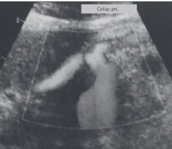
Doppler ultrasonography of the visceral arteries dem-onstrated moderate fusiform dilatation of the celiac artery, (Figure 1) starting 8 mm ater its origin, with an extension of 19 mm, maximum diameters of 18x18 mm and images suggesting dissection (Figure 2). he diameter of the artery proximal do the dissection was 8x8 mm. he superior and inferior mesenteric arteries had normal diameters and low. CT angiography of the abdominal aorta with mul-tislice helical technique and tridimensional reconstruction Abstract
he isolated spontaneous dissection of the celiac artery without the concomitant dissection of the aorta is a rare condition seldomly described in the literature. he objective of the present study is to describe a case of this clinical entity in a 74-year-old male patient, who was asymptomatic, and whose diagnosis was established by means of ultrasound and conirmed using computed angiotomography. he patient has been successfully followed up by means of clinical management for a period of 25 months.
Keywords: Celiac artery, dissection, ultrasonography.
Resumo
A dissecção espontânea isolada do tronco celíaco sem a dissecção concomitante da aorta é uma condição rara, pouco descrita na literatura. O objetivo do presente trabalho é descrever um caso dessa entidade clínica em um paciente masculino, 74 anos, assintomático, cujo diagnóstico foi feito por ultrassonograia e conirmado com angiotomograia computadorizada. O paciente tem sido acompanhado com sucesso mediante observação clínica por um período de 25 meses.
Palavras-chave: Artéria celíaca, dissecção, ultrassonograia.
Paper presented at the International Congress on Endovascular Surgery, São Paulo, April 2009.
1 PhD; Associate professor of Angiology and Vascular Surgery at Universidade Estadual de Londrina (UEL), Londrina, PR. 2 PhD; Associate professor of Angiology and Vascular Surgery at UEL, Londrina, PR.
3 Physician in charge of CT angiography at Ultramed - Diagnóstico por Imagem, Londrina, PR. 4 Physician; Specialist in Ultrasonography at Ultramed - Diagnóstico por Imagem, Londrina, PR. 5 Resident of Angioradiology and Endovascular Surgery, UEL, Londrina, PR.
6 Medical Student at Universidade Estadual do Oeste do Paraná (UNIOESTE), Cascavel, PR. No conlict of interest was declared concerning the publication of this article.
Isolated dissection of the celiac artery - Silvestre JMS et al. J Vasc Bras 2010, Vol. 9, Nº 2 153
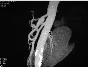
demonstrated indings similar to those of Doppler ultra-sonography, with celiac artery transverse diameters of 17.3x15.9 mm and a dissection lap inside the aneurysm (Figures 3 and 4). he abdominal aorta had calciied and non-calciied atheromatous plaques without signiicant ste-nosis, with normal diameters and no images of dissection. he superior and inferior mesenteric arteries, as well as the renal arteries, were normal (Figure 5).
As the patient was asymptomatic, we chose to follow up without medications. Doppler ultrasonography has been performed every six months and has not shown any chang-es, as there has been no increase in the aneurysm diameter, in the extension of the dissection or occlusion of celiac ar-tery branches.
Discussion
Arterial dissection may be deined as a cleavage of the arterial wall between two elastic layers and local hematoma formation2.
Isolated dissection of an artery which is not associat-ed with aortic dissection has been reportassociat-ed in the carotid and renal arteries, but rarely in the visceral arteries1,3-8. he
extra-aortic dissections include, in decreasing order, renal, coronary, cerebral, carotid, vertebral and visceral arteries9.
It is probable that these dissections were not diagnosed in the past, before the age of imaging methods.
Among visceral arteries dissections, those of the supe-rior mesenteric artery are more frequent10, but those of the
Figure 1 – Color Doppler ultrasound demonstrating celiac artery
dila-tation
Figure 2 – Ultrasonography in B mode: celiac artery dilatation near its
origin in the aorta
Figure 3 – CT Angiography demonstrating dissection lap
Isolated dissection of the celiac artery - Silvestre JMS et al. J Vasc Bras 2010, Vol. 9, Nº 2
154
inferior mesenteric and celiac arteries and its branches are rare. In a series of 19 patients with dissection of these ar-teries, Takayama et al.10 found involvement of the superior
mesenteric artery in 57.9%, of the celiac artery in 36%, of the isolated hepatic artery in 10.5% and of the splenic artery in only 5.3% of the cases.
Atherosclerosis is the most frequent etiologic factor. here are also reports of cystic degeneration, trauma, fun-gal infections, ibromuscular dysplasia, pregnancy, connec-tive tissue diseases and periarterial inlammation accompa-nied by cases of cholecystitis and pancreatitis. However, in more than half of the cases, no deinite cause can be found. It happened in our case, which did not present any of the previously mentioned factors.
Males aging 45 to 85 years are the most frequently afected by splanchnic arteries dissections, accounting for 88% of the cases described by Sparks et al.7 and by
Takayama et al.10.
As in our patient, nearly half of all dissections of viscer-al arteries is asymptomatic2,3,11, being detected by imaging
methods. In cases of symptomatic dissection of the celiac artery, Matsuo et al.3 reported the possibility of jaundice
when the hepatic artery is involved, or of pancreatitis when there the pancreatic arcades are involved and associated with abdominal pain, the most common symptom. Due to the rich collateral circulation originating from the superior mesenteric artery, the thrombotic complications of the ce-liac artery dissection are oten well tolerated.
CT angiography is considered the imaging method of choice. The image most frequently found is the pres-ence of an intimal flap, which confirms the diagnosis of dissection. Other findings are the presence of hematoma in the arterial wall, which generally corresponds to false lumen thrombosis, aneurysm formation and presence of infiltration in the fat tissue surrounding the celiac artery. This last finding, which sometimes is revealed by tomog-raphy during investigation of abdominal pain, may in-dicate the need for CT angiography. D’Ambrosio et al.8
reported that this prognostic factor may suggest dissec-tion extension into the adjacent vases. CT angiography may also enable assessment of dissection extension, of aneurysmal formation and dimensions, and other arter-ies involvement.
Doppler ultrosonography is also a valuable diagnostic modality that may be readily used without radiation expo-sure, besides ofering precise data for diagnosis. Magnetic resonance imaging and catheter angiography may also be used.
Most authors suggest that non-complicated asymp-tomatic lesions must receive conservative treatment, with
monitoring of the patient by imaging examinations. he use of antiplatelet or anticoagulant agents from three to six months to avoid thromboembolic complications, as well as the modiication of cardiac risk factors to limit the spread of the dissection and to reduce the risk of rupture, have been suggested. Our patient did not receive medication and has been asymptomatic for 25 months.
Surgical or endovascular treatment must be carried out only when there is aneurysm formation with progres-sive growth to a transverse diameter of more than 20 or 24 mm, which represents 2.5 to 3.0 times the normal artery diameter.
Intervention is also indicated in the presence of com-plications such as arterial rupture, true lumen thrombosis with hepatic ischemia and multiple lesions compromising the lower digestive tract.
However, in a literature review, we veriied that the conservative treatment may be safely indicated in patients with limited dissection who do not present evidence of rupture or expansion in a serial follow-up with imaging methods.
References
1. Woolard JD, Ammar AD. Spontaneous dissection of celiac artery: A case report. J Vasc Surg. 2007;45:1256-8.
2. Chaillou P, Moussu P, Noel SF, et al. Spontaneous dissection of ce-liac artery. Ann Vasc Surg. 1997;11:413-5.
3. Matsuo R, Ohta Y, Ohya Y, et al. Isolated dissection of celiac artery: a case report. Angiology. 2000;51:603-7.
4. Muller MF, Kim D. Spontaneous dissection of the hepatic artery. Abdom Imaging. 1995;20:462-5.
5. Yasuhara H, Shigematsu H, Muto T. Self-limited spontaneous dis-section of the main trunk of the superior mesenteric artery. J Vasc Surg. 1998;27:776-9.
Isolated dissection of the celiac artery - Silvestre JMS et al. J Vasc Bras 2010, Vol. 9, Nº 2 155
6. Suzuki S, Furui S, Kohtake H, et al. Isolated dissection of the supe-rior mesenteric artery: CT indings in six cases. Abdom Imaging. 2004;29:153-7.
7. Sparks SR, Vasques JC, Ergan JJ, Owens EL. Failure of nonoperative management of isolated superior mesenteric artery dissection. Ann Vasc Surg. 2000;14:105-9.
8. D’Ambrosio N, Friedman B, Siegel D, Katz D, Newatia A, Hines J. Spontaneous isolated dissection of the celiac artery: CT indings in adults. AJR Am J Roentgenol. 2007;188:W506-11.
9. Glehen O, Feugier P, Aleksic T, Delannoy P, Chealier JM. Spontaneous dissection of the celiac artery. Ann Vasc Surg. 2001;15:687-92.
10. Takayama T, Miyata T, Shirakawa M, Nagawa H. Isolated spon-taneous dissection of the splanchnic arteries. J Vasc Surg. 2008;48:329-33.
11. Foord AG, Lewis RD. Primary dissection aneurysm of peripheral and pulmonary arteries: dissecting hemorrhage of media. Arch Pathol. 1959;68:553-77.
Correspondence:
José Manoel da Silva Silvestre Av. Bandeirantes, 901 sala 504 CEP 86010-020 – Londrina, PR Tel: (43) 3339.6347 E-mail: [email protected]
Authors’ contributions
Study conception and design: JMSS Data analysis and interpretation: JMSS, GSS Data collection: MP, BF, FM, GSS Writing of the paper: JMSS, FM, GSS Critical analysis: JMSS, WES Final text approval: JMSS, WES, MP, BF, FM, GSS Statistical analysis: N/I Overall responsibility: JMSS Financing: N/I All authors have read and approved the inal version of the paper submitted to