Functional lung rejuvenation in obese patients after bariatric
surgery
SAULO MAIA DAVILA MELO¹*, PEDRO ALVES ARGENTINO², MURILO MATOSDE SANTANA OLIVEIRA², GABRIELA NABUCO CHAVES MELO2,
GILDO LIMA SOUZA NETO2
1PhD in Health Sciences – Universidade Federal de Sergipe. Full Professor, Medical School, Universidade Tiradentes, Aracaju, SE, Brazil
²Medical Student. – Universidade Tiradentes, Aracaju, SE, Brazil
S
UMMARYStudy conducted at Centro de Estudos e Pesquisas em Medicina Interna,
Aracaju, SE, Brazil
Article received: 6/19/2014
Accepted for publication: 5/4/2015
*Correspondence:
Address: Rua Dr. Moacir Rabelo Leite, 84 Bairro São José
Aracaju, SE – Brazil Postal code: 49020-280
http://dx.doi.org/10.1590/1806-9282.62.02.157
Objective: To determine the lung age (LA) in obese people before and after bar-iatric surgery, compare the LA with the chronological age (CA) before and after the operation, and verify whether there was a functional pulmonary rejuvena-tion after it.
Methods: A prospective longitudinal study including 43 morbidly obese pa-tients who underwent bariatric surgery. The papa-tients underwent clinical and spi-rometric evaluation in two stages, before and after the surgery. In both stages, LA, CA and spirometric variables were measured.
Results: A significant improvement in the spirometric variables (FVC; FEV1; and
FEV1/FVC ratio) was found after the operation (p≤ 0.0001). Comparing the LA
before (50.93±13.36 years) and after the surgery (39.02±12.95 years), there was an important reduction of 11.90±9.12 years (95CI:9.10-14.71; p≤0.0001) in LA after surgery. The difference between LA and CA before surgery was 12.20± 11.71 years (95CI:8.60-15.81) with significant difference (p≤0.0001), and the difference between LA and CA after surgery was -1.95±11.83 years (95CI: -5.59-1.69) with no significant difference (p≤0.28). Regarding LA, we observed a pulmonary ag-ing of 12.20±11.71 years before the surgery and a pulmonary rejuvenation of 11.90±9.12 years after it.
Conclusion: Morbid obesity is responsible for early damage and functional ac-celerated pulmonary aging. After the correction of the body weight by surgery, there is a functional pulmonary rejuvenation demonstrated by the normaliza-tion of LA in relanormaliza-tion to CA.
Keywords: Lung diseases, premature aging, morbid obesity, spirometry, respi-ratory function tests, bariatric surgery.
I
NTRODUCTIONThe consistent and significant growth of obesity in the last two decades, regardless of age, gender and socio-eco-nomic conditions of the population has become a prob-lem of public and economic health for the World Health Organization (WHO).¹
In Brazil, recent data showed that 51% of the popu-lation is overweight and 17% obese (16% of men vs. 18%
women), with a staggering advance in recent years, and with frightening forecasts for all regions of the country.2
Several comorbidities are caused by excess body weight and obesity is a major independent risk factor of prema-ture death.3-5
Obesity is a chronic systemic inflammatory disease that early compromises lung function.6 Lung function
decreases faster when the lungs are attacked by gases, toxic fumes, other pollutants, and inflammatory lung disease.7,8
Chronic inflammatory diseases cause progressive and early decline of cellular homeostasis, predisposing to an accelerated aging of the affected organs.7
Lung age (LA) is increased in obese patients before surgery for morbid obesity, suggesting premature and ac-celerated lung aging.6
To our knowledge, the concept of LA to assess the functional lung rejuvenation in obese patients after weight loss has never been used.
We hypothesized that obese undergoing bariatric sur-gery present preoperatively with early and accelerated functional lung aging when we compare LA with chron-ological age (CA), and that after bariatric surgery, with consequent weight loss, the mechanical effects and the inflammatory process caused by obesity in the lungs will be reduced, providing the pulmonary functional rejuve-nation from the recovery of LA.
The aim of this study was to determine the spiromet-ric LA in obese individuals without pulmonary disease, pre- and post-bariatric surgery; to compare LA and CA before and after bariatric surgery; and to verify whether there was spirometric lung rejuvenation after bariatric surgery.
M
ETHODSThis is a longitudinal, prospective study conducted at the Center for Studies and Research in Internal Medicine in the city of Aracaju, Sergipe, Brazil, between January 2010 and September 2012.
43 morbidly obese patients were selected for the study. They underwent clinical evaluation and spirometry in a private respiratory medicine practice by an assistant pul-monologist to determine lung surgical risk for surgical obesity treatment in the bariatric surgery clinic of two in-stitutions, one public and one private. Obesity was classi-fied according to World Health Organization (WHO) cri-teria for body mass index (BMI), obtained based on the weight/height equation2 (kg/m2). Indications for surgical
treatment followed the guidelines of the WHO and the Brazilian Ministry of Health.1,2
The study was approved by the Ethics Committee on Human Research of the Federal University of Sergipe (CAAE Extension 0050.0,107,000-07) and performed in two stages, before and after bariatric surgery. All partici-pants signed a free and informed consent form. Preoper-atively, patients were referred and evaluated consecutive-ly, according to the demand of the clinic. For the evaluation after surgery, patients were invited by tele-phone to participate in the study as volunteers, random-ly, provided that they had lost 20% or more of body weight and had at least six months of surgery.
Patients with any acute or chronic pulmonary dis-ease were excluded, as well as active smokers and former smokers with any current or previous smoking history,
incapacity to perform spirometry, and patients younger than 20 years according to the original formula for LA.
After clinical evaluation, body weight was obtained with the patient being asked to remove shoes and heavy clothing, and height was obtained using an anthropom-eter coupled to the scale that met the criteria for weight measurement evaluation of the morbidly obese.
Spirometry was performed using a computerized spi-rometer (Microlab-3500, Cardinal Health U.K. 232 Ltd.; England), with the patient in a sitting position and us-ing a nose clip performus-ing at least three forced expirato-ry maneuvers that met the acceptability and reproduc-ibility criteria required by the current recommendations of the SBPT11, being selected the best of them. Forced vital capacity (FVC), forced expiratory volume in one sec-ond (FEV1) and FEV1/FVC ratio were measured before
the bronchodilator (pre-BD), with values expressed in li-ters and in percentage of the normal values calculated ac-cording to the equation by Hankinson et al.12
The patient’s LA was estimated during spirometry
using Pre-BD FEV1. The minimum pre-established LA
was 20 years, the maximum LA being represented by the highest value in the original LA formula (Table 1).13
TABLE 1 Original formula for calculating lung age (years).11
Men: Lung age = 2.87 × height − (31.25 × FEV1 obtained) – 39.375 Women: Lung age = 3.56 × height − (40.00 × FEV1 obtained) − 77.280 Lung age in years; height in inches; 1 inch: 2.54 centimeters; FEV1 in liters.
Statistical analysis was performed with Statistical Pack-age for the Social Sciences software, version 15 (SPSS Inc., Chicago, IL, USA). Categorical data are presented in ab-solute and percentage value, and the numeric data in av-erage and standard deviation. 95CI was calculated for LA and CA in both stages. To determine the differences in continuous variables within one step and between steps, paired Student t test was used. Statistical significance was set at p<0.05, and all statistical tests were two-tailed.
R
ESULTSTable 2 illustrates a comparison of the preoperative and postoperative stages including demographic, anthropo-metric and spiroanthropo-metric characteristics of the population studied.
The average time between bariatric surgery and post-operative evaluation was 14.0±8.7 months.
37.79±44.16), p≤ 0.001. There was a significant reduction in BMI between the two stages of evaluation (44.18±7.4 and 29.06±4.6 kg/m², respectively; p≤0.0001).
As for the evaluated spirometric variables (FEV1, FVC
and FEV1/FVC ratio), a significant improvement after
bar-iatric surgery (p≤0.0001) was observed.
Table 3 compares the CA and LA before and after bar-iatric surgery, displaying the differences between CA and LA in each step and between steps. Comparing the LA be-fore (50.93±13.36 years; 95CI: 46.82-55.04) and after sur-gery (39.02±12.95 years; 95CI: 35.04-43.01), we found a significant reduction in LA after surgery of 11.90±9.12 years (95CI: 9.10-14.71; p≤0.0001).
TABLE 2 Demographic, anthropometric and spirometric characteristics before and after bariatric surgery in the studied population.
Variables Before (n=43) After (n=43) p
Agedb, years 38.72±10.12 40.98±10.35 0.001
Gender femalea 30 (69.8) –
Skin color, Whitea 30 (71) –
BMIb, Kg/m2 44.18±7.45 29.06±4.66 0.0001*
FEV1b, L 2.72±0.59 3.04±0.78 0.0001*
FEV1b, % foreseen
value
82.41±8.13 93.69±9.90 0.0001*
FVCb, L 3.25±0.74 3.51±1.87 0.0001*
FVCb, % foreseen
value
80.53±8.33 87.95±9.87 0.0001*
FEV1/FVCb, % 82.90±7.69 86.69±7.15 0.01* BMI: Body mass index; absolute and relative spirometric values (pre-bronchodilator). FEV1:
Forced expiratory volume in one second. FVC: Forced vital capacity. FEV1/FVC: % of FEV1/FVC
ratio. a: Values expressed as n (%). b: Values expressed as average ± SD. *Student’s t test.
TABLE 3 Comparison between chronological age and lung age before and after bariatric surgery; at each stage and between stages.
Variables Before (n=43)
95CI After (n=43)
95CI p
CA, years 38.72±10.12 35.61 - 4 1.84
40.98±10.35 37.79 - 44.16
0.001
LA, years 50.93±13.36 46.82 - 55.04
39.02±12.95 35.04 - 43.01
0.0001
LA – CA, years
12.20±11.71 8 . 6 0 - 15.81
-1.95±11.83 -5.59 -1.69
0.0001
CA: Chronological age; LA: Lung age; LA - CA: Difference between lung age and chronological age. Values expressed as average ± SD. Student’s t test.
The difference between LA and CA before surgery was 12.20±11.71 years (95CI:8.60-15.81) with a significant dif-ference (p≤0.0001), and the difdif-ference between LA and
CA after surgery was -1.95±11.83 years (95CI: -5.59-1.69) with no significant difference (p≤0.28).
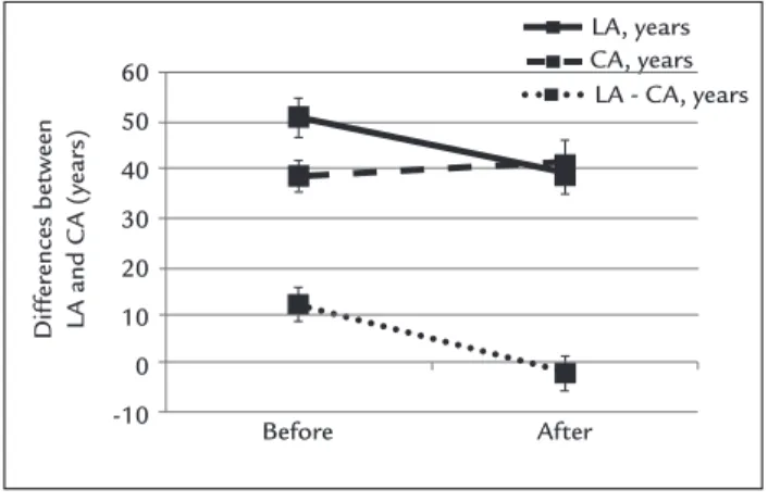
Figure 1 is a graphic demonstration of CA differenc-es before and after bariatric surgery, as well as LA before and after surgery, also displaying the differences between CA and LA at every stage, and between stages. Preopera-tively, the comparison between LA and CA demonstrates an accelerated early functional lung aging 12.20±11.71 years with a significant difference (p≤0.0001). Postoper-atively, the comparison between LA and CA revealed that there was no significant difference between them (p ≤ 0.28), so that the patients showed practically the same chronological and lung ages (LA - CA: -1.95±11.83 years). Thus, comparing LA before and after bariatric surgery, we found functional lung rejuvenation in the study sam-ple of 11.90±9.12 years (95CI 9.10-14.71) after surgery.
D
ISCUSSIONThis study showed that obese patients scheduled for bar-iatric surgery have an early and accelerated functional ag-ing (12.20±11.71 years), and that after bariatric surgery there is a functional lung rejuvenation (11.90±9.12 years) evidenced based on the concept of LA.
In a previous study, we have demonstrated an acceler-ated early lung aging (12.20±2.4 years) in a group of mor-bidly obese patients previously to bariatric surgery compared with a group of non-obese controls,6 which corroborates our
results of early lung aging around a decade.
To our knowledge, there are no studies in the literature evaluating functional lung rejuvenation in obese patients after correction of body weight using the concept of LA.
The segment of obese population is increasing alarm-ingly in both the developed and developing world; how-ever, there are few studies on the impact of obesity on the
FIGURE 1 Comparison between chronological age and lung age before and after bariatric surgery; at each stage and between stages.
LA, years CA, years
LA - CA, years 60
50
40
30
20
10
0
-10
Before After
Dif
fer
ences between
LA and C
A (year
lungs, despite the clear effect of obesity on lung function as a result mainly of the mechanical and inflammatory effects of excessive weight.3,5,14,15
With increasing degree of obesity, the mechanical ef-fects are attributed to increased intra-abdominal pressure and decreased compliance of the rib cage (pulmonary and chest wall) caused by excessive fat tissue in these sites and the impairment of respiratory muscles with decreased di-aphragmatic displacement capacity and overload of the in-spiratory muscles, reducing FVC and FEV1.3,5
FEV1 is an independent factor of cause of death and
a risk factor for sudden death, cancer and cardiovascular disease.5,11
FEV1 is influenced by FVC and is part of the
formu-la of lung age,13 which explains in our study the increased
lung age (accelerated early functional lung aging) justi-fied by the reduction in FEV1 in morbidly obese patients
preoperatively, with FEV1/FVC ratio being preserved.
After bariatric surgery with correction of body weight, there is a recovery of lung function with improvement in FVC and FEV1.16 Our results demonstrate similar results,
with improved FEV1 after bariatric surgery, justifying lung
rejuvenation seen in our sample.
Whenever LA is in disagreement with CA, obese pa-tients may be warned of the early detection of lung dam-age (accelerated early functional aging) that can be pre-vented or controlled with the reduction in body weight (functional lung rejuvenation). However, one must be careful not to translate to the patient the concept of LA as a life expectancy, but only alert of accelerated early pul-monary function damage and the real possibility of func-tional recovery after correction of body weight.
It is now known that adipose tissue is not simply an inert body to store energy. Rather, it is a great source of production of pro-inflammatory mediators, working as an endocrine organ that regulates systemic inflammation producing a variety of acute phase proteins (adipocyto-kines), the main being leptin, which has pro-inflamma-tory action, and adiponectin with anti-inflammapro-inflamma-tory ac-tivity. In obese people, there is a reduction in serum adiponectin and an increase in leptin, establishing a low-grade chronic inflammation with systemic and pulmo-nary repercussions.14,15,17
The aging process is for life; it is incredibly complex and heterogeneous, and is characterized by anatomical and functional changes. However, there are diseases that accel-erate aging.6-8,15,18 The main preventive measure to combat
obesity is to encourage the correction of the patient’s body weight, using every tool available. The concepts of LA and pulmonary functional rejuvenation have this purpose.
This study shows some limitations. In our research, we do not enter into the merits of the mechanisms of ag-ing and senescence involvag-ing molecular biology, chang-es in morphology, and cellular rchang-espiratory function. Our goal is to demonstrate the functional spirometric altera-tions in the obese that, after correction, translates into significant functional recovery. Our attempt is to convey the message functional modification of the lungs in obe-sity, and the possibility of recovery, using the new con-cept of functional lung rejuvenation. This is an idea that can be easily understood by the patient, lay people and health professionals, so that they become aware of the se-rious effect of obesity on the lungs, and use this tool to combat this serious epidemic.
Future research should assess the psychological strength of the concept of LA to demonstrate accelerat-ed early lung aging and spirometric lung rejuvenation, as a warning tool and additional incentive to the treatment of obesity in our country.
C
ONCLUSIONSumming up, morbid obesity causes early damage and accelerated functional lung aging, which is demonstrat-ed by the discrepancy between chronological age and lung age. After correction of the body weight using bariatric surgery, there is functional lung rejuvenation, translated by the normalization of lung age in relation to chrono-logical age.
R
ESUMORejuvenescimento pulmonar funcional em pacientes obe-sos após cirurgia bariátrica
Objetivo: determinar a idade pulmonar (IP) em obesos no pré e pós-operatório de cirurgia bariátrica, comparar a IP com a idade cronológica (IC) antes e após a cirurgia, e verificar se houve rejuvenescimento pulmonar funcio-nal após a cirurgia.
Métodos: estudo longitudinal, prospectivo, envolvendo 43 pacientes obesos mórbidos submetidos à cirurgia ba-riátrica. Os pacientes foram submetidos à avaliação clí-nica e espirométrica antes e após a cirurgia, sendo deter-minadas IP, IC e variáveis espirométricas.
Resultados: observou-se melhora significativa nas variá-veis espirométricas (VEF1, CVF e razão VEF1/CVF) após
e IC no pré-operatório foi de 12,20±11,71 anos (IC 95% 8,60-15,81) com diferença significativa (p≤0,0001). A di-ferença entre IP e IC no pós-operatório foi de -1,95±11,83 anos (IC 95% -5,59-1,69), sem apresentar diferença signi-ficativa (p≤0,28). Quando comparamos a IP antes e após a cirurgia, observamos um envelhecimento pulmonar de 12,20±11,71 anos antes e um rejuvenescimento
pulmo-nar de 11,90±9,12 anos após a cirurgia.
Conclusão: a obesidade mórbida causa dano precoce e envelhecimento pulmonar funcional acelerado. Após a correção do peso corpóreo pela cirurgia, há um rejuve-nescimento pulmonar funcional, mostrado pela norma-lização da IP em relação à IC.
Palavras-chave: pneumopatias, senilidade prematura, obesidade mórbida, espirometria, testes de função respi-ratória, cirurgia bariátrica.
R
EFERENCES1. World Health Organization. Obesity and overweight. Geneva: World Health Organization [cited 2014 Jan 2]. Available from: http://www.who.int/ mediacentre/factsheets/fs311/en/index.html.
2. Brasil. Ministério da Saúde. Mais da metade da população brasileira tem excesso de peso. Brasília: Ministério da saúde, 2013 [cited 2014 Jan 2]. Available from: http://portalsaude.saude.gov.br/portalsaude/noticia/12926/162/mais-da-metade-da-populacao-brasileira-tem-excesso-de-peso.html.
3. Koenig SM. Pulmonary complications of obesity. Am J Med Sci. 2001; 321(4):249-79.
4. Poulain M, Doucet M, Major GC, Drapeau V, Sériès F, Boulet LP, et al. The effect of obesity on chronic respiratory diseases: pathophysiology and therapeutic strategies. CMAJ. 2006; 174(9):1293-9.
5. McClean KM, Kee F, Young IS, Elborn JS. Obesity and the lung: 1. Epidemiology. Thorax. 2008; 63(7):649-54.
6. Melo SM, Melo VA, Melo EV, Menezes Filho RS, Castro VL, Barreto MSP. Accelerated lung aging in patients with morbid obesity. J Bras Pneumol. 2010; 36(6):746-52.
7. Ito K, Barnes PJ. COPD as a disease of accelerated lung aging. Chest. 2009; 135(1):173-80.
8. Papaioannou AI, Rossios C, Kostikas K, Ito K. Can we delay the accelerated lung aging in COPD? Anti-aging molecules and interventions. Curr Drug Targets. 2013; 14(2):149-57.
9. Caruso LB, Silliman RA. Medicina geriátrica. In: Fauci AS, Braunwald E, Kasper DL, Hauser SL, Longo DL, Jameson JL, et al. (eds.). Harrison Medicina Interna. 17.ed. Rio de Janeiro: McGraw-Hill Interamericana do Brasil, 2008. p.53-62.
10. Costa EFA, Galera SC, Porto CC, Cipullo JP, Martin JFV. Semiologia do idoso. In: Porto CC (ed.), Porto AL (co-ed.). Semiologia médica. 6.ed. Rio de Janeiro: Guanabara Koogan, 2011. p.159-93.
11. Sociedade Brasileira de Pneumologia e Tisiologia. Diretrizes para testes de função pulmonar. J Pneumol. 2002; 28(3):S2-S238.
12. Hankinson JL, Odencrantz JR, Fedan KB. Spirometric reference values from a sample of the general U.S. population. Am J Respir Crit Care Med. 1999; 159(1):179-87.
13. Morris JF, Temple W. Spirometric “lung age” estimation for motivating smoking cessation. Prev Med. 1985; 14(5):655-62.
14. Mancuso P. Obesity and lung inflammation. J Appl Physiol. 2010; 108(3):722-8. 15. Sood A. Obesity, adipokines, and lung disease. J Appl Physiol (1985). 2010;
108(3):744-53.
16. Santana AN, Souza R, Martins AP, Macedo F, Rascovski A, Salge JM. The effect of massive weight loss on pulmonary function of morbid obese patients. Respir Med. 2006; 100(6):1100-4.
17. Ali Assad N, Sood A. Leptin, adiponectin and pulmonary diseases. Biochimie. 2012; 94(10):2180-9.