Analysis of skeletal maturation in patients
aged 13 to 20 years by means of hand wrist
radiographs
Yasmine Bitencourt Emílio Mendes*, Juliana Roderjan Bergmann**, Marina Fonseca Pellissari**, Sérgio Paulo Hilgenberg***, Ulisses Coelho****
Objectives: Evaluate an alternative and simplified radiographic method that will enable implantologists and orthodontists to keep track of bone growth completion as well as dif-ferences between genders in a sample of individuals from 13 to 20 years of age. Methods:
A hand wrist radiograph was acquired with the use of occlusal radiographic film to assess the maturation of the radius bone. One hundred and sixty patients of both genders were divided into groups of 10 individuals. The radiographs were examined by five raters using applicable rating standards, all specialists in orthodontics. Results: The results showed that there was a positive correlation between the chronological age of the patients and their biological age, whereas female bone maturation occurs earlier than in males. Conclusion:
This method can determine the stage of maturation of the radius bone, allowing an afford-able means of diagnosis and rapid determination of bone age for correct installation of an implant and/or orthodontic appliance.
Abstract
Keywords: Maturity. Tooth movement. Dental implant.
* Master’s candidate in Dentistry; area of concentration Restorative Dentistry, Universidade Estadual de Ponta Grossa (UEPG). ** Surgeon Dentists.
*** Master of Dentistry; area of concentration Integrated Clinical Practice – UEPG. Postgraduate student specializing in Orthodontics - (UEPG).
**** Adjunct Professor of Orthodontics - UEPG. Orthodontics Specialist from Universidade de São Paulo (USP-Bauru); Master and Doctor of Orthodontics from Unesp - Araraquara/SP.
INTRODUCTION
Understanding the events of craniofacial growth is of paramount importance in dental prac-tice.10 Knowing the patient’s exact stage of
matu-rity and the period of occurrence of the pubertal growth spurt (PGS) may influence not only diag-nosis and progdiag-nosis, but also the development of a treatment plan.24 Thus, this knowledge is
consid-ered convenient for orthodontic treatments that
require the use of devices influenced by the stage of maturation of the craniofacial complex.28 This
growth stage also helps in preparing the treatment plan, especially in defining surgical procedures.15
current chronological age.9 Therefore,
chronolog-ical age is not always a good parameter for the correct evaluation of the body’s maturation stage because, for example, the PGS does not occur at the same time in all individuals.20 Biological age is
more reliable since it is reported as the body’s de-velopment towards maturity, which encompasses anatomical changes, dental and skeletal matura-tion, development of secondary sexual charac-teristics, hormonal function and enzyme activity, even if those mechanisms are influenced by ge-netic factors, socio-economic, environmental, nu-tritional and gender-related conditions.4
According to Rigertz and Eklöf,8 Caffey (1961)
consider that the ideal method for assessing skel-etal maturity should include a study of the whole skeleton. However, these autors8 affirm thatdue
to practical and economic reasons this is not only impossible but also incompatible with the efforts to reduce exposure to radiation. The difficulties in finding a procedure that combines a simple and accurate definition of normal variations with rea-sonable statistical confidence have surfaced in the design of different methods.1,3,5,8,19,27
In contemporary Orthodontics, early correc-tion of various types of malocclusion is rendered easier when it is possible to take advantage of the moments of maximum increment in an individu-al’s facial and overall growth occurring during the PGS.21 A patient’s growth stage, therefore, proves
essential in establishing accurate diagnoses and reliable prognoses.21 The stage of bone
develop-ment can be estimated by means of hand wrist radiographs and through skeletal maturation as visualized in lateral cephalometric radiographs of the cervical vertebrae.21
Orthodontic treatment should be performed preferably in very young patients or during their PGS, as it is during this period that facial struc-tures respond more efficiently to the stimuli provided by orthodontic mechanics.23 Moreover,
cases that are treated in later stages do not usu-ally undergo significant skeletal changes as a result
of treatment, with the exception of surgical inter-ventions in the bone bases.23
The hand wrist has been widely studied and used as an area that helps to determine bone maturation with scientifically proven efficacy. Furthermore, it is a complementary exam that can be easily obtained with an X-ray and used for diagnosis and orthodontic treatment plan-ning.12,27 Several investigations have shown that
these areas represent the overall maturity of the skeleton and thus are suitable for such evalua-tion.12 Similarly, other specialties require
knowl-edge of skeletal maturation, especially Implan-tology since the aesthetic and functional success of an implant is associated with complete bone maturation.13
Therefore, considering the importance of PGS, this study aims to establish an affordable radio-graphic methodology that will allow dental pro-fessionals to assess skeletal maturation and moni-tor craniofacial growth and the alveolar process, focusing on adolescents aged 13 to 20 years. Ad-ditionally, it also seeks to establish the best time for dental implant placement and the most con-venient moment to begin orthodontic treatment in this age group.
MATERIALS AND METHODS
This study was approved by the Ethics Com-mittee of the Universidade Estadual de Ponta Grossa - COEP – UEPG, Protocol No. 03569/06, Opinion No. 16/2006.
Hand wrist radiographs were performed with the use of occlusal films on 80 male patients and 80 female patients 13 to 20 years of age, further divided into 10 subjects for each age group toting 8 groups for each gender. The radiographs al-lowed an assessment of the degree of maturation and the fusion of the epiphysis with the diaphysis of the radius bone.
40 cm cone was positioned perpendicular to the position of the hand and the film, and the radio-graphs were acquired. The films were processed using the time and temperature method and, after complete drying and identification, were stored for evaluation.
The evaluation was performed by five special-ists in Orthodontics using applicable rating stan-dards, who observed and scored (Fig 1) the stage of maturation of the radius bone. When the epiphysis and diaphysis of the radius bone were either small-er or equal in width, a zsmall-ero score was assigned; a score of 1 was given when the epiphysis was larger than the diaphysis; a score of 2 when the epiphysis was starting to merge with the diaphysis; a score of 3 when the epiphysis and diaphysis were bound together by a slight radiolucent line; and a score of 4 to indicate the complete and final maturation of bone growth, with no significant difference be-tween epiphysis and diaphysis.
The results obtained by each rater were ana-lyzed by Kappa statistics to establish the Kappa coefficient of agreement. The non-parametric Mann-Whitney test was performed to see if both genders had the same distribution of results. The Spearman correlation test was used to assess the correlation between chronological age and the relative gender scores.
RESULTS
A statistical analysis of the data showed that the average level of inter-rater agreement was con-sidered good (kappa = 0.66) (Table 1).
The nonparametric Mann-Whitney test yield-ed a p value of 0.0011, which shows a statistically significant difference between groups due to the fact that the distribution of scores occurred differ-ently in females, with men showing faster results.
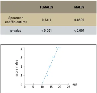
The Spearman correlation test showed a val-ue of p < 0.001 for both females and males, i.e., scores increased with age (Table 2).
Whereas there was good inter-rater agreement and a statistically significant difference between males and females, and considering that as chron-ological age advances, the scores will also increase, the next step consisted in evaluating—for both genders—at what age stage 4 (complete fusion of the epiphysis with the diaphysis of the radius bone) occurred significantly. For this the authors used the median values for each age considering the analysis of all raters. The occurrence of stage 4 is significant in females aged 16 years and in males aged 18 years (Graphs 1, 2).
DISCUSSION
The planning and clinical conduct for rehabili-tation of patients with partial anodontia, especially
FIGURE 1 - Kappa inter-rater agreement coefficient.
SCORE 0 SCORE 1 SCORE 2 SCORE 3 SCORE 4
The complete maturation and final bone growth are indicated with no signifi-cant difference between the epiphysis and diaphysis A slight radiolucent line
appears between the epiphysis and diaphysis
of the radius bone The epiphysis is
start-ing to merge with the diaphysis of the radius The epiphysis is larger
than the diaphysis of the radius bone The epiphysis and
multiple partial anodontia, is hampered by the de-cision of when to start treatment and what tech-niques to adopt.13 Thus, as the patient is born with
missing teeth, prosthetic rehabilitation should be undertaken as soon as the patient’s development is complete.
The patient’s bone development can be tracked in various ways, but the use of hand wrist radio-graphs can provide information on the patient’s growth and maturity, which are clinically impor-tant in the application of dentofacial diagnosis and orthopedic therapy,10,11,14 and more recently, the
installation of dental implants. Another way to as-sess skeletal maturation is accomplished through an analysis of the cervical vertebrae in lateral ra-diographs, which are part of the orthodontic doc-umentation.26 However, it is suggested that
pro-fessionals exercise caution in considering cervical vertebrae examination as an absolute method to evaluate skeletal maturation in growing patients for as long as professionals have not yet developed enough familiarity with this method.1,7 Thus, it
is believed that carpal index is the most reliable resource for bone assessment given the fact that most professionals are very comfortable using the method, which is not only easy to obtain but in-volves less radiation exposure.
The patient’s exact stage of maturation has been the target of research, which has sought to facilitate its assessment, reduce costs and re-duce the exposure of patients to ionizing radia-tion.1,5,18,19,24 Silva Filho et al27 evaluated a
simpli-fied alternative method consisting of an X-Ray of the metacarpophalangeal joint area of the thumb, using a radiographic periapical film. In this film the presence of the ulnar sesamoid bone can be observed, in a clear indication that PGS onset will occur within a 1-year period, thereby signaling the correct time for the installation of the orth-odontic appliance.
In this study, the authors have attempted to find a fast and secure way for dentists to detect, in their own office, the patient’s stage of maturation for the placement of a dental implant and assess
RATER KAPPA VALUE
rater 1 x rater 2 0.63
rater 1 x rater 3 0.71
rater 1 x rater 4 0.66
rater 1 x rater 5 0.66
Mean Kappa value 0.66
TABLE 1 - Kappa inter-rater agreement coefficient. TABLE 2 - Representation of the Spearman correlation test results for
males and females.
FEMALES MALES
Spearman
coefficient(rs) 0.7314 0.8599
p-value < 0.001 < 0.001
GRAPH 2 - Graphical representation of the relationship between chron-ological age and male scores.
GRAPH 1 - Graphical representation of the relationship between chron-ological age and female scores.
score females
4
3
2
1
0
0 5 10 15 20 25 age
score males
4
3
2
1
0
the most suitable time to begin orthodontic treat-ment of patients aged 13 to 20 years. This was achieved by acquiring an X-ray—using occlusal film—of the radius bone region, which indicates the end of the alveolar bone growth. It should be underscored that in establishing the bone age diagnosis of a patient who will undergo a dental implant surgery, the exclusion of the carpal and metacarpal bones does not significantly influence bone age determination.27 Moreover, this method
readily addresses a clinical issue while ensuring less patient exposure to radiation.25
A literature review has revealed a growing concern by orthodontists to establish parallels between a patient’s chronological and biological ages.9,15 As can be observed in this study, there is
a positive correlation between biological age and chronological age, i.e., in this sample, chronologi-cal age progressed in tandem with biologichronologi-cal age.
Regarding gender differences in terms of bone maturation, the results endorse other studies6,16,19,22 which concluded that all events
and stages of skeleton growth occur earlier in female than in male individuals.
In this study, most women’s readiness to un-dergo dental implant installation or orthodontic treatment depended on the time of maturation, on their chronological age (16 years) and on com-plete ossification (score 4) of the radius bone, while most men were able to undergo the same
procedure only at 18 years of age. However, we must consider that some of the individuals—al-though being the same age—had not completed their growth. Thus, this finding should not be gen-eralized, requiring the mandatory implementation of radiographic examination in all patients who are subjected to such treatment. Nevertheless, the method used in this study has proven an effec-tive alternaeffec-tive to address routine clinical issues related to skeletal maturation.
CONCLUSION
Based on the results and the methodology em-ployed, the authors have concluded that:
1) The method is effective in predicting the skeletal age of the patient. Furthermore, it consists of a practical, quick and accessible method that can assist in the planning of dental implants and orthodontic treatment.
2) There are individual differences for each group, indicating that each individual—regardless of their chronological age—may have a different biological bone age.
1. Armond MC, Castilho JCM, Moraes LC. Estimativa do surto de crescimento puberal pela avaliação das vértebras cervicais em radiografias cefalométricas laterais. Ortodontia. 2001;34(1):51-60.
2. Björk A, Helm S. Prediction of the age of maximum puberal growth in body height. Angle Orthod. 1967 Apr;37(2):134-43. 3. Bosco VL, Silva RHH. Relação entre crescimento e erupção
dentária. RGO. 1991;39(3):189-90.
4. Carvalho PL, Ando T, Reis HSM, Pannunzio E. Considera-ções sobre metodologias de avaliação das idades dental e óssea, em pacientes com idades cronológicas de 3 a 14 anos. JBP – Rev Ibero Am Odontopediatr Odontol Bebê. 2005;8(45/46):312-20.
5. Castriota-Scanderbeg A, Sacco MC, Emberti-Gialloreti L, Fraracci L. Skeletal age assessment in children and young adults: comparison between a newly developed sonographic method and conventional methods. Skeletal Radiol. 1998 May;27(5):271-7.
6. Chaves AP, Ferreira RI, Araújo TM. Maturação esquelética nas raças branca e negra. Ortodontia Gaúcha. 1999 jan/ jul;3(1):45-52
7. Damian MF, Woitchunas FE, Cericato GO, Cechinato F, Moro G, Massochin ME, Castoldi FL. Análise da confiabilidade e da correlação de dois índices de estimativa da maturação esque-lética: índice carpal e índice vertebral. Rev Dent Press Ortod Ortop Facial. 2006 set/out;11(5):110-20.
8. Eklöf O, Ringertz H. A method for assessment of skeletal matu-rity. Ann Radiol. 1967;10(3-4):330-36.
9. Fishman LS. Chronological versus skeletal age, an evaluation of craniofacial Growth. Angle Orthod. 1979;49(3):181-89. 10. Fishman LS. Radiographic evaluation of skeletal maturation.
A clinically oriented method base on hand-wrist films. Angle Orthod. 1982;52(2):88-112.
11. Fishman LS. Maturational patterns and prediction during ado-lescence. Angle Orthod. 1987 Jul;57(3):178-93.
12. Flores-Mir C, Nebbe B, Major PW. Use of skeletal matura-tion based on hand-wrist radiographic analysis as a predictor of facial growth: a systematic review. Angle Orthod. 2004 Feb;74(1):118-24.
13. Francischone CE, Vasconcelos lW. Osseointegração e as próte-ses unitárias. 1a ed. São Paulo: Artes Médicas; 1998. 14. Franco AA, Santana AH, Santana IS, Melo MFB, Santos Júnior
JH. Determinação radiográfica da maturidade esquelética e sua importância no diagnóstico e tratamento ortodôntico. Ortodontia. 1996;29(1):53-9.
15. Guzzi BSS, Carvalho LS. Estudo da maturação óssea em pacientes jovens de ambos os sexos através de radiografias de mão e punho. Ortodontia. 2000;33(3):49-57.
REFERENCES
16. Hägg U, Taranger J. Skeletal stages of the hand and wrist as indicators of the pubertal growth spurt. Acta Odontol Scand. 1980;38(3):187-200.
17. Hägg U, Taranger J. Maturation indicators and the pubertal growth spurt. Am J Orthod. 1982 Oct;82(4):299-309. 18. Hamui T, Prata C. Estudo do crescimento maxilar e
man-dibular na fase de aceleração do surto de crescimento puberal. Rev Dent Press Ortod Ortop Facial. 2001 jul/ ago;6(4):19-31, 2001.
19. Hilgenberg S, Pinto SCS, Pinheiro JC, Jimenez EEO, Coelho U. Comparação entre as idades óssea, dentária e cronológica por meio de método radiográfico simplificado. Rev Odonto. 2008 jul/dez;16(32):31-8.
20. Lejarraga H, Guimarey L, Orazi V. Skeletal maturity of the hand and wrist of healthy Argentinian children aged 4-12 years, as-sessed by the TW II. Ann Hum Biol. 1997;24(3):257-61. 21. Martins EG, Simone JL, Reis RRB. Estudo comparativo de dois
métodos de avaliação da maturação esquelética utilizando radiografias carpais e telerradiografias em norma lateral. RGO. 2006;54(4):322-27.
22. Moraes MEL, Medici Filho EM, Moraes LC. Surto de crescimen-to puberal. Relação entre mineralização dentária, idade crono-lógica, idade dentária e idade óssea – método radiográfico. Rev Odontol UNESP. 1998;27(1):111-29.
23. Moraes, MEL, Morosolli ARC, Moraes LC, Castilho JCM. Com-paração dos métodos de Martins & Sakima e de Fishman para avaliação do surto de crescimento puberal. J Bras Ortodon Ortop Facial. 2005;10(57):255-62.
24. Prata THC, Moraes Filho E, Moraes LC, Moraes MEL. Estudo do crescimento maxilar e mandibular na fase de aceleração do surto de crescimento puberal. Rev Dent Press Ortod Ortop Facial. 2001;6(4):19-31.
25. Santos SCBN, Almeida RR. Estudo comparativo de dois mé-todos de avaliação da idade esquelética utilizando telerradio-grafias em norma lateral e radiotelerradio-grafias carpais. Ortodontia. 1999;3(2):33-45.
26. Schusterchitz T, Haiter NF. Estudo comparativo entre a matura-ção óssea das vértebras cervicais e a região carpal. Ortodontia. 2002;35(3):33-41.
27. Silva Filho OG, Sampaio LL, Freitas JAS. Avaliação de um método simplificado para estimar a maturação esquelética. Ortodontia. 1992;25(1):21-36.
28. Vieira CL, Oliveira AEF, Ribeiro CCC, Lima AASJ. Relação entre os índices de maturação das vértebras cervicais e os estágios de calcificação dentária. Rev Dent Press Ortod Ortop Facial. 2009 mar/abr;14(2):45-53.
Contact address