THE ESTUARINE BENTHIC
DIATOM
Margaritum terebro
(BACILLARIOPHYT A, HYALODISCACEAE):
MORPHOLOGYANDTAXONOMY
Roseli M. de Souza-Mosimann1; Luciano F. Fernandes2 & Thelma V. Ludwig 2
lUniversidade Federal de Santa Catarina Horto Botânico - Campus Universitário, Trindade (Caixa Postal 476, 88040-900 Florianópolis, SC, Brasil)
2Universidade Federal do Paraná, Setor de Ciências Biológicas Departamento de Botânica, Centro Politécnico
(Caixa Postal 19031, 81531-970 Jardim das Américas, Curitiba, PR, Brasil)
.
Abstract: Margaritum terebro is an estuarine centric diatom recorded along the southeast coast of Brazil, the Atlantic coast of Aftica, and in regions close to the Galapagos Islands. Previous works only give a very limited information on the morphology and taxonomy of the genus based on light microscope. In this work, the species is described with scanning electron microscope. Results show a valvar surface with specific structures such as external projections of the rimoportulae bearing a central pore and convex sides. The internal openings are sessile. The term rimoportulae "papilliformis" is proposed for this new structure. The margin of the valve is undulated and presents a ring of sessile rimoportulae and spines irregularly arranged. Comparisions between the monospecific genusMargaritum and other related genera(PodosiraandHyalodiscus),and its systematic positioning are made..
Resumo: Margaritum terebroé uma diatomácea marinha estuarina registrada apenas para a costa sul-brasileira, costa atlântica da África e adjacências das Ilhas Galápagos. Os trabalhos publicados até o momento referem-se apenas à morfologia da valva sob microscopia ótica, pouco esclarecendo sobre a posição taxonômica do gênero. No presente trabalho, a espécie é descrita sob microscopia eletrônica de varredura a partir de amostras de plâncton e perifiton de águas neríticas do sul do Brasil. Os resultados revelam uma superficie valvar provida de rimopórtulas características com projeções externas robustas, poro central e lados convexos; na superficie interna sua abertura é séssil. O termo rimopórtula "papilliformis" é proposto para este tipo inédito de rimopórtula. As estrias são constituídas por estruturas semelhantes a espinhos. A margem valvar é ondulada e apresenta um anel de rimopórtulas sésseis e espinhos irregularmente arranjados. Comparações entre o gênero monoespecíficoMargaritum e gêneros próximos(PodosiraeHyalodiscus)permitiram posicioná-Io convenientemente na Família Hyalodiscaceae Crawford.·
Descriptors: Margaritum, Diatom, Hyalodiscaceae, Taxonomy, Estuarine, Periphyton, Southern Brazil.46 Rev. bras. oceanogr., 45(1/2), 1997
Introduction
The centric marine diatom Margaritum terebro. (Leuduger-Fortmorel) H. Moreira originally described as Podosira terebro by Leuduger-Fortmorel (1898), was transferred to the new genus Margaritum by Moreira-Filho (1968) based on the presence of "pearl shaped" structures or "hemispheres" on the external surface of the valve; they do not occur in other genera, including those closely related, such as Hyalodiscus, Melosira and Podosira. In 1969, Dr. G. Dallas Hanna from the California Academy ofSciences and Dr. N.!. Hendey sent letters to Dr. Moreira-Filho to express the acceptance of Dr. Moreira-Fi1ho's proposal of the new genus. As pointed out by Hendey (1971), the specific epithet was misspelled by Moreira-Filho as
"tenebro".
Margaritum terebro is an estuarine species that has been recorded a10ngthe southeast coast of Brazil (Moreira-Filho, 1968; Souza-Mosimann, 1988; Fernandes et aI., 1990), the Atlantic coast of Africa (Leuduger-Fortmorel, 1898; Hendey, 1958) and in the proximity of the Galapagos Islands (Hendey, 1971). These fmdings suggest that the species is restricted to tropical regions.
Some research on this species has been done through light microscopy, but failed to detail. the morphology and taxonomy of the genus. Until now, its position in the classification of Round et aI. (1990) was uncertain, because the internal valvar surface and the cingulum structure were not described.
Recently, periphyton samples collected in Paranaguá Bay (Paraná State, Brazil) contained a reasonable amount ofMargaritum terebro cells and permitted a more detailed study of its morphology based on scanning electron microscopy observations, elucidating the taxonomic affinities between Margaritum and other genera, besides placing it conveniently in higher taxonomic categories. The relationships between Margaritum and related genera such as Podosira and Hyalodiscus are also presented and a new type of rimoportula is described.
Material and methods
Samp1es containing the species Margaritum terebro were obtained from periphyton on glass slides and from plankton samples in Paranaguá Bay, Paraná state, Southern Brazil (25°27'S, 48°20'W). Permanent slides and the original material examined were deposited in the FLOR Herbarium at Federal
University of Santa Catarina (UFSC), number 12.759.
Samples were preserved with buffered formaldehyde (2%) and prepared for light and electron microscopy according to the method described by Hasle & Fryxell (1970). Permanent slides were prepared using Permount as the mounting media. Light microscopy (LM) was performed with an Olympus BX40 microscope using 100x oil immersion objective. For scanning electron microscopy (SEM) samples were air-dried onto coverslips and mounted on aluminium stubs with conductive paint, coated with gold 16-20 nm thick and examined at 15-30 Kv acce1erating vo1tage in a Phillips model XL30 scanning e1ectronmicroscope.
The descriptive termino10gy followed that of Rosset ai.(1979) and Roundet aI. (1990).
Results
Division Bacillariophyta
Class Coscinodiscophyceae Round & Crawford Subclass Coscinodiscophycidae Round & Crawford
Order Melosirales Crawford Family Hyalodiscaceae Crawford
GenusMargaritum H. Moreira
SpeciesM. terebro(Leuduger-Fortmorel)
H. Moreira
Description LM: Cells are spherical; plastids are disk-like and circular. Va1ves circular, 30llm to 50 11min diameter (120 valves measured); valvar suÍ'face deeply convex, with hemispheric projections arranged in diagonal rows (Fig. 1). Spines are scattered on the surface, visible as refringent small spots forming concentric striae (Fig. 1). Radial rows of areolae are visible under Phase-contrast illumination (Fig. 2). Valvar margin thick, forming an undulation separated bya concavity (Fig. 3).
rimoportula "papilliformis". The valvar margin presents a thickened undulation projected outward and includes a ring of rimoportulae lacking external projections (Figs 4,6,9). The internal valvar surface possesses openings of rimoportulae and foramina (Figs 11-13). The foramina are circular with slight1y thickened edges, and arranged in radial rows (Figs
10-13). As the basal siliceous layer is thickened, the foramina clearly form elongated channels through it
(Fig. 14). Areolae loculate lacking rotae, non-bululate (Fig. 14). The valvar edge is thickened, slight1y concave, and its borders show a small groove. Cingulum is composed of 4-5 narrow bands. Each copula is formed by a longitudinal smooth ring and numerous longitudinal pores (Figs 8-9); ligulae were not observed.
Figs 1 - 3.Margaritum terebro, Light microscope, valve
48 Rev. bras. oceanogr., 45(1/2), 1997
Figs. 10 - 14.Margaritum terebro, SEM, internal view. Fig. 10. General view of the valve. Scale bar= 10 ~m. Fig. 11.
Detail ofthe valvar margin with three rimoportulae aligned to form a ring (arrowheads). Note areolae in radial rows. Scale bar= 2 ~m. Fig. 12. Internal structure of a rimoportula. Scale bar= 500 um. Fig. 13. Disposition of the rimoportulae on the surface (except for the valvar margin; see Fig. 11). Scale bar = 2 ~m. Fig. 14. Broken valve showing non-bululate areolae. Arrowhead índicates an elongated foramen. Scale bar = 1~m.
Discussion and "wart-like structures" by Hendey (1958, 1971). Its morphology does not fit into any of the known types of rimoportulae (Hasle, 1972; Ross et aI., 1979; Round et ai., 1990), and therefore we propose the term rimoportula "papilliformis" for this new type. In addition, the The main feature ofMargaritum terebro is the
50 Rev. bras. oceanogr., 45(1/2), 1997
marginal ring of rimoportulae, and the undulation of the valve make the species well distinct trom others.
In order to better circumscribe the genus
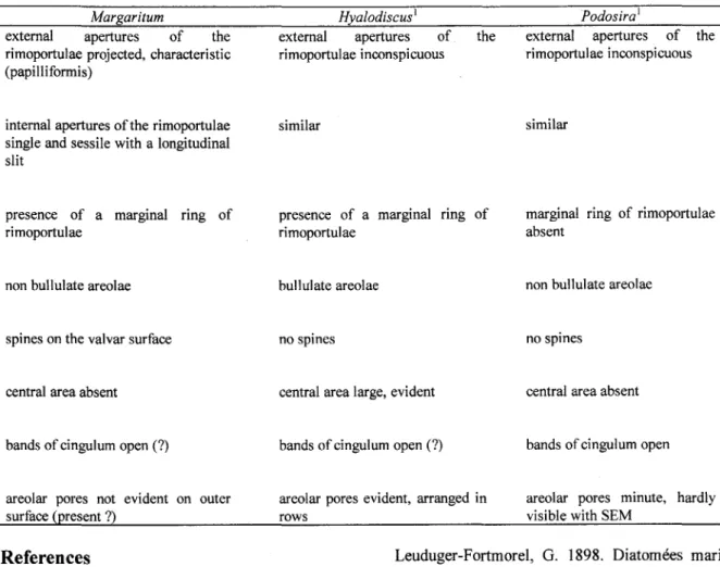
Margaritum some comparisons with related genera were made. Table 1 summarizes the main features of the genusMargaritum compared with the generaHyalodiscus andPodosira.
The genusPodosira does not poses a marginal ring of rimoportulae, as seen in the genus Margaritum. On the valvar surface ofPodosira the rimoportulae occur in greater numbers and are not ordered. Moreover, their external openings are sessile, i.e., lacking projections. InMargaritum the rimoportulae are ordered and characteristic (papilliformis). The foramina of the areolae are arranged in radial rows and lack rotae. Observations of the areolar structure inPodosira by Round et ai. (1990) showed rotae occ1uding foramina. Furthermore, these authors recorded no defined pattern in the areolar disposition, and c10sely packed areolae. The minute external pores of the areolae, as cited by Round et aI. (op. cit.) for Podosira were not present, or were not discernible in our samples of Margaritum with SEM. In
relation to the spines, these structures have not yet been found in Podosira, although they have been observed inMargaritum.
On the other hand, both genera show non-bululate areolae that, as suggested by Round et aI. (op. cit.), could be a potential criterion of separation between Hyalodiscus, Margaritum and Podosira. Copulae are also similar, although less developed in Margaritum, with 4-5 bands. Judging by the location of the copulae and the structure of the margin edge, it is likely that the species presented 2 cells joined by the cingulum, as in the case of Podosira. However, we were not able to find any specimen forming cell diplets in preserved samples.
The genus Hyalodiscus shows characteristics that are not present inMargaritum as a conspicuous central area and bullulate areolae (c1osely packed).
Inaddition, the rimoportulae of Hyalodiscus show differences in morphology regarding toMargaritum, although a marginal ring of rimoportulae is present in both genera. Stidolph (1993) describes the species H pustulatus A. Schmidt under SEM reporting ligulate open bands, spinules, peculiar baciliform structures on the inner side of the valve, and microlabiate processes. However, the species shows dissimilarities when compared with more typical representatives like H subtilis and H scoticus. Indeed, it seems to bear characteristics ofMelosira Agardh, as special loculate areolae with external ridges encirc1ing the pores, granules, spines and bands of cingulum (see figures 12, 13 and 23 of Stidolph, 1993).
We decided to place the genus
Margaritum within the Family Hyalodiscaceae, because its trustule morphology is more c10sely related to those ofPodosira andHyalodiscus. For instance, in all of them the loculate areolae and/or the disposition of the rimoportulae are similar.
As demonstrated trom the SEM study in the present work, we support the maintenance of the genus Margaritum as first proposed by Moreira-Filho (1968), due to the presence of typical rimoportulae papilliformis, marginal ring of rimoportulae (as in Hyalodiscus) and the non-bullulate areolae and bands of the cingulum (as inPodosira).
Margaritum terebro has been found in estuarine environments trom tropical regions of the World. It was collected trom the stomachs of benthic invertebrates (Leuduger-Fortmorel, 1898; Valente-Moreira et aI., 1994), epiphytic on macroalgae (Moreira-Filho & Valente-Moreira, 1980, 1981; Valente-Moreira et ai., 1980; Moreira-Filho et ai., 1990), and trom the sand gravels of intertidal zones (Hendey, 1958 and 1971). In the present study the
species was fairly common on glass slides used to analyze the seasonal dynamics of the periphyton in the Paranaguá Bay (Brandini, pers. com.*). All of these fmdings lead us to believe that M terebro grows in a benthic habitat, an environment that could be a unifYing characteristic among the representatives of the Family Hyalodiscaceae. Other authors have encountered M terebro in plankton samples, but always in shallow, turbulent regions of the sea (Fernandes et ai., 1990; Moreira-Filho et ai., 1975; Souza-Mosimann, 1984, 1985 and 1988; Souza-Mosimannet ai., 1989).
Acknowledgements
We thank COPEL (Companhia Paranaense de Energia Elétrica), and to Dr. Maurício P. Cantão (LAC) and Dra. Daura R. Stofella (CME!UFPR) for the use ofthe SEM; to Dr. J. P. Kocioleck (California Academy of Sciences) who kindly sent some papers. Our gratitude to Jones J. Bastos for his photographic work. The anonymous referees improved the manuscript of this paper.
Table 1. Comparative morphology between the GenusMargaritum and the related GeneraHyalodiscus andPodosira.
(1) ITomRoundet ai. (1990). Margaritum
external apertures of the rimoportulae projected, characteristic (papilliformis)
Hyalodiscusl
external apertures of the rimoportulae inconspicuous
Podosiral
external apertures of the rimoportulae inconspicuous
internal apertures of the rimoportulae single and sessile with a longitudinal slit
similar similar
presence of a marginal ring of rimoportulae
presence of a marginal ring of rimoportulae
marginal ring of rimoportulae absent
non bullulate areolae bullulate areolae
spines on the valvar surface no spines
non bullulate areolae
no spines
central area absent central area large, evident central area absent
bands of cingulum open (?) bands of cingulum open (?) bands of cingulum open
evident on outer areolar pores evident, arranged in
rows areolar pores minute, hardlyvisible with SEM
References
Femandes, L. F.; Souza-Mosimann, R. M. de & Felício-Femandes, G. 1990. Diatomáceas (Bacillariophyceae) do Rio Ratones, Florianópolis, SC, Brasil: baixo curso e estuário. Insula, 20: 11-112.
Hasle, G. R. & Fryxell, G. A. 1970. Diatoms: cleaning and mounting for light and electron microscopy. Trans. Am. micros. Soe., 89:469-474.
Hasle, G. R. 1972. Two types of valve processes in centric diatoms. Nova Hedwigia, 39:55-78.
Hendey, N. 1. 1958. Marine diatoms from some west African ports. 1. R. microsc. Soe., 77(1/2):28-85.
Hendey, N. L 1971. Some marine diatoms from the Galapagos Islands. Nova Hedwigia, 22:(1/2), 371-422.
Leuduger-Fortmorel, G. 1898. Diatomées marines de Ia cote occidentale d"Afrique. Mém. Soe. Emula1. Côtes Nord, p. 1-41. 8 pls.
Moreira-Filho, H. 1968. Marf{aritum (Podosira) tenebro (Leuduger-Fortmorel) Nov. Genus et nova comb. Bolm Univ. Fed. Paraná, Bot., 20: 1-4.
Moreira-Filho, H., Valente-Moreira, L M. & Tripia-Cecy, L 1975. Diatomáceas da Baía de Paranaguá, Estado do Paraná, Brasil (Chrysophyta-Bacillariophyceae). Bolm Mus. Bo1.Munic., 20:1-25.
Moreira-Filho, H. & Valente-Moreira, 1. M. 1980. Diatomáceas epífitas em Uiva fasciata Delile. Bolm Mus. Bot. Munic., 41: 1-10.
52 Rev. bras. oceanogr., 45( 1/2), 1997
Moreira-Filho, H. & Valente-Moreira, I. M.; Souza-Mosimann, R M. de & Cunha, J. A. 1990. Avaliação florística e ecológica das diatomáceas (Chrysophyta-Bacillariophyceae) marinhas e estuarinas nos Estados do Paraná, Santa Catarina e Rio Grande do Sul. Est. Biol.,25:5-48.
Ross, R; Cox, E. 1.; Karayeva, N. 1.; Mann, D. G.; Paddock, T. B. B. ; Simonsen, R. & Sims, P. A. 1979. An emended terminology for the siliceous components ofthe diatom cell. Nova Hedwigia, 64:513-533.
Round, F. E.; Crawford, R M. & Mann, D. G. 1990. The diatoms. Biology and morphology of the genera. Cambridge, Cambridge University Press.747p.
Souza-Mosimann, R. M. de 1984. Levantamento preliminar das diatomáceas (Chrysophyta-Bacillariophyceae) na região de Anhatomirim, Santa Catarina, Brasil. Insula, 14:2-46.
Souza-Mosimann, R M. de 1985. Contribuição ao conhecimento das diatomáceas (Chrysophyta-Bacillariophyceae) em algu-mas estações localizadas na Baía Norte, Florianópolis, Santa Catarina, Brasil. Insula, 15:3-31.
Souza-Mosimann, R M. de 1988. Estudo das diatomáceas (Chrysophyta Bacillariophyceae) da Baía Sul, Florianópolis, Santa Catarina, Brasil. Insula, 18:23-74.
Souza-Mosimann, R M. de; Felício-Femandes, G. & Femandes, L. F. 1989. Contribuição ao conhecimento das diatomáceas da Baía de Tijucas, Santa Catarina, Brasil. Insula, 19:95-122.
Stidolph, S. R 1993. A light and electron microscopical study of Hyalodiscus pustulatus A. Schmidt (Bacillariophyceae) &om New Zeland marine habitats. Bot. Mar., 36(2):79-86.
Valente-Moreira, I. M.; Moreira-Filho, H.; Cunha, J. A. & Ludwig, T. A. V. 1980. Diatomáceas epífitas em Padina vickersiae Hoyt ex Howe. Trib. Farmac., 48 (1-2):114-122.
Valente-Moreira, I. M.; Moreira-Filho, H. ; Cunha, 1. A. & Nakamura, I. T. 1994. Diatomáceas (Chrysophyta-Bacillariophyceae) no conteúdo estomacal de peixes e crustáceos do manguezal do rio Perequê, Pontal do Sul, Estado do Paraná, Brasil. Est. Biol., 3:99-114.