Universidade Federal de Minas Gerais – UFMG
Mailing address: Luiz Oswaldo Carneiro Rodrigues - Laboratório de Fisiologia do Exercício – Campus da Pampulha – UFMG - Av. Presidente Carlos Luz, 4664 – 31310-250 - Belo Horizonte – MG - Brazil
Purpose – To compare peak exercise oxygen con-sumption (VO2peak) of healthy individuals with
asympto-matic individuals with probable heart disease.
Methods – Ninety-eight men were evaluated. They were divided into two groups: 1) 39 healthy individuals (group N) with an age range of 50±4.6 years; and 2) 59 asymptomatic individuals with signs of atherosclerotic and/or hypertensive heart disease (group C) with an age range of 51.9±10.4 years. In regard to age, height, body surface area, percentage of fat, lean body mass, and daily physical activity, both groups were statistically similar. Environmental conditions during the ergometric test were also controlled.
Results – Maximal aerobic power (watts), VO2peak,
maximal heart rate, and maximal pulmonary ventilation were lower in group C (p<0.01) than in group N; weight, however, was lower in group N (p=0.031) than in group C. Differences in the respiratory gas exchange index, heart rate at rest, and the maximal double product of the two groups were not statistically significant.
Conclusion – Signs of probable heart disease, even though asymptomatic, may reduce the functional capacity, perhaps due to the lower maximal cardiac output and/or muscle metabolic changes.
Key words - functional capacity, oxygen consumption,
heart disease
Arq Bras Cardiol, volume 73 (nº 1), 6-10, 1999
Luiz Oswaldo Carneiro Rodrigues, Emerson Silami-Garcia, Maria da Consolação Vieira Moreira, Giane Amorim Ribeiro
Belo Horizonte, MG - Brazil
Assessment of Functional Capacity through Oxygen Consumption
in Patients with Asymptomatic Probable Heart Disease
Maximal oxygen consumption (VO2max) has been considered the best indicator of the human capacity for tolerating prolonged effort 1. Its assessment, however, has some technical difficulties, especially in individuals with lower physical fitness or those limited by heart disease 2. Therefore, the greatest oxygen consumption during exercise, called peak exercise oxygen consumption (VO2peak), is considered an objective indicator of functional capacity. It may also provide an indirect and accurate assessment of cardiac reserve 3. VO
2peak is considered particularly reliable in functional assessment when associated with measurement of anaerobic metabolism4, either through measurement of blood lactate 5,6 or through recording ventilatory variables 2, in young and middle-aged individuals 7.
Several authors have shown a strong correlation be-tween exercise capacity assessed through VO2peak and the prognosis and severity of congestive heart failure (CHF): the one-year mortality rate is higher in patients with VO2peak below 10 mL/kg.min; this group of patients can benefit from heart transplantation 8,9. VO
2peak may also reveal changes in exercise capacity due to the use of medicines, reflecting, more precisely, the compensatory adjustments and the prognosis of the CHF than any other hemodynamic and clinical indicator 2,10-14.
VO2peak, therefore, has become an important prognostic tool in the evaluation and selection of candidates for cardiac transplantation. All patients with stable terminal CHF should have their functional capacity evaluated through the direct measurement of oxygen consumption, as part of the mandatory procedures for selection for cardiac transplan-tation 15. Most of the patients with VO
2peak higher than 14mL/ kg.min-1 can wait longer for transplantation even though VO2peak is only one of the criteria to be considered in each patient 13.
Although VO2peak provides an indicator for cardiac output (CO) and cardiac reserve, factors, such as age, sex, level of physical fitness, muscle mass, and angina, can influence its results 2. For example, a VO
capacity expected in an active 60-year-old man, but only 30% of the capacity of an active 20-year-old man; such a low exercise capacity would be an unacceptable quality of life for a young adult 16.
On the other hand, despite the recognized usefulness of the VO2peak in measuring symptomatic heart diseases, its value in asymptomatic or poorly symptomatic (NYHA class I) heart diseases is still being discussed 17. This study aimed to approach this question through a comparison of healthy individuals with asymptomatic individuals with signs of atherosclerotic and/or hypertensive heart disease.
Methods
This study was approved by the Ethics Committee of the Hospital das Clínicas of the Federal University of Minas Gerais (UFMG). The sample comprised 98 male volunteers, who signed a consent form, agreeing with the methodology of the study. They were divided into two groups: one with 39 healthy individuals (group N) and the other with 59 asymptomatic individuals with indicative (57.6%) or suggestive (43.2%) electrocardiographic signs of heart disease (group C). The indicative electrocardiographic signs of heart disease were the following: depression of the ST segment >2mm, during effort; complete bundle-branch block; left ventricular overload; and frequent ventricular extrasystoles triggered by effort. The electrocardiographic signs suggesting heart disease were the following: straigh-tening and depression of the ST segment >1mm and <2mm, during effort; unspecific disorders of intraventricular conduction; unspecific alterations of repolarization; arrhythmias, such as nonsustained paroxysmal supraven-tricular tachycardia and isolated vensupraven-tricular extrasystoles. In addition, group C also showed evidence of heart disease on coronary angiography (13.5%) and/or echocardiogram (11.8%). Group C, as a whole, comprised individuals with probable atherosclerotic and/or hypertensive heart disease; there was no suspicion of Chagas’ cardiomyopathy, valvar disease nor any other disease.
Both groups underwent direct assessment of the VO2peak through the direct method of spirometry with the use of an Ergopneumotest metabolimeter. Indirect determi-nation of the anaerobic metabolism was also performed, as well as the determination of other metabolic, cardiovascular and ventilatory variables during standard exercise until exhaustion 12, in a electronically braked bicycle ergometer (Monark), at a speed of 18 km/h. All patients underwent an ergometric test until exhaustion, characterized as the mo-ment at which the predetermined potency and/or velocity could no longer be maintained. The examiners did not interrupt the tests. The maximal aerobic power (POW) was equivalent to the highest value observed during at least 3 minutes of exercise and VO2peak was obtained during this period. Exercise was subjectively evaluated through the rating of perceived exertion (REP) (from 0 to 10) scale 18. Electrocardiogram (ECG) and heart rate (HR) were conti-nuously monitored during rest and exercise until exhaustion
and also for 10 minutes during recovery. Blood pressure (BP) was obtained through a sphygmomanometer gra-duated in mmHg. Bicycle ergometetric tests were performed in an environment with air conditioning to maintain tempe-rature (between 21 and 24°C) and relative humidity (between 50 and 75%) levels so that heat stress was unlikely 19. The impact of heat stress was calculated through the wet bulb globe temperature index (WBGTI) 20,21. Tests comparing the mean of variables with normal distribution were performed, as well as nonparametric tests, when necessary, adopting the significance level of p<0.05.
Results
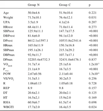
Table I shows that groups N and C were similar in re-gard to the following variables: age, usual physical activity, HR at rest, maximal cardiac work, ventilatory equivalent for oxygen, BP variation during exercise, respiratory gas ex-change index, REP at the potency of 100 watt, dry environ-mental temperature, humid environenviron-mental temperature, relative humidity of the air.
Table I also shows that group C had greater weights,
Table I – Values (mean ± standard deviation) obtained in the group of healthy individuals (group N) and the group of asymptomatic individuals with signs of probable heart disease (group C) for the
following parameters: age, weight, usual physical activity (UPA), systolic blood pressure (SBP), diastolic blood pressure (DBP), heart rate
(HR) at rest and during exercise, peak exercise oxygen consumption (VO2peak), ventilatory equivalent (VE/VO2), respiratory gas exchanges index (R), rating of perceived exertion scale (REP), double product (DP),
maximal aerobic power (POW), dry environmental temperature (DT), humid environmental temperature (HT), relative humidity of the air
(RHA), and the wet bulb globe temperature index (WBGTI).
Group N Group C p
Age 50.0±4.6 51.9±10.4 0.221
Weight 71.5±10.1 76.4±12.1 0.031
UPA 3.5±1.9 4.1±2.6 0.207
HRrest 68.4±11.1 71.0±11.4 0.273
SBPrest 125.9±11.1 147.7±17.5 <0.001
DBPrest 81.6±6.0 94.1±12.0 <0.001
DPrest 8612.1±1597.1 10533.8±2341.6 <0.001
HRmax 165.0±11.9 150.3±16.8 <0.001
SBPmax 195.1±26.1 215.3±29.2 <0.001
DBPmax 92,9±11,7 107.7±17.1 <0,001
DPmax 32203.4±4732.3 32431.0±6176.1 0.837
VO2peak 31.7±7.4 25.1±5.6 1.2x10⁻⁵
Poxy 21.1±4.9 16.7±3.5 <0.001
POW 2,67±0.56 2.11±0.44 1.3x10⁻⁶
VE/VO2 31.5±5.1 30.2±5.5 0.256
R 1.06±0.13 1.05±0.18 0.728
REP 8.9 9.8 0.157
DT 20.6±2.1 20.0±2.1 0.125
HT 16.5±2,1 15.9±2.0 0.169
RHA 60.9±6.7 61.5±7.4 0.698
WBGTI 17.7±2.0 16.4±3.5 0.024
Where: weight in kg; age in years; UPA in multiples of the basal metabolism, MET; SBP and DBP in mmHg; DBP in mmHg; HR in min-1; DP in mmHg.min-1;
VO2peak in ml/kg.min-1; VE/VO2 and R in absolute figures; REP in the power of 100
higher BP at rest and during exercise, and a higher cardiac work at rest than the healthy individuals of group N. On the other hand, group C shows a lower maximal HR, lower peak exercise oxygen consumption, lower oxygen pulse (Poxy) and lower POW during exercise. In addition, the environ-mental heat stress of group C was lower during exercise.
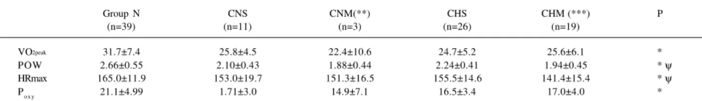
Table II shows that, when group C was subdivided in regard to the presence or absence of hypertension and use of medication there were no differences in the peak exercise oxygen consumption and oxygen pulse. In the subgroup of hypertensive patients, however, the use of medication reduced the maximal aerobic power and maximal HR.
Discussion
The measure of VO2peak in healthy individuals in this study showed results consistent with those obtained in previous studies 22 in regard to the age of the volunteers 2,16,23. The lower VO2peak in group C, when compared with that of healthy individuals in the same age range, suggests that the presence of signs of heart disease, even though the patient is asymptomatic, can reduce functional capacity 24. In the present study, this decrease was 2 METs, represen-ting a significant reduction in the physical capacity for work, leisure and daily activities, corresponding to 14 to 16 years of early aging16. A decrease of 3 METs was also observed in a group of individuals with Chagas’ heart disease, when compared with healthy individuals in the same age range 17. Findings of the present study also suggest that both the direct measure of oxygen consumption (VO2peak, in mL/ kg.min-1) and the measure of the maximal aerobic power (POW, in watt/kg) can identify the reduction in functional capacity, even for those asymptomatic individuals with signs of heart disease (Table I). POW, however, was statis-tically 10 times more sensitive than VO2peak for detecting a reduction in the functional capacity of group C participants, when compared with the healthy individuals. In addition, POW revealed some significant differences due to hyper-tension and use of medication in group C, which did not occur with VO2peak (Table II). These findings are not in accordance with that which has been postulated for the diagnosis of the functional capacity in patients with heart disease 25 and also for the prescription of exercise 7. However,
the highest sensitivity of POW in the assessment of the functional capacity has also been observed for women 26.
Some mechanisms may be involved in the decrease of the functional capacity in group C, for example a reduction in the maximal CO (Qmax) due to the probable heart disease, hypertension or the use of medication; muscle metabolic alterations; and the iatrogenic sedentary lifestyle caused by the diagnosis.
At the beginning, the simple presence of signs of heart disease as a criterion of inclusion in group C could suggest that the limiting factor in the functional capacity would be a reduction in Qmax. Even though this study did not assess Qmax directly, the maximal cardiac work of group C (Table I) was similar to that of the healthy group, and this similarity of the maximal double product (DPmax) occurred due to a lower HRmax in group C, resulting from a higher BP in this group. This implies that when the same maximal cardiac work is reached with a lower HR, there is a smaller Qmax in group C, even though the stroke volume is kept constant 27.
When group C, however, is subdivided into normo-tensive and hypernormo-tensive individuals (Table II), one obser-ves that peak exercise oxygen consumption (VO2peak) and oxygen transportation per cardiac beat (Poxy) are not diffe-rent in the two groups, indicating that the presence of hy-pertension is not the main cause of the lower capacity of oxygen uptake and transportation among individuals in group C. Hypertension, however, caused a reduction in POW in this group, suggesting the existence of a peripheral reducing factor.
Observation of Poxy shows that less oxygen was con-sumed and/or less blood was transported per cardiac beat in group C (Table I), which could result from the lower stroke volume or from the smaller peripheral extraction of oxygen. It is known that chronic heart failure renders the skeletal muscle function difficult, independently from blood flow and from the availability of oxygen 28,29. It is also known that individuals with symptomatic heart diseases can have metabolic dysfunctions of the skeletal muscles, which reduce their functional capacity 30. Observation of the respiratory gas exchange index and of the ventilatory equivalent for oxygen (VE/VO2) shows that group C reached the same relative level of lactic metabolic acidosis, but with absolute lower efforts than healthy individuals. This
Table II – Values (mean ± standard deviation) of peak exercise oxygen consumption (VO2peak ), maximal aerobic power (POT), maximal heart rate (HRmax) and
oxygen pulse (Poxy) obtained for groups N and C, the latter subdivided into two subgroups: CNS- normotensive individuals without medication;
CNM-normotensive individuals with medication; CHS- hypertensive individuals without medication; CHM- hypertensive individuals with medication.
Group N CNS CNM(**) CHS CHM (***) P
(n=39) (n=11) (n=3) (n=26) (n=19)
VO2peak 31.7±7.4 25.8±4.5 22.4±10.6 24.7±5.2 25.6±6.1 *
POW 2.66±0.55 2.10±0.43 1.88±0.44 2.24±0.41 1.94±0.45 * ψ
HRmax 165.0±11.9 153.0±19.7 151.3±16.5 155.5±14.6 141.4±15.4 * ψ
Poxy 21.1±4.99 1.71±3.0 14.9±7.1 16.5±3.4 17.0±4.0 *
Where: VO2peak in ml/kg.min-1; POW in watt/kg; HR
max : maximal heart rate, in min-1; Poxy: in ml O2. min-1; (*): significant differences (p<0.05) comparing the
finding indicates the precocious predominance of the anaerobic glycolysis, strengthening the hypothesis that the best functional capacity can result either from the lo-west blood supply to the muscles (smaller stroke volume) or from the smallest peripheral extraction of oxygen (muscle metabolic dysfunction) in group C.
The use of some medicines, especially those blockers of the sympathetic activity upon the heart, may decrease the capacity of adequately adjusting CO during exercise, reducing the blood supply to tissue, resulting in a smaller physical capacity in group C. In this group, 57% of the patients used medicines that were potential cardiac capa-city reducing agents, resulting in some degree of chrono-tropic block and reduction in the maximal power in the hypertensive patients during exercise. There were, however, no significant differences in regard to VO2peak and no significant repercussion on the response of BP during exercise (Table II). These data do not support the hypo-thesis that group C would have a smaller Qmax due to the use of medicines, even though the drugs caused a reduction in maximal power.
Lack of physical fitness is one of the most important factors in the reduction of functional capacity at any age 23. Medical and/or cultural factors could contribute to resis-tance of patients to becoming involved in physical activities more effective in promoting fitness. In the present study, group C had an usual mean physical activity similar to that of healthy individuals (Table I), i.e., they do not have a more sedentary lifestyle than healthy individuals, not justifying the smallest functional capacity observed in group C. The
1. Sutton JR. VO2max. New concepts on an old theme. Med Sci Sport Exerc 1992; 24:
26-9.
2. Itoh, H, Taniguchi K, Koike A, Doi M. Evaluation of severity of heart failure using ventilatory gas analysis. Circulation 1990; 81(suppl II): 31-7. 3. Wilson JR, Mancini DM. Factors contributing to the exercise limitation of heart
failure. J Am Coll Cardiol 1993a; 22(suppl A): 93-8.
4. Romano A, Silva PRS, Ramirez JAF, Mady C, Yasbek Jr P. Exercício físico na insuficiência cardíaca crônica estável. In: Yasbek P, Battistella LR. Do Atleta ao Transplantado. Condicionamento Físico. São Paulo: Sarvier 1994: 191-211. 5. Jennings GL, Esler MD. Circulatory regulation at rest and exercise and the
functional assessment of patients with congestive heart failure. Circulation 1990; 81(suppl II): 5-13.
6. Consenso Nacional de Ergometria. Arq Bras Cardiol 1995; 65: 191-211. 7. Rondon MAPB, Forjaz CLM, Nunes N, Amaral SL, Barreto ACP, Negrão CE.
Comparação entre a prescrição de intensidade de treinamento físico baseada na avaliação ergométrica convencional e na ergoespirometria. Arq Bras Cardiol 1998; 70: 159-66.
8. Mancini DM, Eisen H, Kussmaul W, Mulkl R, Edmunds LH, Wilson JR. Value of peak exercise oxygen consumption for optimal timing of cardiac transplantation in ambulatory patients with heart failure. Circulation 1991; 83: 778-86. 9. Cohn JN, Johson GR, Shabetai R, et al. Ejection fraction, peak exercise oxygen
consumption, cardiothoracic ratio, ventricular arrythmias and plasma norepinefrine as determinant of prognosis in heart failure. Circulation 1993; 87(suppl VI): 5-16.
10. Weber KT, Janicki JS. Respiratory gas exchange during exercise in the noninvasive evaluation of the severity chronic cardiac failure. In: Weber KT,
coefficient of variation of usual physical activity in this study, however, was very high, indicating the need for fur-ther studies.
The REP, measured in the power of 100 watt and rea-ched by all the individuals, was not statistically different between the two groups N and C, showing that the REP scale was not sensitive to the reduction in the functional capacity. Perhaps this may occur because the individuals in group C were still at a higher functional level than that described as a threshold (18 mL/kg.min-1) for the subjective perception of their own dysfunction 31.
Finally, the room temperature during the tests was slightly cold 19. In such an environment, possible cardio-vascular overload resulting from heat stress was statis-tically higher than that of environmental conditions in which the healthy individuals underwent ergometric tests. This would not explain, however, the reduction in the functional capacity observed in group C. Nevertheless, the magnitude of the increase observed in the WBGTI between the two environments does not seem to cause significant variations in performance during physical activity 31.
In conclusion, the lower VO2peak and POW found in group C indicate that signs of heart disease, even though asymptomatic, can significantly decrease functional capacity. The measure of POW seems to be more sensitive than that of VO2peak for detecting a reduction in functional capacity caused by the asymptomatic probable heart di-sease. The mechanisms of this decrease have not yet been well clarified and reduction in the maximal CO and/or peripheral metabolic changes are suggested as hypotheses.
References
Janicki JS, (eds). Cardiopulmonary Exercise Testing: Physiologic Principles and Clinical Applications. Philadelphia: WB Saunders, 1986: 221-35. 11. Mady C, Cardoso RHA, Barretto ACP, Luz PL, Bellotti G, Pileggi F. Survival
and predictors of survival in patients with congestive heart failure due to Chagas’ Cardiomyopathy. Circulation 1994; 90: 3098-102.
12. Smith RF, Johnson, G, Ziesche S, Bath G, Blankenship K, Cohn JN. Functional capacity in heart failure. Comparison of methods for assessment and their relation to other index of heart failure. Circulation 1993; 87(suppl VI): 88-93. 13. Moreira MCV, Figueroa CCS. Transplante cardíaco. In: Pereira AW. Transplantes
de Órgãos e Tecidos. Rio de Janeiro: Medsi, 1995: 235-59.
14. DeGroote P, Millaire A, Decoulx E, Nugue O, Guimier P, Ducloux G. Kinetics of oxygen consumption during and after exercise in patients with dilated cardiomyopathy. J Am Coll Cardiol 1996; 28: 168-75.
15. Bethesda Conference. Guideline for heart transplantation. J Am Coll Cardiol 1993; 22: 21-30.
16. Stamford BA. Exercise and the elderly. Exerc Sport Sci Rev 1988; 16: 341-79. 17. Mady C, Cardoso RHA, Ianni BM, et al. Capacidade funcional máxima “normal”
em pacientes com insuficiência cardíaca congestiva por miocardiopatia chagásica. Arq Bras Cardiol 1996; 67: 1-4.
18. Borg G, Hassmen P, Lagstroem M. Perceived exertion related to heart rate and blood lactate during arm and leg exercise. Eur J Appl Physiol Occup Ther 1987; 56: 679-85.
19. Haymes EM, Wells LL. Environment and Human Performance. Illinois: Cham-paign Human Kinectics Publishers, 1986: 120-5.
21. McCann DJ, Adams WC. Wet bulb globe temperature index and performance in competitive runners. Med Sci Sports Exerc 1997; 29: 955-61.
22. Hansen JE, Sue DY, Wasserman K. Predicted values for clinical exercise testing. Am Ver Resp Dis 1984; 129(suppl): S49-S55.
23. Kenney WL. Thermoregulation at rest and during exercise in healthy older adults. Exerc Sports Sci Ver 1997; 25: 41-76.
24. Mady C, Barreto ACP, Nacruth R, Mesquita ET, Bellotti G, Pileggi F. Maximal functional capacity in patients with cardiomyopathy due to Chagas’ disease without congestive heart failure. J Am Coll Cardiol 1993; 21: 103A. 25. Young DR, Steinhardt MA. The importance of physical fitness for the reduction of
coronary artery disease risk factors. Sports Med 1995; 19: 303:10.
26. Bishop D, Jenkins DG, Mackinnon LT. The relationship between plasma lactate parameters, Wpeak and 1-h cycling performance in women. Med Sci Sports Exerc
1998; 30: 1270-5.
27. Semigran MJ, Thaik CM, Fifer MA, Boucher CA, Palacios IF, William GD -Exercise capacity and systolic and diastolic ventricular function after recovery from acute dilated cardiomyopathy. J Am Coll Cardiol 1994; 24: 462-70. 28. Massie BM, Conway M, Rajagopalan B, Yonge R, Frostick S, Ledingham J.
Skeletal muscle metabolism during exercise under ischemic conditions in congestive heart failure. Evidence for abnormalities unrelated to blood flow. Circulation 1988; 78: 320-6.
29. Xu L, Poole DC, Musch TI. Effect of heart failure on muscle capillary geometry: implications for O2 exchange. Med Sci Sports Exerc 1998; 30: 1230-7.
30. Wilson JR, Mancini DM. Skeletal muscle metabolic dysfunction. Implications for exercise intolerance in heart failure. Circulation 1993b; 87(suppl VII): 104-9. 31. Morey MC, Pieper CF, Cornoni-Huntley J. Is there a threshold between peak