1
Arquivos Brasileiros de Cardiologia - Volume 84, Nº 4, Abril 2005
Case Report
Percutaneous Transluminal Coronary Angioplasty in a
Patient with Idiopathic Thrombocytopenic Purpura
Luis Gustavo M. Marques, Murillo Kenji Furukawa, Thenyson Pereira Leitão,
José Luis A. Quiñones, Fernando César de Queiroz, Rogerio Felippe Tiossi,
Virgílio Ribeiro Franco Jr, Carlos Eduardo M. Domingues, Decio Salvadori Jr
São Paulo, SP - Brazil
Real e Benemérita Associação Portuguesa de Beneficência, São Paulo Mailing address: Murillo Kenji Furukawa - Rua Haddock Lobo, 1459/ 102 - Cep 01414-003 - São Paulo, SP, Brazil E-mail: [email protected]
Sent for publication: 04/30/2004 Accepted for publication: 08/03/2004 English version by Stela Maris Costalonga
The association between coronary heart disease and throm-bocytopenic purpura is rare and poses some difficulties when myocardial revascularization is necessary. We report a case with this association and significant coronary impairment, which was percutaneously treated with stent implantation.
The incidence of idiopathic thrombocytopenic purpura, also known as Werlhof’s disease, is low in the general population. It occurs in all age groups, in acute and chronic forms. Children usually have the acute form, with a similar incidence in both sexes. Adults, on the other hand, usually have the chronic form, with a higher incidence in the female sex (3:1)1. The pathophy-siology of both forms is the production of anti-platelet antibodies, usually of the IgG class, that lead to accelerated destruction of platelets by the reticuloendothelial system.
The association of purpura and coronary heart disease is also rare. We report a case of that association, which was percuta-neously treated with intracoronary stent implantation.
Case Report
The patient is a 54-year-old male, complaining of typical pre-cordial pain for 4 months associated with dyspnea on medium exertion, with rapid progression. He also reported that in the week preceding hospitalization, the pain was triggered by minimum exertion, appearing even at rest.
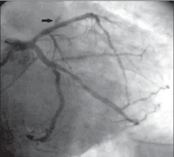
As antecedents, the patient reported hypertension and diabe-tes, and the use of captopril (75 mg/dL) and glibenclamide (10 mg/dL). He also reported having thrombocytopenic purpura and being on regular hematological follow-up in his city, and was being treated with prednisone (60 mg/dL), with no pathological bleeding in recent months. He denied familial antecedents of co-ronary heart disease, tobacco smoking, and hypercholesterolemia. While still in his city, he underwent cine coronary angiography, which showed coronary circulation with left dominance and severe lesions in the following arteries: anterior descending (AD), cir-cumflex (Cx), and the first left marginal branch (MB-1) (fig. 1).
The patient also had anomalous origin of the right coronary artery (RC) from the proximal third of the AD, close to the lesion in the latter, and the RC had a significant lesion in its origin.
The patient was referred for coronary artery bypass grafting. On admission, his hemogram was as follows: Hb, 10.4 g/dL; Ht, 28.8%; and very low platelet count (8,000 plat/mm3). No other laboratory alterations were observed.
The patient was assessed at the hematology unit. Initially, he had his anemia and low platelet count treated. Methylprednisolone and immunoglobulin were also administered, and the patient was indicated for surgery only after achieving a platelet count of at least 90,000 plat/mm3. However, even after several transfusions for 20 days, the maximum count achieved was 86,000 plat/mm3 (fig. 2). During that period, the anginal pain worsened, and the patient was transferred to the coronary unit and was administered high doses of intravenous nitroglycerin (Tridil). Because his platelet count did not improve, assessment by an interventional cardiologist was requested, aiming at performing percutaneous transluminal coronary angioplasty.
The previous electrocardiogram showed sinus rhythm, with diffuse and unspecific alterations in ventricular repolarization.
Then the patient underwent dissection of the right brachial artery, which was uneventful. On the day of the procedure, the patient had a platelet count of 15,000 plat/mm3. After dissection of the brachial artery, intravenous heparin was administered at the dose of 70 U/kg. A 6F Wiseguide guiding catheter, FL 3.5 SH model, and a 0.014” guidewire, 182 cm, Choice PT Extra Support model (both from Boston Scientific – SCIMED), were used. Initially, the lesion in the Cx artery was approached and easily surpassed by the guidewire. Then, a Driver-RX stent, 3.5x15 mm (Medtronic AVE), was directly implanted and deployed at 12 atm. The guide-wire was then redirected towards the AD artery. The lesion was, once again, surpassed without great difficulty. Another Driver-RX stent, 3.5x18 mm, was directly implanted, then deployed at 14 atm, with a good final result (fig. 3). Although the RC artery had an anomalous origin from the AD artery, and the first left marginal branch originated close to the lesion in the Cx artery, no occlusion was observed. A significant reduction in the RC artery flow was observed (TIMI I), but it was not accompanied by precordial pain or hemodynamic instability.
2
Arquivos Brasileiros de Cardiologia - Volume 84, Nº 4, Abril 2005
Percutaneous Transluminal Coronary Angioplasty in a Patient with Idiopathic Thrombocytopenic Purpura
Fig. 1 - A) Caudal RAO view of the LC artery; B) LAO view of the LC artery. The arrows show the anomalous origin of the RC artery.
Plat/mm3
100.000
80.000
60.000
40.000
20.000
0 2
0/1 21/1 22/1 23/1 24/1 25/1 26/1 27/1 28/1 29/1 30/1 31/1 1/2 2/2 3/2 4/2 5/2 6/2 7/2 8/2 9/2
Days
Fig. 2 - Evolution of platelet count during hospitalization. Fig. 3 - Caudal RAO view of the LC artery after PTCA; the arrow shows the flow
reduction in the RC artery.
CKMB (U/I)
50
40
30
20
10
0
PRE 8 18 24 36 60
Hours after PTCA
Fig. 4 - Curve with the CK-MB values (NV: up to 25 U/L). During hospitalization, the patient evolved well, with
impro-vement of the anginal pain. The CK-MB level before percutaneous transluminal coronary angioplasty (PTCA) was 42 U/L, with nor-malization within the first 24 hours (fig. 4). The electrocardiogram after PTCA showed no alterations as compared with that preceding PTCA.
Although the hematology team did not recommend the use of anti-platelet agents, we chose to use enoxaparin, subcutaneously, at the dosage of 1 mg/kg/day during the in-hospital period. However, within the first 24 hours of drug administration, 3 episodes of epistaxis occurred, and the new platelet count was 11,000/mm3.
Enoxaparin was suspended, and the patient was maintained with no type of heparin or anti-platelet agent. After 2 days, the patient was discharged from the hospital, and evolved with no precordialgia or bleeding. He was instructed to undergo strict hematological control in his city, and advised to use at least one anti-platelet agent when his platelet count allowed it.
During recent telephone contact, 4 months after the procedure, the patient reported having no angina. During that period, he had not begun using any anti-platelet agent, due to the recommendation of his hematologist, because of a persistent significantly low platelet count, which achieved a maximum of 20,000 plat/mm3, but most values were below 10,000. The last platelet count (06/22/04) was 3,000 plat/mm3. The patient continues to use corticoids and immunoglobulins, and waits for improvement so that he can un-dergo a splenectomy. He is still using captopril (50 mg/dL), atenolol (100 mg/dL), and isosorbide mononitrate (30 mg/dL).
Discussion
3
Arquivos Brasileiros de Cardiologia - Volume 84, Nº 4, Abril 2005 Percutaneous Transluminal Coronary Angioplasty in a Patient with Idiopathic Thrombocytopenic Purpura
1. Burns TR, Saleem A. Idiopathic thrombocytopenic purpura. Am J Med 1983; 75:
1001-7.
2. Oda K, Fujimura K, Kuramoto A. ITP (Idiopathic Thrombocytopenic Purpura).
Nitijo Shinryo To Ketsueki 1995; 5: 25-30.
3. Mathew TC, Vasudevan R, Leb L et al. Coronary artery bypass grafting in immune
thrombocytopenic purpura. Ann Thorac Surg 1997; 64: 1059-62.
References
4. Thompson LD, Cohen AJ, Edwards FH, Barry MJ. Coronary artery bypass in
idi-opathic thrombocytopenia without splenectomy. Ann Thorac Surg 1989;48:721-2.
5. Fuchi T, Kondo T, Sase K et al. Primary Percutaneous Transluminal Coronary
An-gioplasty Performed for Acute Myocardial Infarction in a Patient With Idiopathic Trombocytopenic Purpura. Jpn Circ J 1999;63:133-6.
Our patient had an aggressive form of idiopathic thrombocy-topenic purpura associated with severe obstructive coronary heart disease. A possible explanation for the significantly low platelet count, with an inexpressive therapeutic response, could be the concomitance of important levels of stress, generated by precor-dialgia and the imminence of intricate surgery.
Although systems for local hemostasia of the femoral puncture, such as the Perclose – AbbotVascular Devices, were available, the possibility of occurrence of accidents during arterial puncture was considered, which could cause, due to the significantly low platelet count, hemorrhagic complications even before initiating the interventional procedure. The radial approach was not used because the necessary material was not available, and our profes-sional team lacked experience with that technique. Therefore, we chose to dissect the right brachial artery, which allowed direct visualization of the artery and better control of hemostasia. Although the patient had already undergone a previous cine coro-nary angiography by use of that technique, his brachial artery was easily palpable above the first incision, with no decrease in the radial pulse.
Although the patient had a significantly low platelet count, we considered it prudent to use at least enoxaparin, because 2 stents were implanted in the AD and CX arteries, which were dominant. However, the occurrence of epistaxis caused the sus-pension of that drug, and the patient was maintained without any type of heparin or antiplatelet agent.
It is worth noting that some type of antiplatelet agent should be introduced as soon as possible. Fuchi et al 5 performed a primary percutaneous transluminal coronary angioplasty in a patient with
idiopathic thrombocytopenic purpura and acute myocardial in-farction, who had subocclusion of the AD artery in its proximal third and another lesion, of 50%, in its middle third. Although percutaneous transluminal coronary angioplasty of the proximal lesion was successfully performed, 9 hours later, the patient ex-perienced a new episode of acute myocardial infarction with oc-clusion in the middle third of the DA artery, close to the 50% lesion. The authors considered the possibility of an iatrogenic lesion caused by the guidewire during the first percutaneous trans-luminal coronary angioplasty, associated with an increase in blood viscosity caused by the contrast medium and platelet replacement. They emphasized the importance of carefully performing transfu-sions, as less frequently as possible, to avoid thrombogenic com-plications after percutaneous transluminal coronary angioplasty in patients with thrombocytopenic purpura.
We recommended a careful hematological follow-up for our patient, with the introduction of at least one antiplatelet agent as soon as the platelet count was acceptable, and that agent should be maintained for a period sufficient to allow stent endothelialization.