ABSTRACT
http://dx.doi.org/10.1590/1678-775720140243
Inluence of the magnetic ield on microorganisms
in the oral cavity
Snezana BRKOVIC1, Srdjan POSTIC1, Dragan ILIC2
1- Clinic for Prosthodontics, School of Dental Medicine, Rankeova 4, University of Belgrade, Belgrade, Serbia. 2- Clinic for Endodontics, School of Dental Medicine, Rankeova 4, University of Belgrade, Belgrade, Serbia.
Corresponding address: Snezana Brkovic - Clinic for Prosthodontics, School of Dental Medicine, Rankeova 4, University of Belgrade - Beograd - Serbia - Phone: +381 11 319 33 07/063 13 02 963 - e-mail: [email protected]
Submitted: July 11, 2014 - Modiication: October 27, 2014 - Accepted: October 30, 2014
S
ince the beginning of their lives, all living organisms are exposed to the inluence of geomagnetic ields. Objectives: With respect to the positive effects that magnetic ieldshave on human tissues, especially the bactericidal effect, this investigation aimed to assess their inluence on the reduction of oral microorganisms. Material and Methods: In order
to obtain adequate specimens of dental plaque deposit, microbes such as Streptococcus
parasanguinis, Staphylococcus epidermidis, Rhodococcus equi and Candida albicans
were isolated from the human mouth. To establish the intensity of microbial growth on the basis of the modiied optical density (OD) of agar turbidimetry assay, microbial count and spectrophotometry were applied. The study was carried out with two microbial concentrations (1 and 10 CFU/ml) after periods of incubation of 24 and 48 h and using micromagnets. Results: A positive effect of the magnetic ield, resulting in the reduction
of dental plaque microbes in vitro, was found. In the irst 24 hours of exposure to the
magnetic ield, the number of all isolated microbes was signiicantly reduced. The most potent inluence of magnets and the most intensiied reduction after 24 hours were evident in Candida albicans colonies. The decrease in the inluence of magnets on microbes in vitro was also detected. Conclusions: Magnets reduce the number of microbes and might be recommended as a supplement in therapy for reduced periodontal tissues. This is important because periodontal tissues that are in good conditions provide prolonged support to the oral tissues under partial and supradental denture.
Keywords: Microbes. Oral cavity. Magnetic field. Overdenture. Candida albicans.
Streptococcus parasanguinis.
INTRODUCTION
Living creatures have been exposed to the inluence of geomagnetic and electric ields since the beginning of their lives on Earth. There is a lot of evidence and studies about magnet applications in medicine8,12,13,14,23. One of them, using lab rats,
has been performed in the conditions of 60 times the power of the geomagnetic ield. Growth on these animals has been reported on the 5th, 13th and 26th
days of the postnatal period, prior to euthanasia. The animal mortality increased by 30% when compared with control groups. Moreover, it has been observed that young animals showed a state of hyper mobility which has been followed by paralysis. Yellow smear have been found on their livers, and
,
cirrhosis was frequent among rats22. According to
the information learned, living organisms create a reaction of effectors that generates changes in the inner electromagnetic ield. These changes have been modiied by periodical transformations, occasionally related to the non-periodic luctuations of the outer electromagnetic ield of the earth20.
In experiments in which E. coli has been added
to the water running between two magnetic polarities with a speed of 0.5 L per hour and
intensity of magnetic ield of 39.9 KA/m, a number of bacteria have been reduced from 105 to 0. This
present3,4,22.
In favor of antibacterial effect, it could be stated that in the case of the laboratory animals with infected fractured bones, the magnetic ield increased the effect of antibiotics with reduction of posttraumatic oedema and acceleration of fracture healing11,20,22.
Electromagnetic fields in the region of the edentulous ridge signiicantly decrease and slow down the residual ridge reduction (RRR)22.
A relation between inluence of the magnetic ield and reduction of microbes in natural water has also been evidenced. Microbes act in a “magnetic-sensitive” manner. This means that microbes have been oriented and distributed along force lines within environments rich in ferric and magnetic oxides. This orientation with transition of microbes has been designated “magnetic-vowing”, which means microbe orientation to the magnetic ield. Magnet-sensitive microbes have been evaluated in early 1970s. During changes of direction within the ield itself, microbes turn around to reach the direction of the magnetic ield. This phenomenon has been added to a number of studies on the various microbes with two common attributes, all of them were anaerobic and their magnetic osmolarity due to the intracytoplasmic structure of Fe3O4.
A number of studies report the inluence of the electromagnetic ield on the inhibition of growth of microbes and fungi, as well as slowing down their multiplication19.
There are controversial opinions about microbe growth. For example, the results of experiments with the well-studied Vibrio campbellii strain
BB120 (originally classified as Vibrio harveyi)
and derivative mutants unable to synthesize autoinducers suggest that the effects of magnetic ields on quorum sensing may be mediated by AI-2, the interspecies quorum sensing signal molecule21.
Kohno, et al.15 (2000) has stated that growth under
static magnetic ield depends on the experimental conditions.
In 1992 Grossmann, et al. investigated the effect of magnetic ields on the microbes Escherichia coli
(E. coli) and Staphylococcus aureus (Sta. aureus)16.
Microorganisms were exposed to a static magnetic ield of 0.5-4 T (Tesla unit) power for 30-120 minutes. No signiicant effects on the growth and biochemical activity of microbes were noted, as with antibiotics.
Many researchers have advocated the fact that magnetic ields inactivate only pathogenic microbes1,2,3.
MATERIAL AND METHODS
The materials used in this study are samples of dental plaque deposits under in vitro conditions
originated from the surfaces of the teeth of patients of the Clinic for Prosthodontics – Belgrade School of Dental Medicine and Belgrade Military Medical Academy. The magnetic ield has been obtained from AKMA micromagnet pieces created by Institute Mihajlo Pupin, Belgrade, Serbia, and certiied by the Military Medical Academy, Belgrade, Serbia, certiication number 1600-2/8312.
The magnets used are based on a ceramic barium ferrite (BaF). The magnets were planconvex discs with a diameter of 3 mm, height of 1.4 mm and the radius of action of 5–8 mm. Considering all of that determined data, we were able to accurately apply it to the parts of dentures where we wanted to achieve a homogeneous magnetic ield. Since the magnet is ixed on the overdenture base in the problem area, as the one treated zone that does not change the oscillation of the ield in the tissue and which is applied to this, the treated ield was considered the homogeneous one.
The magnetic ield was static. The intensity on the surface of the magnet was 60 mT. The power of the magnet decreases as the distance from the magnet in the proportion 1/r3 up to 1mT when there
is no inluence on the tissue metabolism.
According to the World Health Organization (WHO) standards, this ield was classiied as “safe” for human tissue treatments with no consequences, for an indeinite period, i.e., more consecutive months.
The advantages of static magnetic ields consist in the fact that the user clearly knows how the ield continuously treated some of the tissue. Variable magnetic ields are very questionable in terms of frequency of the ield, ield strength, and diameter and depth of ield effects. Static magnetic ields are easy to control and it is possible to use them with no restrictions, in continuum. Adversely,
variable magnetic fields are restricted to the laboratory environment and the patients must be constantly charged to current, which makes them non-appropriate for everyday dental use. In our experiment, magnet discs were oriented on the overdenture base in a way that the plain surface of the disc (south pole) was oriented inward and the convex surface (north pole) to the outward – to the gingival tissue – therapeutic pole.
Material used in microbiological in vitro
researches
It was necessary to obtain adequate samples (smears of dental deposits or specimens of dental plaque) wherefrom certain microbes have been isolated for the scope of the in vitro investigation
School of Veterinary Medicine, Belgrade University). Microorganisms identified as Streptococcus parasanguinis, Sta. epidermidis and Rhodococcus equi strains have been isolated from pure cultures.
Preservation of isolated strains was enabled by an assay including the reference strain of C. albicans
ATCC 23344 (Becton Dickinson). The selection of bacterial species intended to refer to an assay based upon bibliographic data proving that Str. mutans and C. albicans play a crucial role due to
their ability to adhere onto materials (teeth, acrylic) and simultaneously colonize epithelized surfaces. The rest of the referred species also accelerate plaque formation but much less than the previous ones. Rhodococcus equi was selected for being an
opportunistic pathogenic member of the group of the so-called coryneform bacterial culture that is well-known for its inluence on plaque formation. Blood agar was enriched by adding 6% of sterile sheep blood. Nutritional substrate was used for isolation of the referred bacterial strains (BioLab). Lyophilized rabbit plasma diluted at 1:5 ratio (Becton Dickinson), purpur agar (Oxoid), DNA agar (Becton Dickinson), “Staph-ident” system for latex agglutination tests (BioMerieux), SlidexStrepto (BioMerieux) system for latex agglutination tests and automatic identiication system BD Crystal Gram Positive ID (Becton Dickinson) were used for standardization, i.e., for inal strain identiication. Semi-liquid agar media of 0.5% and BH I agar (Merck) with the addition of 30% glycerol (ICN) were used for preservation of referred bacterial strains. Dextrose agar (BioLife) and Sabouraud agar (BioLab) were applied for assaying the inluence of the magnetic ield upon bacterial number reduction. Sterile physiological solution was used for the scope of preparation of inoculums of assayed bacterial strains. Initial inoculums density of 1×10⁸ bacteria/
ml was achieved by comparison of the dilution of a 0.5 McFarland standard solution with a Becton Dickinson scale.
Material for in vivo experiment
This material was obtained from 38 men and women with similar oral status and oral treatment: conventional upper denture and lower overdenture on a small number of remaining teeth.
An accumulation of dental plaque under in vivo
conditions was found in the remaining teeth under overdentures. Firstly, the samples of dental plaque from the determined surfaces were taken before incorporation of magnets in the denture base on the seventh day. Patients were asked to follow oral a hygienic regime without the use of tooth brushes and to rinse their mouths using only water. These microbes have been smeared in vivo on microscope
slides using two or three drops of distilled water.
A) Methods of in vitro examination into the
inluence of AKMA micromagnets on the dental plaque bacteria
The precondition for carrying out this in vitro investigation enabled the application of
microbiological investigation methods which necessitated the isolation and identiication of evaluated bacteria to analyze the inluence of AKMA micromagnets-related magnetic ields upon reduction of the number of microbes.
Isolation and identiication of examined bacterial strains
Isolation and identiication of examined bacterial strains was achieved with the application of conventional microbiological methods. Finally, afirmative identiication has been carried out by the application of automatic identiication systems. Target isolation of Sta. epidermidis, a subgroup
of Str. milleri (Str. sanguinis, Streptococcus parasanguinis, Str. intermedius), Str. mutans, Rhodococcus equi, Prevotella species, C. albicans
etc. was completed. Those microbes substantially hindered work activities and prolonged evaluation time since all of the abovementioned strains, of signiicance in dental plaque formation, are present in small numbers and were outnumbered many times by present fast-growing bacteria. Non-haemolytic coagulase-negative staphylococci and DNA belonging to Str. epidermidis species were
selected for further investigation.
When referring to streptococci, further investigations were only performed using alpha-haemolytic specimens. The circumstances were additionally hindered by the fact that all
Streptococcus specimens are responsible for
dental plaque formation and belong to the Alpha viridans haemolytic streptococci group that
includes numerous opportunistic pathogen strains continually present in the mouth (Str. pneumoniae, Enterococcus specimens). All abovementioned
bacterial strains are facultative anaerobe, unlike
Str. Mutans, which is necessarily anaerobe; it
means that all referred bacterial strains, resulting from processed samples, were growing quite well in anaerobic conditions of cultivation that practically prevented isolation and identiication of the required
Str. mutans. The reason to apply conventional
bacteriological methods is due to the fact that those species are justiiably responsible for dental plaque formation. Afterwards, after applying the BD Crystal Gram Positive ID set, the referred strains were standardized up to the specimen.
Regarding Rhodococcus equi identification,
CAMP was performed on blood agar employing
S t a p h y l o c o c c u s a u r e u s ATCC 6538P and
Examination of the influence of AKMA micromagnets ield on bacteria reduction
Turbidimetric assay has been applied for this type of investigation, which involves spectrophotometric measuring, i.e., indirect determination of the growth intensity of seeded microorganism strains on the basis of the modiied optical density (OD) of the agar.
Sterile dextrose agar and Sabouraud dextrose agar were standardized as the “zero” value of OD on spectrophotometer. Reading was made at a 546 nm wavelength (recommended value). Bacterial cultures have been seeded in Erlen-Mayer lasks (EM) with 100 ml dextrose agar, as well as the C. albicans yeast strain.
The effect of AKMA magnets was evaluated in agar media with different numbers of bacteria and yeasts.
The 103 microbes were totally inoculated in the
irst series into 100 ml of nutritional agar media. Inoculation was done in 1 ml of previously prepared suspension containing the evaluated strains in a 103 microbial density (1.000 CFU/ml). Thus, the
initial count of agar-contained microbes amounted to 10 CFU/ml. The inoculated agars with and without magnetic inluence had the same number of microbes.
In the second series, a hundred bacteria were inoculated in agars containing magnets (inoculation of 1 ml of previously prepared suspension of the evaluated microbes in a 100/mldensity – 100 CFU/ ml). Thus, the initial count of microbes in agar amounted to 1 CFU/ml. Inoculated agars without AKMA magnets have been arranged for comparison.
The referred densities of microbe suspensions have been obtained by the double dilution of previously prepared initial suspensions of 100 CFU/ ml bacterial/yeast density. Agars containing the referred number of inoculated microbes did not show different OD values on the spectrophotometer in comparison to sterile agars serving as zero standards. Agars that contained initial bacterial count of 1 and 10 CFU/ml were “clouded” and spectrophotometrically recorded as a modiication of the OD value in comparison to the standard. That is why the initial values of inoculum density of the evaluated bacteria could amount up to 10 CFU/ml. Incubation has been set at 37°C and spectrophotometric readings have been done after 24 and 48 hour periods.
B) In vivo methods of investigation on the
inluence of AKMA micromagnets on dental plaque bacteria
The dental plaque samples were initially taken from the areas already determined in the in vivo
conditions. Plaque accumulation was monitored on the remaining teeth at the experimental and at the
control sides. The material collected from the teeth (plaque deposits) was not cultivated, but prepared directly onto microscopic slides. The smeared plaque material was prepared by adding of 2-3 drops of distilled water, which were subsequently dried in the air. Afterwards, the microscope slide was passed three times through a lame to ix the sample. Later on, the samples have been colored by Gram stain. Their identiication was completed by means of a microscope with homogenous immersion. The total bacterial number was recorded in three visible ields on the basis of their respective morphological-tintorial characteristics within each observation period on individual samples.
After a 3-month screening, the patients were told to behave the same way as prior to the inclusion in the study, i.e., they bore common dentures with inserted magnets.
Regarding microbe strains, it was not possible to achieve standardization of the prevalence of some of them in dental plaque.
The ethics committee certiicate was issued by the School of Dental Medicine, Belgrade, Serbia (certiicate number 572/1 issued on April 14th,
2006).
RESULTS
Statistics
The results of measurements of OD in experimental groups of microbes have been calculated as average values ±SD (standard deviation). Student’s t test has been used for comparison among the groups with a statistically signiicance level of 0.05.
R e s u l t s o f m i c r o b i o l o g i c i n v i t r o
investigations
A) Test results of series with an inoculum density of 1 CFU/ml
Based upon the OD values obtained by spectrophotometer reading, it could be observed that AKMA magnets inluence the growth reduction of all investigated bacteria and yeast under in vitro
conditions. OD values of inoculated agars with and without AKMA magnets are both shown in the igures.
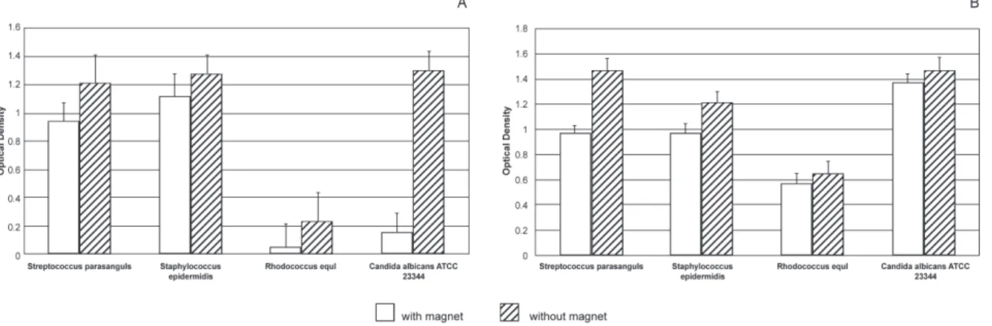
Figure 1 data point out to the reduced number of all investigated microbes following 24 hours of AKMA magnet effect. The greatest magnet inluence, i.e., the most severe microbe reduction after 24 h of incubation, was noted in the C. albicans
was 4.27 times lower in comparison to magnet-free agar.
AKMA magnets showed the least influence on microbial reduction in the Str. parasanguinis
strain, which is noted in the lowest difference in the OD values obtained. There was only 1.28 times more intense growth in magnet-free agar than in magnet-containing agar. Figure 1 data indicate that the OD values of agar after 48 h upon incubation with or without magnets turned out to be very close themselves, i.e., AKMA magnets reduced the studied bacteria/yeast strains much less signiicantly in the second day of incubation. The OD values of reference C. albicans strain in
magnet-containing agar increased 8.9 times following 24 h incubation (Figure 1).
Regardless of the signiicant increase of OD values in magnet-containing agar following 48 h of incubation, the value of OD in AKMA-magnet-free agar was only 1.07 times higher in comparison with the values of magnet-containing agar. It is possible to conclude that magnets signiicantly inluence the reduction of the number of C. albicans even after
48 h (Figure 1).
R h o d o c o c c u s e q u i behaves similarly.
Micromagnets inluenced their reduction even 48
h later, while the recorded values of OD of magnet-free agar amounted to 0.649 (only 1.126 times greater than the values of magnet-containing agar samples).
As for Str. parasanguinis, the OD values were
achieved through an increased reduction of the total number of bacteria under the inluence of AKMA magnet. That was noted on the second day of incubation. The OD values of the agar only increased to 1.01, i.e., 0.007 times, calculated on the basis of the OD values obtained with magnets throughout the irst 24 hours. In other words, streptococci almost failed to multiply in dextrose magnet-containing agar in the course of the second day of incubation.
B) Test results for the series with inoculums density of 10 CFU/ml
On the basis of the obtained OD values having been read by the spectrophotometer it may be noted that AKMA micromagnets influence the reduction of the number of all tested bacterial and yeast strains under in vitro conditions. The resulting
OD values of inoculated agars with and without AKMA magnets after 24 h and 48 h are shown numerically in Figure 2.
Figure 1- Optical density values upon 24 and 48 h of incubation with a inoculum density of 1 CFU/ml
Results of microbiologic in vivo investigations
Results of in vivo investigation and analysis
were assessed for every subject (patient). The distribution of all studied microbes is shown in Figure 3, in which the decrease in the number of all studied plaque microbes is conirmed. There were no statistical signiicant differences between microbial counts because more precise evaluation was conducted within observation periods (0, 3, 6 and 12 months) (Figure 3).
DISCUSSION
The turbidimetry method has been widely discussed in the pharmacopeia of European countries, as well as the Yugoslavian pharmacopeia, with regard to application of bacteria to the nutrient media and antibiotic effect. On the basis of our study, OD values obtained by the spectrophotometer and the degree of reduction of microbial growth could be also applied to the exact inluence of antibiotics on microbes. In our study, magnets were used instead of antibiotics, with a speciic method of application adapted to the purpose of the study. The modiications concerned the type and the amount of nutrient media as well as the initial number of microbes. Dextrose nutrient media was favored because it is completely clear when sterile, showing even minimal discoloration and opalescence.
Under in vitro conditions, AKMA micromagnets
have shown a certain influence on microbial decrease in our study, but in the course of time this inluence weakened and the number of microbes began to rise. Since the initial numbers of microbes were 10 CFU/ml and 1 CFU/ml, and considering numerical OD values in the amount of 0.100-0.120 as related to the total number of 1-2x10⁸/
ml of microorganisms, it could be presumed that the increase in the total number of microbes was
evident after 24 hours. Some authors showed that magnets act less effectively under in vitro
conditions, i.e., only slightly reducing the number of microbes19.
Since the in vitro reduction of microbial growth
by magnetic ield was conirmed, the next task of our study was to verify their reduction in the oral cavity of the patients (in vivo). For that purpose,
the safety of application of magnets inside the mouth had been assessed in our pilot study9, in
which it was determined that static magnets (of BaF content) do not expose the oral environment to corrosive effects, contrary to the indings of other authors9,16,17.
Speciically, for in vitro conditions, magnets
influence the reduction of the number of microorganisms, but in the course of time, which could be seen in the obtained results, the inluence is decreased and the number of microbes increases signiicantly again. The initial numbers of microbes in this assessment were 10 CFU/ml and 1 CFU/ ml. Knowing that the OD value of inoculated agar or other media of 0.100-0.120 corresponds to a total number of microbes of 1-2x10⁸/ml, it
could be concluded that after the initial 24 hours of incubation in agar with magnets, there was a signiicant increase in the total number of microbes, despite the evident inluence of magnets on its reduction compared with agar without magnets. In other words, it is not the same to assess whether magnets have effects on 100 microbes or on 106
microbes in 1 ml of agar.
On the basis of the obtained results, it could be discussed how magnets affected the reduction in the number of microbes under in vitro conditions
if the initial number was not great. When the total number of microbes is massively multiplied, magnets express less inluence on their growth and differentiation. For in vivo, it is a prerequisite that
the initial number of microbes is always at a low
level, but when magnet has to be inserted into the dentures, it could continually inluence the reduction in the number of microbes. Consequently, the detrimental inluence of the low level of microbes is to be expected in the oral cavity. It could be speculated that magnet discs are more effective in the in vivo then in the in vitro experiments due to
the oxygen level.
The obtained results obtained by the statistical method of ANOVA showed that magnets inserted into dentures provoke a signiicant reduction in the number of all detected microbes in dental plaque (p<0.05). The p values for only spiral microbes were at the low level of statistical signiicance (P=0.05).
The effect of magnetic ield is still controversial in literature data. Potenza, et al.17 (2004) stated that
the magnetic ield improves the E. coli proliferation,
although Talà, et al.21 (2014) noted no differences
in proliferation for a V. harveyi related strain.
Our in vitro investigation conirmed the effects
of magnets on the reduction in the number of microbes. Additionally, in vivo investigation showed
that for identical samples and without conducting the regime of oral and teeth hygiene, as well as for correct oral-hygienic procedures, a reduction in the number of microbes occurred. In both conditions, reduction in the number of microbes in staining smear isolated from dental plaque was documented at the beginning of the experiment. Literature data1,2,19 conirm those indings too,
with a claim that magnets20,22 cannot replace the
beneicial action of antibiotics but deinitely could improve their bactericidal effects, as well as other healing methods5,17,16. Apparently, magnets cannot
substitute periodontal treatment (surgery) but might be an excellent supplement5.
Considering the effects of magnetic ields, it could be advocated that magnets have to be used as a supplement to conventional therapies3,18.
CONCLUSION
The positive effect of magnetic ields on the reduction in the number of dental plaque bacteria was conirmed in vitro. The inluence of magnetic
field was recognized in the first 24 hours of exposure. The count of all isolated microbes was signiicantly reduced. Over time, a decrease in the inluence of magnets versus in vitro microlora
density was noted. Beneicial inluence of magnetic ields on the reduction of the number of dental plaque microbes was also proved in vivo. A
reduction in the number of microbes was noted under overdentures containing inserted magnets in all observation periods (3, 6 and 12 months). Static magnetic ields applied in the area of the remaining teeth have beneicial effects on the alveolar bone and other periodontal tissues.
ACKNOWLEDGEMENTS
The author thanks Prof. Asanin Ruzica, specialist of microbiology at Belgrade University, School of Veterinary Medicine, for her part in this investigation.
REFERENCES
1- Abdel-Kader HM, Aref MI, Yousef SW. The biological effects of static magnetic ield of commercial samarium-cobalt (SmCo5) orthodontic magnets on cultured Escherichia coli and staphylococci
aureus. Int J Clin Dent. 2012;5(3):201-10.
2- Bajpai I, Saha N, Basu B. Moderate intensity static magnetic ield has bactericidal effect on E. coli and S. epidermidis on
sintered hydroxyapatite. J Biomed Mater Res B Appl Biomater. 2012;100(5):1206-17.
3- Brković-Popović S, Stamenković D, Stanisić-Sinobad D, Rakocević Z, Zelić O. The inluence of continuous magnetic ield on periodontal tissues under overdentures. Srp Arh Celok Lek. 2009;137(7-8):363-70.
4- Creanga DE, Poiata A, Morariu VV, Tupu P. Zero-magnetic ield effect in pathogen bacteria. J Magn Magn Mater. 2004; 272:2442-4.
5- Darendeliler MA, Zea A, Shen G, Zoellner H. Effects of pulsed electromagnetic ield vibration on tooth movement induced by magnetic and mechanical forces: a preliminary study. Aust Dental J. 2007;52(4):282-7.
6- Esmekaya MA, Acar SI, Kiran F, Canseven AG, Osmanagaoglu O, Seyhan N. Effects of ELF magnetic field in combination with iron(III) chloride (FeCl3) on cellular growth and surface morphology of Escherichia coli (E. coli). Appl Biochem Biotechnol.
2013;169(8):2341-9.
7- Faraj KA, Muhamad DA. Effect of high magnetic ield on gram negative bacteria. Eur J Sci Res. 2012;74(2):240-3.
8- Feychting M. Health effects of static magnetic ields-a review of the epidemiological evidence. Prog Biophys Mol Biol. 2005;87(2-3):241-6.
9- Fojt L, Strasak L, Vetterl V, Smarda J. Comparison of the low-frequency magnetic ield effects on bacteria Escherichia
coli, Leclercia adecarboxylata and Staphylococcus aureus.
Bioelectrochemistry. 2004;63(1-2):337-41.
10- Grosman Z, Kolár M, Tesaríková E. Effects of static magnetic ields on some pathogenic microorganisms. Acta Univ Palacki Olomuc Fac Med. 1992;134:7-9.
11- Hsu SH, Chang JC. The static magnetic ield accelerates the osteogenic differentiation and mineralization of dental pulp cells. Cytotechnology. 2010;62(2):143-55.
12- IMP-PIEZOTEHNOLOGIJA DOO [homepage]. Subotica: NovaMedia; c2009 [cited 2014 July 7]. Available from: http:// novamedia.rs/showCategory/13203/mikromagneti-akma. 13- Janković BD, Marić D, Ranin J, Veljić J. Magnetic ields, brain and immunity: effect of magnetic ield. Int J Neurosci. 1991;59(1-3):25-43.
14- Jovanovic-Nesic K, Eric-Jovicic M, Spector NH. Magnetics stimulation of the brain increase Na+, K+-ATPase activity decreased by injection of AlCl3 into nucleus basalis magnocellularis
of rats. Int J Neurosci. 2006;116(6):681-95.
15- Kohno M, Yamazaki M, Kimura I, Wada M. Effect of static magnetic ields on bacteria: Streptococcus mutans, Staphylococcus
aureus, and Escherichia coli. Pathophysiology. 2000;7(2):143-8.
16- Morrow AC, Dunstan RH, King BV, Roberts TK. Metabolic effects of static magnetic ields on Streptococcus pyogenes.
Bioelectromagnetics. 2007;28(6):439-45.
18- Steffensen B, Caffesse RG, Hanks CT, Avery JK, Wright N. Clinical effects of electromagnetic stimulation as an adjunct to periodontal therapy. J Periodontol. 1988;59(1):46-52.
19- Strasak L, Vetterl V, Fojt L. Effects of 50 Hz magnetic ields on the viability of different bacterial strains. Electromagn Biol Med. 2005;24(3):293-300.
20- Strasak L, Vetterl V, Smarda J. Effects of low-frequency magnetic ields on bacteria Escherichia coli. Bioelectrohemistry
2002;55(1-2):161-4.
21- Talà A, Delle Side D, Buccolieri G, Tredici SM, Velardi L, Paladini F, et al. Exposure to static magnetic ield stimulates quorum sensing circuit in luminescent Vibrio strains of the Harveyi clade.
PLoS One. 2014;9(6):e100825.
22- Van der Kuij P, Vingerling PA, Sillevis Smitt PA, de Groot K, de Graaf J. Reducing residual ridge reduction. In: van Rens TJ, ed. Electric and electromagnetic stimulation of bone growth. New York: S. Karger; 1985. p. 98-105.