www.scielo.br/aabc
The surface charge of trypanosomatids*
THAÏS SOUTO-PADRÓN
Laboratório de Biologia de Protozoários, Instituto de Microbiologia Prof. Paulo de Góes Centro de Ciências da Saúde, Bloco I, Universidade Federal do Rio de Janeiro, Ilha do Fundão
21941-590 Rio de Janeiro, RJ, Brazil
Manuscript received on August 16, 2002; accepted for publication on August 20, 2002; presented byJulio Scharfstein
ABSTRACT
The surface charge of trypanosomatids was evaluated by means of the binding of cationic particles, as visualized by electron microscopy and by direct measurements of the electrophoretic mobility of cells. The results obtained indicate that most of the trypanosomatids exhibit a negatively charged surface whose value is species specific and varies according to the developmental stages. Sialic acids associated with glycoproteins, glycolipids and phosphate groups are the major components responsible for the net negative surface charge of the trypanosomatids.
Key words:trypanosomatids, cell surface charge, electrophoretic mobility, cytochemistry.
INTRODUCTION
The Trypanosomatidae family comprises a large group of flagellated protozoa separated in 12 genera, which comprise monogenetic (Angomonas, Blas-tocrithidia,Crithidia,Herpetomonas,Leptomonas, Rhynchoidomonas and Wallaceina) and digenetic (Endotrypanum,Leishmania, Phytomonas, Sauro-leishmaniaandTrypanosoma) parasites with ability to parasitise a very diverse range of hosts, including animals, plants and other protists.
The parasites from the genusTrypanosomaare responsible for diseases of great medical and veteri-nary importance such as Chagas’ disease or Amer-ican Trypanosomiasis, sleeping sickness or AfrAmer-ican Trypanosomiasis. The several species of the Leish-mania genus are agents of visceral and cutaneous leishmaniasis.
Species from the generaHerpetomonas,
Lep-*Dedicated to the memory of Dr. Hertha Meyer on the centenary of her birth.
E-mail: [email protected]
tomonas, Crithidia and Blastocrithidia are found only in insects. Phytomonasspp is pathogenic for plants. The generaAngomonas,Rhynchoidomonas, Wallaceina, Endotrypanum and Sauroleishmania will not be discussed here because of the lack of data about cell surface charge.
Trypanosomatids have a complex life cycle, ex-hibiting a number of differentiation stages that ap-pears as a result of transformations involving both structural and physiological changes. Previous stud-ies have shown that the occurrence of differentiated cell stages is usually associated with changes in cell surface components and that changes in the net sur-face charge can also be found as a result of cell dif-ferentiation. Such changes directly influence the parasite-host cell interaction.
SURFACE CHARGE
membrane and the electrokinetic potentials. The first, also denominated trans-membrane potential, is due to the semi-permeable characteristic of the bio-logical membranes and is maintained by an energy-dependent mechanism. The second, also denomi-nated cell surface charge, is due to a complex interac-tion between the polar groups residing at the cell sur-face with different ions of the surrounding medium (James 1979, Mehereish 1972, Weiss 1969).
Pro and eukaryotic cells bear a net negative electrical charge which influences the adhesiveness and selective affinity between cells, playing an im-portant role in morphogenesis during the embry-onic development (Schaeffer et al. 1973), in the malignancy and ability of the cells to metastasize (Haeffner et al. 1988, Carter et al. 1989) and in parasite-host cell interaction (Meirelles et al. 1984, Araújo-Jorge et al. 1992, Monteiro et al. 1998).
Previous studies indicate that the candidates most likely for anionic sites in the cell membrane are sulfate groups, found in acid mucopolysaccha-rides, ionized phosphate groups, found in phospho-lipids, and charged carboxyl groups largely due to the presence of sialic acid and from carboxyl groups from acidic amino acids of proteins (Ambrose 1966, Burry and Wood 1979, Eylar et al. 1962, Seaman and Uhlenbruck 1963, Gasic et al. 1968).
The surface charge of trypanosomatids has been studied by the use of the following techniques: a) passage of cells through diethylaminoethyl (DEAE)-cellulose columns; b) visualization, by electron microscopy, of cationic particles bound to the cell surface; c) determination of cell surface charge by Atomic Force Microscopy associated to surface potential spectroscopy; d) determination of the cellular electrophoretic mobility; and e) use of sialic acid-binding components.
USE OF DEAE-CELLULOSE COLUMNS
The application of anion exchange columns to the study of Trypanosomatids started with the neces-sity to develop an efficient method to separate trypo-mastigote forms of trypanosomes from blood cells or from other evolutive forms for biological,
chemi-cal or immunologichemi-cal studies (Lanham 1968, 1971, Lanham and Godfrey 1970, Al-Abbassy et al. 1972, Howells and Chiari 1973, Jackson 1975, Goldberg et al. 1976, Kreier et al. 1977, Gutteridge et al. 1978, Villalta and Leon 1979, Alvarenga and Brener 1979, Selden and Baker 1980, Langenbach 1985). On passing infected blood throughout a DEAE-cellulose column the blood cells and platelets, which are very negative, were completely adsorbed while the parasites could be easily eluted (Lanham 1968, 1971, Lanham and Godfrey 1970). A standard tech-nique was established which successfully separated different species of Salivarian trypanosomes, con-firming previous studies which showed that the sur-face of bloodstream forms ofT. bruceiwas not very negative. However, it was not possible to separate T. cruzitrypomastigotes of the Y strain. According to Lanham (1968), the bloodstream forms of try-panosomes belonging to different species could be arranged in the following order of negative surface T. cruzi>T. lewisi>T. vivax>T. congolense>T. rhodesiense,T. gambiense,T. bruceiandT. evansi.
DEAE-cellulose columns have also been used for the purification ofT. cruzitrypomastigotes from tissue and axenic culture (Al-Abbassy et al. 1972, Goldberg et al. 1976, Kreier et al. 1977, Selden and Baker 1980, Langenbach 1985) and from feces of triatomines (Alvarenga and Brener 1979) where they are mixed with epimastigote forms. As these metacyclic trypomastigotes have a negative surface similar to that of bloodstream trypomastigotes and epimastigotes have a less negative surface it would be expected that during the process of separation the first forms to be eluted should be the epimastigotes. However, trypomastigotes are initially eluted and the epimastigotes remain associated with DEAE-cellulose. It was showed that passage of metacyclic trypomastigotes through a DEAE-cellulose column alters the kinetics of arginine and lysine transport by the plasma membrane (Goldberg et al. 1976) and re-moves some negatively-charged constituents of the parasite surface as shown by the decrease of the sur-face charge, determined by cellular electrophoretic mobility (De Souza et al. 1977) and the change in the attachment of parasites to chitosan (Kleffmann et al. 1998).
To avoid the effects of DEAE-cellulose column Pinho and coworkers (1986) developed a method of purification of metacyclic trypomastigotes using glass wool columns. As it occurs with DEAE -cellulose column, trypomastigotes are the first to be eluted while the epimastigote forms remain ad-sorbed on the glass wool. Mortality studies in mice showed that the biological activity of the glass wool purified trypomastigotes remained unchanged. No estimation of cell surface charge of these trypomas-tigotes was reported.
There are no reports about the use of DEAE-cellulose columns to isolate other trypanosomatids or evolutive forms.
ULTRASTRUCTURAL DETECTION OF CELL SURFACE ANIONIC SITES
Electrically charged electron stains may be bound electrostatically by inorganic groups of the cell sur-face. Two cationic labels have been widely used: the colloidal iron hydroxide (CIH) and the
cation-ized ferritin (CF) (Danon et al. 1972, Gasic et al. 1968). There are some important differences be-tween these two labels: only pre-fixed cells can be labeled with CIH since the positive colloid was used at pH 1.8. Cationized ferritin is a derivative of na-tive ferritin that can be used at physiological pH ei-ther in pre-fixed or even in living cells (Danon et al. 1972). Treatments such as methylation and the use of neuraminidase prevent the staining by the CIH indicating that the iron micelles are bound almost exclusively to sialic acid (Gasic et al. 1968). Nowa-days, poly-L-lysine-gold probes have also been used as markers for anionic sites (Goode et al. 1992). However, this technique has not been applied to try-panosomatids.
Labeling ofT. bruceiwith CIH and CF was de-scribed by De Souza (1989) as results of unpublished observation in collaboration with Benchimol, Tetley andVickerman. According to De Souza (1989), CIH and CF bind to the surface of epimastigote forms obtained from axenic culture. There is no binding of CIH to the surface of bloodstream trypomastig-otes. A weak labeling with CF, indicated by a single layer of particles, is only observed on the surface of glutaraldehyde fixed trypomastigotes. No label was seen on parasites, which were incubated in the presence of CF before fixation or after fixation in formaldehyde. The same effect of glutaraldehyde fixation was described for the binding of antibodies against VSG on the surface ofT. brucei(Masterson et al. 1988). According to Masterson and coworkers (1988) and De Souza (1989), there are at least two explanations of this phenomenon: a) fixation may denature the VSG, producing new epitopes which then should be recognized by antibodies; and b) that glutaraldehyde may induce conformational changes on the surface components of trypomastigote forms, leading to the exposure of cryptic epitopes and acidic amino acids which bind to CF but not to CIH (Mas-terson et al. 1988, De Souza 1989).
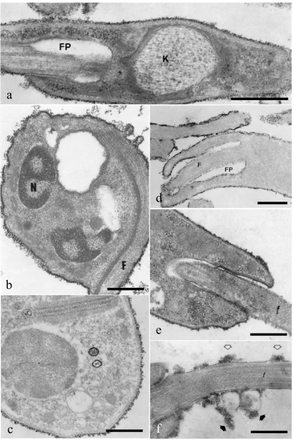
extent of the binding is represented by the number of layers of particles on the surface of the different evolutive forms and could be arranged in the follow-ing decreasfollow-ing order: bloodstream trypomastigotes > cell culture trypomastigotes > metacyclic trypo-mastigotes > atrypo-mastigotes > epitrypo-mastigotes. In epi-mastigotes both cationic particles appeared uni-formly distributed over the cell surface and the flag-ellum, in the flagellar pocket and in the region be-tween the flagellum and the cell body (Martinez-Palomo et al. 1976). In bloodstream trypomastig-otes and amastigtrypomastig-otes, CIH and CF particles were seen distributed homogeneously throughout the cell surface and flagellar membranes. However, no cationic particles were seen associated with the small portion of the flagellum which remains inside the flagellar pocket, with the membrane which lines the flagellar pocket or in the region of adhesion be-tween cell body and flagellar membranes (Figs. 1a-1b). A special array of integral proteins is involved in the adhesion of the flagellum to the cell body im-pairing the diffusion of such cationic labels to the flagellar pocket (De Souza et al. 1977, Carvalho et al. 1985).
Treatment of T. cruzi with neuraminidase or trypsin completely abolishes the binding of CIH par-ticles to the parasites surface and almost completely abolishes the binding of CF particles. This suggests that the cationic particles bind to sialoglycoproteins exposed on the surface of amastigote, epimastiogote and trypomastigote forms (Carvalho et al. 1985, Souto-Padrón and De Souza 1986).
There are few studies concerning the cytochem-ical localization of negative surface charge on try-panosomes of rodents (Dwyer 1975, Dwyer and D’Alessandro 1976a). Large quantities of cation-ized ferritin were bound to the cell body and flagellar membranes ofT. lewisiandT. musculibloodstream forms (Dwyer 1975, Dwyer and D’Alessan-dro 1976a). As described for trypomastigotes ofT. cruzi, no particles were seen in the region of adhe-sion between the cell body and the flagellum and in the flagellar pocket (Dwyer 1975, Dwyer and D’A-lessandro 1976a). Trypsin treated trypomastigotes fromT. lewisiandT. musculihad 3 and 4 times less
ferritin bound / 100µ2of cell surface area than intact bloodstream forms, respectively. Culture forms ofT. lewisipresented 3 times less cationic particles than bloodstream forms and the labeling was observed on the cell and flagellar membranes, as well as in the region of adhesion between cell body and the flagellum and in the flagellar pocket (Dwyer 1975). Promastigote and amastigote forms of some species ofLeishmaniaalso presented negative sur-face charge detected by cationic particles (Mühlp-fordt 1975, Pimenta and De Souza 1983, Ayesta et al. 1985, Saraiva et al. 1989).
Cationized ferritin binds to the surface of pro-mastigotes ofL. donovani,L. tropicaandL. brazi-liensis(Mühlpfordt 1975, Dwyer 1977, Ayesta et al. 1985). InL. braziliensis CF binding was used to identify promastigotes, which belong to pathogenic and non-pathogenic strains. Only pathogenic pro-mastigotes were strongly agglutinated by CF and presented an abundant CF surface labeling (Ayesta et al. 1985). Cationized ferritin and colloidal iron hydroxide bind to the surface of amastigote and pro-mastigote forms ofL. mexicana amazonensis (Pi-menta and De Souza 1983). An intense labeling with both CF and CIH was observed on the surface of all evolutive forms analysed. There are no differ-ences in the amount of cationic particles bound to the surface of promastigotes which had been passed 5 or 176 times in axenic culture, thus suggesting that in the case of the strain ofL. mexicana amazo-nensisstudied there is no relationship between cell surface charge and its ability to induce lesion in ham-sters. In the case of amastigotes, labeling was more intense when the membrane of the endocytic vac-uole surrounded amastigotes isolated from lesions in hamsters. Treatment with neuraminidase did not interfere with the binding of cationic particles to the cell surface (Pimenta and De Souza 1983). Treat-ment of promastigotes ofL. mexicana amazonensis with trypsin, alkaline phosphatase and phospholi-pase C, but not with neuraminidase, reduced the CF binding (Silva Filho et al. 1990).
par-a
b
c
d
e
f
Fig. 1 – Binding of cationic particles to the surface ofTrypanosoma cruzi(a-c) andLeishmania mexicana amazonensis(d-f). (a)
asites with neuraminidase reduced the binding of CF and CIH particles only on the surface ofPhytomonas davidi(Esteves et al. 1988).
Cytochemical studies on the surface charge of monogenetic trypanosomatids were made in differ-ent species of the genera Crithidia and Herpeto-monas. Crithidia deanei is known to harbor en-dosymbiotic bacterium-like organism that is inte-grated into the physiology of the host cell (McGhee and Cosgrove 1980). Although symbiote-bearing and symbiote-free strains ofC. deaneipresented a negative cell surface charge due to the presence of sialic acid, no significant binding of CIH was ob-served and binding of CF to the surface of both strains was not observed (Oda et al. 1984). Bind-ing of CF particles was observed on the cell sur-face ofCrithidia luciliae and of both wild and drug-resistant mutant strains ofCrithidia fascicu-lata. Enzymatic treatment with phospholipase C and neuraminidase showed that phosphate groups and sialic acid significantly contributed to the neg-ative surface charge (Motta et al. 1991, Matta et al. 1992).
Cationic particles bind to the surface of Her-petomonas samuelpessoai(Soares et al. 1988), Her-petomonas muscarum muscarum(Lopes et al. 1989) andHerpetomonas megaseliae(Fiorini et al. 1991). In these studies parasites were incubated in the pres-ence of substances which induce cell differentiation since it has been observed that the change in form is either accompanied by or preceded by changes in the cell surface composition. Dimethylsulphox-ide (DMSO) treatedH. samuelpessoaipresented a more intense labeling compared with untreated cells (Soares et al. 1988). Differentiation of H. mus-carum musmus-caruminduced by propranolol caused a significant increase in the negative surface charge. In cells incubated in the presence of the drug the CF particles were not uniformly distributed on the cell body and flagellar membranes as described for con-trol cells (Lopes et al. 1989). Previous results onH. megaseliaeshowed that lipopolysaccharides (LPS) trigger the process of cell differentiation from pro-mastigotes to both para and opisthomastigote forms and affect the composition of membrane-associated
polysaccharides (Fiorini et al. 1985). LPS treatment caused a marked decrease in binding of CF, CIH and Sendai virus particles to the cell surface (Fiorini et al. 1991).
More recently the Atomic Force Microscopy (AFM) was introduced in basic studies in biology and medicine. The AFM is based on the principle of scanning the surface of a sample with an atom-ically sharp tip of silicon on a very soft cantilever. AFM records the repulsive forces generated by the overlap of the electron cloud at the silicon tip with the electron clouds of surface atoms. According to the intensity of the repulsive forces the distance be-tween the tip and the specimen varies. The force of deflection can be converted to a surface image of the specimen, using a computer. AFM resolution is limited only by the fineness of the silicon tip and not by any wavelength (Binning et al. 1986). Bi-ological substances are resolved nearly atomic de-tail. The major advantage of AFM for biological research is that it can be used for imaging in solu-tion. Atomic force microscopy associated to sur-face potential spectroscopy (SPS) was introduced on the study of cell surface charge (Aikawa et al. 1996,1997). SPS analysis uses a Si probe coated with CoCr, in order to obtain a high electronic con-ductivity of the tip surface, associated to a reso-nance frequency of about 70-90 kHz. This method-ology provided the possibility to measure net sur-face charges on specific domains of cell membranes (Yokoyama and Inoue 1994).
CELLULAR ELECTROPHORETIC MOBILITY
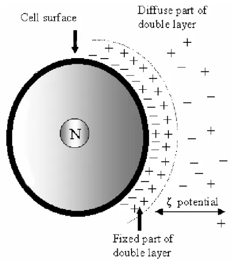
When a charged particle is suspended in an ionic medium, the ionic cloud of opposite charge that is attracted by the particle reduces the surface poten-tial. The resultant potential, i.e. the sum of the potential on the surface of the particle and the po-tential of the ions in the neighborhood of the particle, is the Zeta potential(ζ )(Fig. 2). Theζ potential is evaluated by the mobility of cells under the influ-ence of an electric current of known intensity and it appears to depend entirely on the composition of the suspending medium and is independent of the size, shape or the nature of the particle itself. The tech-nique which measures the ζ potential was named cell electrophoresis or electrophoretic mobility. The mobility (V) of the cells and particles is expressed asµm.sec−1.V−1.cm, i.e. as velocity per unit field strength, and was converted intoζpotential by use of Helmoltz-Smoluchowski equationζ =(4π η/D)V whereηand D are the viscosity and the dielectric constants of the suspending medium, respectively.
Several instruments were developed for deter-mining the electrostatic charge (zeta potential) of small solid particles dispersed in water. In some of them, particles or cells are observed with a mi-croscope. It is common to refer to this method as microelectrophoresis. Particles to be measured are placed in an electrophoresis chamber consisting of two electrode compartments and a connecting cham-ber, which can be cylindrical or rectangular. A volt-age is applied between the two electrodes producing a uniform electric field in the connecting chamber. Cells respond by moving toward one or the other electrode according to the sign of their charges. The speed of a particle or cell is directly proportional to the magnitude of the surface charge or Zeta poten-tial. Measurements must be made in the so-called stationary planeswhere the electrophoretic mobil-ity of the particles measured is not affected by the circulation of the liquid inside the chamber. In some instruments the measuring chamber is in the interior of a temperature-controlled chamber connected to the circulating thermostat.
There are different kinds of
microelec-trophoresis apparatus. The common feature of all conventional instruments has been the measurement of one particle at a time. The velocity of the parti-cles is measured by timing them over a given num-ber of graticule squares in the eyepiece with the aid of a stopwatch, and at a given potential gradi-ent. The electrophoretic mobility is expressed as µm.s−1.V−1.cm whereµm=the distance covered
during the measurement by the particle the particle under study, s = time (sec) required by the parti-cle to cover the distanceµm, V = the final voltage and cm = the distance between the two electrodes. This method allows the direct observation of par-ticles displacement and the analysis of individual values of the electrophoretic mobility. In such con-ventional analysis time of measurement is relatively large inducing to errors such as: a) convection cur-rents caused by heating of the sample; b) contamina-tion of the sample by diffusion of electrode reaccontamina-tions products; c) flocculation of particles; d) polarization of the electrodes and others (Figs. 3a-b).
Fig. 2 – - The surface charge of a particle or cell influences the distribution of nearby ions in the polar medium. Ions of opposite charge (counter-ions) are attracted towards the surface and ions of like charge (co-ions) are repelled away from the surface. This leads to the formation of an electric double layer made up of the charged surface and a neutralizing excess of counter-ions over co-ions distributed in a diffuse manner in the polar medium. The electric double layer can be regarded generally as consisting of two regions: an inner or fixed region which may include adsorbed ions and a diffuse region in which ions are distributed according to the influence of electrical forces and random thermal motion. Electrokinetic orζ(zeta) potential is measured on an imaginary plane (Stern plane) between the fixed part of double layer and the electrolyte solution (diffuse part of double layer).
The third method most used in the determina-tion of cell surface charges was the free flow trophoresis. The basic principle of free-flow elec-trophoresis is that the mixture of cells to be sep-arated is injected in a fine stream into a solution which is flowing perpendicular to the lines of force of an electric field. Electrically charged particles are deflected from the direction of flow at an angle de-termined by a combination of the flow velocity and the electrophoretic mobility of the particle. Thus cells with different electrophoretic mobility move in
different directions, and can be collected separately in a fraction collector after leaving the separation chamber (Fig. 3c).
2
4
1
3
3
9
5
6
7
8
1
1
2
3
4
5
6
a
b
c
Fig. 3 – Principle and construction diagrams of microelectrophoresis (a-b) and free-flow electrophoresis. Fig. 3a – 1) Electrode compartment in electrical contact with the measuring chamber via a membrane; 2) Temperature controlled chamber connected to the circulating thermostat; 3) Funnel-cells in normal saline solution were introduced into the measuring chamber through plastic tubes. The liquid stream is controlled by a cock (4); 5) Measuring chamber; 6) Viewing window-place where the front lens of the microscope objective had to be connected to the temperature-controlled chamber. Fig. 3b – One of the focusing eyepieces of the light microscope contains a special reticule which serves as micrometer. The time it takes a cell edge to pass two or more whole micrometer intervals is measured with the aid of a stopwatch. Fig. 3c – 1) Separation chamber; 2) Chamber buffer; 3) Electrode compartment in electrical contact with the separation area via a membrane; 4) Sample pump; 5) Multitube pump; 6) Cooled fraction container with vials for sample collection; 7) Electrode buffer circulation; 8) Electronically regulated cooling system. 9) Optical window for analytical measurements.
Hollingshead and coworkers (1963) using the same methodology showed that bloodstream forms ofT. lewisi,T. cruziandT. rhodesiensedid not attach to normal erythrocytes whereas under the same condi-tions bloodstream forms ofT. equinum,T. vivaxand T. congolensewere completely covered by erythro-cytes. In the same study Hollingshead and cowork-ers (1963) determined the electrophoretic mobility (EPM) of some trypanosome species and observed that the random orientation of trypanosomes is
analyz-ing the EPM ofT. brucei, the causative agent of Na-gana in livestock and which is non infective to man, showed that bloodstream forms presented a mean electrophoretic mobility of−0.68µm.s−1.V−1.cm while epimastigotes presented a mean EPM of −0.83µm.s−1.V−1.cm (Table I). An important observation is that the mean electrophoretic values for bloodstream forms of both species are less neg-ative than those observed for epimastigote forms. It is important to mention that according to Broom and coworkers (1936) bloodstream form ofT. gam-biense, another specie of parasite causing human Sleeping Sickness, also presented positive or slight negative surface charge, as mentioned forT. rhode-siense.
T. bruceiandT. rhodesiensebelong to the sub-genusTrypanozoonwhich is the most homogeneous group of Salivarian trypanosomes, represented by species which are morphologically indistinguish-able but differing in biological features. The possi-bility to identify small differences on surface charge suggests that cell electrophoresis, under controlled conditions, would help to distinguish morphologi-cally identical trypanosomatids.
Trypomastigote forms ofT. equinum, another member of the Salivarian trypanosomes, had an electrophoretic mobility which varied from 0 to
+0.4µm.s−1.V−1.cm. The cell surface charge in-creases in magnitude as the parasite was kept in culture medium for 1h at room temperature, reach-ing a mean EPM value of −0.95µm.s−1.V−1.cm. This change in surface charge accompanies loss of motility and death. The same behavior was de-scribed for bloodstream forms of T. rhodesiense. According to Hollingshead and coworkers (1963), the loss of surface component may be responsible for the change with time to a more negative elec-trophoretic mobility.
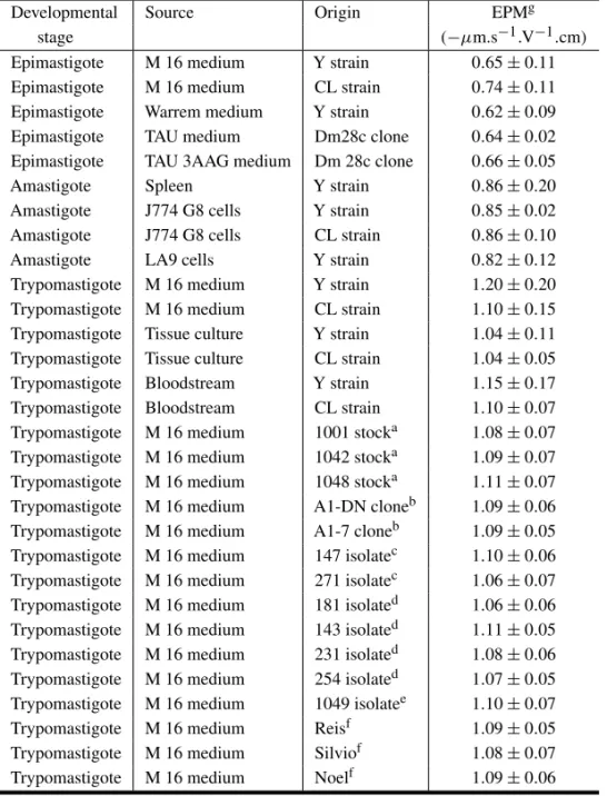
Cell surface charge ofTrypanosoma cruzihas been analyzed in more detail (De Souza et al. 1977, Kreier et al. 1977, Souto-Padrón et al. 1984, 1990, Carvalho et al. 1985, Souto-Padrón and De Souza 1985, Bonaldo et al. 1988). The electrophoretic mo-bility analysis of the different developmental stages ofT. cruzi showed that each evolutive form has a
characteristic mean EPM which does not vary in the different strains or isolates of the parasite, is in-dependent of the medium in which the cells were grown and do not change after glutaraldehyde fix-ation (Table II) (Souto-Padrón et al. 1990). Epi-mastigotes present the smallest negative sur-face, with a mean EPM of−0.65µm.s−1.V−1.cm. Amastigotes obtained from cell culture infected with parasites of the Y or CL strains, or isolated from the spleen of mice infected with the Y strain of the parasite, present similar mean EPM, which is of about−0.85µm.s−1.V−1.cm (Carvalho et al. 1985). Trypomastigotes present a mean EPM of
−1.15µm.s−1.V−1.cm which is the most negative surface charge compared with the other evolutive forms. As observed for the epimastigote and amastigote forms, the mean EPM of trypomastig-otes obtained from the bloodstream of infected mice, from the supernatant of cell cultures, from axenic culture in complex or defined media does not dif-fer significantly (Souto-Padrón et al. 1984, 1990, Bonaldo et al. 1988). Some results are not in agree-ment with those reported by Kreier and cowork-ers (1977) and Murray and coworkcowork-ers (1982). Us-ing free-flow electrophoresis, Murray and cowork-ers (1982) found that amastigotes have the low-est charge, epimastigote the highlow-est and the charge of trypomastigotes law in between. Kreier and coworkers (1977) reported that trypomastigotes had less negative charge than epimastigotes and that amastigotes had the same charge as epimastigote. The results obtained by Kreier and coworkers (1977) and Murray and coworkers (1982) do not explain the difficulties found for the separation of erythrocytes and bloodstream forms using DEAE-cellulose column, which is probably due to the ionic strength of the solution in which the parasites were suspended, as will be discussed below.
TABLE I
Mean electrophoretic mobility of trypanosomatids.
Species Strain/ Developmental Mean EPM References Isolate form (µm.s−1.V−1.cm)
T. brucei Epimastigote –0.83 Souto-Padrón et al. (1990)
Trypomastigotea –0.68
T. cruzi Y Epimastigote –0.65 Souto-Padrón et al. (1984)
Trypomastigoteb –1.15 Carvalho et al. (1985)
Amastigote –0.85
T. conorhini Epimastigote –1.73 Souto-Padrón et al. (1990)
T. dionisii Epimastigote –1.05 Souto-Padrón et al. (1990)
Trypomastigotec –1.90
T. equinum Trypomastigote +0.40 Hollingshead et al. (1963)
T. lewisi Trypomastigote –0.61 Hollingshead et al. (1963)
T. myoti 1 Epimastigote –1.03 Souto-Padrón et al. (1990)
3 Epimastigote –1.03
4 Epimastigote –1.06
5 Epimastigote –1.14
T. rangeli Epimastigote –0.62 Souto-Padrón et al. (1990)
T. rhodesiense Epimastigote –0.91 Hollingshead et al. (1963) Trypomastigoted –0.10
T. vespertilionis Epimastigote –0.61 Souto-Padrón et al. (1990)
Subgenus M5 Epimastigote –0.57 Pinto et al. (1996)
Schizotrypanume M29 Epimastigote –0.56
Leishmania Josefa Amastigotef –1.58 Pimenta and
mexicana Amastigoteg –1.14 De Souza (1983)
amazonensis Promastigoteh –1.14
Promastigotei –1.49 Promastigotej –1.47
Phytomonas davidi –0.98 Esteves et al. (1988)
Phytomonassp* Isolate 1 –1.22
Isolate 2 –1.09 Vommaro et al. (1989)
Isolate 3 –1.08
Isolate 4 –1.03
Herpetomonas Promastigote –0.76 Soares et al. (1988)
samuelpessoai
Herpetomonas Promastigote –0.69 Lopes et al. (1989)
muscarum muscarum
Herpetomonas Promastigote –0.98 Fiorini et al. (1991)
megaseliae
Herpetomonas Promastigote –0.73 Faria-e-Silva et al. (1999)
TABLE I (continuation)
Species Strain / Isolate Developmental Mean EPM References form (µm.s−1.V−1.cm)
Crithidia deanei Symbiote free Choanomastigote –0.99 Oda et al. (1984) Symbiote bearing Choanomastigote –0.85
Crithidia luciliae Choanomastigote –0.80 Motta et al. (1991)
Crithidia fasciculata Choanomastigote –0.97 Matta et al. (1992)
aBloodstream trypomastigotes isolated using DEAE cellulose column;bBloodstream trypomastigotes isolated by differential
centrifu-gation;cTrypomastigotes obtained in axenic culture medium and isolated using DEAE-cellulose column;dBloodstream trypomastig-otes;eTrypanosomes of the subgenusSchizotrypanumisolated from the batPhyllostomus hastatus;fAmastigote forms surrounded by
the membrane of the endocytic vacuole;gamastigote forms without the membrane of endocytic vacuole;hPromastigote forms in the 1stpassage in axenic culture;iPromastigote forms in the 5thpassage in axenic culture;jPromastigote forms in the 176thpassage in axenic culture; *Isolate 1, fromEuphorbia hyssopifolia; Isolate 2, fromEuphorbia pinea; Isolate 3, fromEuphorbia characias; Isolate 4, fromManihot esculenta.
al. 1988). Transitional forms, which have a rod-like kinetoplast localized beside the cell nucleus, presented a mean EPM similar to the trypomastig-ote forms indicating that during metacyclogenesis, morphological changes are preceded by alterations in some physiological characteristics (Bonaldo et al. 1988).
The pH of the solution in which the parasites are suspended interferes directly on the mean EPM in-dicating that the surface ofT. cruzimust contain pos-itively and negatively charged dissociating groups. At higher pH values the negative charge increases, probably due to the increase in the dissociation of the carboxyl groups. At low pH values the negative charge decrease and below a certain value which corresponds to the isoelectrophoretic point, the sur-face become positive. The isoelectrophoretic point is around 2.0 for amastigotes and epimastigotes, and around 3.0 for trypomastigotes (Souto-Padrón et al. 1984, Carvalho et al. 1985).
The ionic strength of the solution in which the parasites are suspended also interferes with the mean EPM. In a solution with an ionic strength of 0.072 mol.dm–3 epimastigotes have a mean EPM
of−1.19µm.s−1.V−1.cm, a value similar to that of trypomastigote forms suspended in a solution with an ionic strength of 0.145 mol.dm–3.
Cell electrophoresis, in association with
treat-ment of parasites with cationized ferritin, enzymes, lectins and inhibitors of protein synthesis and pro-tein glycosylation, was used to analyze the nature of surface components which contribute to the net negative cell surface charge (Meirelles et al. 1984, Souto-Padrón et al. 1984, Carvalho et al. 1985, Souto-Padrón and De Souza 1985, 1986). Binding of cationized ferritin to the surface of trypomastigote forms reduced the mean EPM by about 53% while the same treatment did not interfere with the mean surface charge of epimastigote forms (Meirelles et al. 1984).
TABLE II
Mean electrophoretic mobility ofTrypanosoma cruzi*.
Developmental Source Origin EPMg
stage (−µm.s−1.V−1.cm)
Epimastigote M 16 medium Y strain 0.65±0.11
Epimastigote M 16 medium CL strain 0.74±0.11
Epimastigote Warrem medium Y strain 0.62±0.09
Epimastigote TAU medium Dm28c clone 0.64±0.02
Epimastigote TAU 3AAG medium Dm 28c clone 0.66±0.05
Amastigote Spleen Y strain 0.86±0.20
Amastigote J774 G8 cells Y strain 0.85±0.02
Amastigote J774 G8 cells CL strain 0.86±0.10
Amastigote LA9 cells Y strain 0.82±0.12
Trypomastigote M 16 medium Y strain 1.20±0.20
Trypomastigote M 16 medium CL strain 1.10±0.15
Trypomastigote Tissue culture Y strain 1.04±0.11
Trypomastigote Tissue culture CL strain 1.04±0.05
Trypomastigote Bloodstream Y strain 1.15±0.17
Trypomastigote Bloodstream CL strain 1.10±0.07
Trypomastigote M 16 medium 1001 stocka 1.08±0.07
Trypomastigote M 16 medium 1042 stocka 1.09±0.07
Trypomastigote M 16 medium 1048 stocka 1.11±0.07
Trypomastigote M 16 medium A1-DN cloneb 1.09±0.06
Trypomastigote M 16 medium A1-7 cloneb 1.09±0.05
Trypomastigote M 16 medium 147 isolatec 1.10±0.06
Trypomastigote M 16 medium 271 isolatec 1.06±0.07
Trypomastigote M 16 medium 181 isolated 1.06±0.06
Trypomastigote M 16 medium 143 isolated 1.11±0.05
Trypomastigote M 16 medium 231 isolated 1.08±0.06
Trypomastigote M 16 medium 254 isolated 1.07±0.05
Trypomastigote M 16 medium 1049 isolatee 1.10±0.07
Trypomastigote M 16 medium Reisf 1.09±0.05
Trypomastigote M 16 medium Silviof 1.08±0.07
Trypomastigote M 16 medium Noelf 1.09±0.06
For details see Souto-Padrón et al. 1984, Carvalho et al. 1985, Bonaldo et al. 1988, Souto-Padrón et al. 1990. aIsolated from the opossumDidelphis albiventris;bIsolated from a chronic patient and which belong all to zymodeme A;cIsolated from a chronic patient and which belong all to zymodeme B;dIsolated from a chronic patient and which belong all to zymodeme C;eIsolated from triatomine
Panstrongylus megistus;fIsolated from patients with the acute phase of Chagas’ disease;gMean EPM plus standard deviation.
Treatment ofT. cruziwith trypsin reduces the mean EPM of epimastigote, amastigote and trypo-mastigote forms by 11, 32 and 35%, respectively, suggesting that sialoglycoproteins contribute to the surface charge of T. cruzi. Neuraminidase and trypsin-treated parasites recover their normal mean
neuraminidase-treated parasites (Souto-Padrón et al. 1984, Souto-Padrón and De Souza 1985, 1986). From these results it was suggested that in trypo-mastigotes ofT. cruziabout 37% of the sialic acid residues exposed on the surface are associated with proteins, mainly to N-glycosylated proteins, and about 63% with glycolipids. Interestingly, the in-cubation of trypomastigote forms in the presence of the lectinLimulus polyphemus(LPA), which binds to the 4-hydroxyl and carboxyl groups of glycosidi-cally linked sialic acid (Schauer 1982) decreased by about 50% the negative surface charge of try-pomastigote forms which is very similar to that ob-tained when trypomastigote forms were incubated in the presence of cationized ferritin (Meirelles et al. 1984) or neuraminidase (Souto-Padrón et al. 1984, Souto-Padrón and De Souza 1985). More recently, it was observed that LPA not only binds to sialic acids but also to galNAc and glcNAc residues.
Trypanosomatids are unable to synthesize sialic acids. The acquisition of sialic acid is due by a sialic acid metabolizing enzyme namedtrans -sialidase (Colli 1993, Cross and Takle 1993, Schenk-man et al. 1994, Frasch 2000). This enzyme is found in epimastigote and trypomastigote forms and catalyzes the transfer of sialic acid from host gly-coconjugates to a terminal Galβ of an appropri-ate molecule on the parasite surface (Colli 1993, Schenkman et al. 1994). The major acceptor of sialic acid of the cell surfacetrans-sialidase is GPI-anchored glycoproteins rich in threonine, serine and proline. Sialic acid is incorporated into O-linked oligosaccharides via N-acetylglucosamine and is called mucin-like molecules or TcMUC (Previato et al. 1994). TcMUC resembled the mammalian mucins and were firstly described by Alves and Colli in 1975. There are some evidences that sialic acid residues present on the surface of trypomastigote forms ofT. cruziplay important roles on the inva-sion of host cell, on the circulation of parasites in the extracellular matrix or bloodstream and on the resistance of parasite lysis by the alternative path-way of complement (Kipnis et al. 1981, Araújo-Jorge and De Souza 1984, Schenkman and Eichinger 1993). Besides the epimastigote and
trypomastig-ote forms ofT. cruzi trans-sialidase is also found in insect forms ofT. brucei(Pontes de Carvalho et al. 1993, Montagna et al. 2002) andEndotrypanum (Medina-Acosta et al. 1994).
Treatment of epimastigote and trypomastigote forms with phospholipase C was only effective af-ter previous treatment with trypsin. According to Souto-Padrón and De Souza (1985), phospholipase sensitive phosphate groups account for 17% of the negative surface charge.
Trypanosomes of the subgenus Schizotry-panum are morphologically almost indistinguish-able from each other and those from bats have cos-mopolitan distribution. Parasites isolated from bats are able to infect vertebrate cells where they re-produce as amastigotes, with an intracellular be-havior characteristic of members of the Schizotry-panumsub-genus. In Latin AmericaTrypanosoma (Schizotrypanum) cruzi can be found in sev-eral mammalian orders, including bats (Pinto and Costa Bento 1986). Thus, in this region it is of con-siderable importance to public health to determine whether isolates from bats are distinct fromT. cruzi. The data presented in Table I show that it is pos-sible, using the electrophoretic mobility, to distin-guishT. cruzifrom other members of the sub-genus Schizotrypanum, such asT. dionisii,T. myoti and M5 and M29 stocks isolated from the batP. has-tatusin Minas Gerais, Brazil (Souto-Padrón et al. 1990, Pinto et al. 1996). In the case ofT. dionisii differences could be observed both in epimastigote and tripomastigote forms. Trypomastigote forms of T. dionisiihave a very negative surface charge, with a mean EPM of−1.90µm.s−1.V−1.cm. It was not possible to distinguish epimastigotes ofT. cruziand T. vespertilionis, since they present a similar mean EPM. The same was observed for epimastigotes of T. dionisiiandT. myoti(Souto-Padrón et al. 1990). In spite of the presence of several common antigens in these trypanosomes at least one antigen ofT. my-oti,T. vespertilionisandT. dionisiiwas distinct from those of the other species (Bower and Woo 1982).
1988), occur in the same mammalian hosts and have common Triatomine vectors. As the second known American trypanosome of man, it is of medical im-portance since mixed infections may occur in both vertebrate and invertebrate hosts (Hoare 1972). Al-beit infection byT. rangeliis apparently harmless for the human host, the parasite induces a humoral response with cross-reacting antibodies againstT. cruzi, causing misleading serodiagnosis of Chagas’ disease. It was not possible to distinguish epimastig-ote forms ofT. cruziandT. rangeliusing cell elec-trophoresis since they present a similar mean EPM (Table I). However, they can be distinguished using other methods such as sialic acid content (Schot-telius 1984), the presence of neuraminidase in the supernatant of cultures (Schottelius 1987) and PCR assays for the amplification of a specific DNA se-quence forT. rangeli(Vargas et al. 2000).
There are few data about the electrophoretic mobility of trypanosomatids parasite of rodents (Hollingshead et al. 1963, Souto-Padrón et al. 1990). Trypomastigote forms ofT. conorhini pre-sented a high negative surface charge with a mean EPM of−1.73µm.s−1.V−1.cm while epimastigote forms of T. lewisi presented a mean EPM of
−0.61µm.s−1.V−1.cm (Table I).
Among the several Leishmania species only Leishmania mexicana amazonensiswas analyzed by cell electrophoresis (Pimenta and De Souza 1983, Saraiva et al. 1989, Silva Filho et al. 1990). All evo-lutive forms ofL. mexicana amazonensisstudied ex-hibit negatively charged surfaces. Amastigotes iso-lated just after homogenization of lesion experimen-tally induced in hamsters, and which is surrounded by a membrane from the endocytic vacuole of the macrophage, present a very high negative surface charge, with a mean EPM of−1.58µm.s−1.V−1.cm. When this parasite is incubated for 4h in a culture medium, the endocytic membrane that surrounds the parasites is eliminated and the cell sur-face becomes less negative, with a mean EPM of
−1.14µm.s−1.V−1.cm. When amastigote forms obtained from lesions were incubated in axenic med-ium and maintained at 25◦C they gradually trans-form into promastigote trans-forms. Freshly transtrans-formed
promastigotes, which have been obtained in the first 48h of culture, and those passed 5-7 times through this medium, were considered infective since they are able to induce lesions when incubated into ham-sters. Those promastigotes, which have been trans-ferred 175 or more times in the same axenic medium, are considered noninfective promastigotes (Saraiva et al. 1986).
Promastigotes in the first passage in vitro had a mean EPM of−1.14µm.s−1.V−1.cm.
Grad-ually the cell surface charge of promastigote forms became more negative and after five pas-sages parasites presented a surface charge, which varied from −1.10 to−1.79µm.s−1.V−1.cm with mean EPM of−1.49µm.s−1.V−1.cm (Table I). No infective promastigotes presented a mean EPM of
−1.47µm.s−1.V−1.cm. indicating that there is an increase in the negativity of the cell surface during amastigote-promastigote transformation and that there is no relationship between cell surface charge of promastigote forms and its ability to induce le-sions in hamsters (Pimenta and De Souza 1983). Interestingly, promastigotes ofL. mexicana amazo-nensisthat had undergone more than 450 passagesin vitropresented a mean EPM varying between 1.14 and 1.22µm.s−1.V−1.cm (Saraiva et al. 1989, Silva Filho et al. 1990).
Electrophoretic mobility of promastigote and amastigote forms ofL. mexicana amazonensisvaries according to the pH of the solution in which the cells are suspended, indicating the presence of both neg-atively and positively charged cell surface dissoci-ating groups (Pimenta and De Souza 1983).
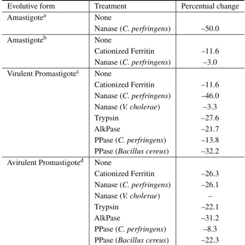
(Ta-ble III). Neuraminidase treated promastigotes re-cover their mean EPM when incubated for 8h in fresh medium by a process that is inhibited by puromicin. Neuraminidase fromV. choleraedid not alter the EPM of any evolutive form analyzed. Trypsin reduced in about 28 and 22% the cell sur-face charge of virulent and avirulent promastigotes, respectively. Treatment of promastigotes with al-kaline phosphatase (AlkPase) and phospholipase C (PPase) showed the importance of phospholipids as one of the surface anionic species in L. mexicana amazonensis (Silva Filho et al. 1990). Alkaline phosphatase treatment reduced by about 30% the cell surface charge of avirulent promastigotes (Table III). Phospholipase C fromBacillus cereus signifi-cantly reduced the cell surface charge of both viru-lent and aviruviru-lent promastigotes. This effect can be explained by the selective action of phospholipase C fromBacillus cereuson Lipophosphoglycan (LPG), the major surface glycoconjugate of promastigote forms of all analyzedLeishmaniaspecies that has a highly anionic nature and is phosphatydilinositol-anchored (Handman and Goding 1985, Turco 1988). Parasites of the genusPhytomonasare respon-sible for important diseases in plants of economical interest such as coconut (Parthasarathy and Slobbe 1978), oil palm (Slobbe et al. 1978) and cassava (Kitajima et al. 1986). They are transmitted to the plants through the saliva of phytophagous insects. Most of the parasites occur in plants and insects as promastigote forms presenting a very twisted cell body.
Phytomonas davidipromastigotes, which were isolated from the milkweed plant and have been maintained for several years in axenic medium, present a negative cell surface charge with a mean EPM of−0.99µm.s−1.V−1.cm. (Table I) (Esteves et al. 1988). Another four isolates were also ob-tained from milkweed plants such as Euphorbia hyssopifolia, Euphorbia pinea, Euphorbia chara-cias and Manihot esculenta (Dollet et al. 1982, McGhee and Postell 1976, Attias and De Souza 1986, Vainstein and Roitman 1986, Vainstein et al. 1987). All of them presented negative cell sur-face charge with a mean EPM varying from -1.03 to
−1.22µm.s−1.V−1.cm (Table I). Despite biochem-ical and ultrastructural evidences of the presence of sialic acid on the surface of all analyzed iso-lates of Phytomonas, treatment with neuraminidase only slightly reduced the mean EPM of P. davidi and did not reduce the mean EPM of the isolates from E. hyssopifolia, E. pinea, E. characias and M. esculenta (Esteves et al. 1988, Vommaro et al. 1989).
In addition toPhytomonasit was shown that both plants and phytophagous insects can harbor parasites from generaCrithidia,Herpetomonasand Leptomonas. Since some of them share indistin-guishable promastigote forms, various approaches have been proposed to classify plant trypanoso-matids such as the production of monoclonal an-tibodies, the analysis of enzymatic activities and the use of molecular markers (Camargo 1999). In this context the determination of cell electrophoresis mobility associated to enzymatic treatments would be useful to help the identification of those parasites. The genusCrithidiacomprises parasites of in-sects that were originally characterized by the pres-ence of amastigote and choanomastigote forms in their life cycle (Hoare and Wallace 1966). Cell sur-face charge of three species of the genusCrithidia was analyzed by determining the cellular electro-phoretic mobility, ultrastructural cytochemistry, thin layer and gas liquid chromatography, lectin aggluti-nations assays and enzymatic treatments.Crithidia deanei, C. fasciculata and C. luciliae have a net negative cell surface charge (Table I) ( Oda et al. 1984, Motta et al. 1991, Matta et al. 1992). Treat-ment of these strains with neuraminidase fromC. perfringensonly reduced significantly the EPM of C. deanei. Enzyme treatment ofC. fasciculataand C. luciliaeshowed that phosphate groups, but not sialic acid, contribute to the negative surface charge of these parasites (Motta et al. 1991).
Blast-TABLE III
Effect of cationized ferritin and enzymatic treatments on the mean electrophoretic mobility ofLeishmania mexicana amazonensis*.
Evolutive form Treatment Percentual change
Amastigotea None
Nanase (C. perfringens) –50.0
Amastigoteb None
Cationized Ferritin –11.6 Nanase (C. perfringens) –3.0 Virulent Promastigotec None
Cationized Ferritin –11.6 Nanase (C. perfringens) –46.0 Nanase (V. cholerae) –3.3
Trypsin –27.6
AlkPase –21.7
PPase (C. perfringens) –13.8 PPase (Bacillus cereus) –32.2 Avirulent Promastigoted None
Cationized Ferritin –26.3 Nanase (C. perfringens) –26.1 Nanase (V. cholerae) –
Trypsin –22.1
AlkPase –31.2
PPase (C. perfringens) –8.3 PPase (Bacillus cereus) –22.3
*For details see Pimenta and De Souza 1983, Saraiva et al. 1989, Silva Filho et al. 1990.
aAmastigote forms surrounded by the membrane of the endocytic vacuole;bAmastigote forms
without the membrane of the endocytic vacuole;cObtained by in vitro culture of skin biopsies from hamster lesions;dPromastigote forms which had passed 500 times in axenic cultures.
ocrithidia culicis(Novy et al. 1907),Herpetomonas roitmani(Faria-e-Silva et al. 1991) and Trypano-soma cobitis(Lewis and Ball 1980). The possibil-ity of elimination of the endosymbiont by the use of antibiotics has increased the interest in the study of endosymbiont-harboring species. The available data indicate that the presence of the endosymbiont induces morphological changes, as the lack of para-flagellar structure located in the flagellum (Frey-müller and Camargo 1981) interferes with the metabolism of the trypanosomatid (Newton 1957, Mundim et al. 1974, Alfieri and Camargo 1982, Salzman et al. 1985, De Souza and Motta 1999), diminishes the secretion of proteolytic enzymes
(d’Avila-Levy et al. 2001), interferes with surface properties of the protozoan such as exposition of carbohydrate residues (Dwyer and Chang 1976, Es-teves et al. 1982, McLaughlin and Cain 1985, Faria e Silva et al. 1994) and on the surface charge (Oda et al. 1984). Symbiont-freeC. deaneipresents a mean EPM of−0.99µm.s−1.V−1.cm, which is 15% more negative than the symbiont-free strain. Treatment ofC. deaneiwith neuraminidase reduced in about 45% the EPM of the protozoan, irrespective of the presence of the endosymbiote (Oda et al. 1984).
composition of the plasma membrane and the sur-face coat are of primary importance in cell response to environmental stimuli and in the interaction of parasites with their hosts (Soares et al. 1988, Lopes et al. 1989, Fiorini et al. 1991, Faria-e-Silva et al. 1999). Besides these aspects,Herpetomonassp presents four developmental stages (promastigotes, paramastigotes, opisthomastigotes and opistho-morphs), are non pathogenic for men and show close antigenic similarities toTrypanosoma cruzi (Souza and Roitman 1971, Souza et al. 1974).
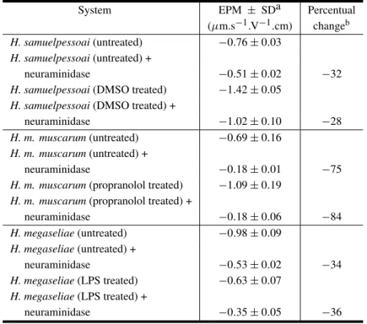
In most species of the genusHerpetomonas, cultures in the logarithmic phase of growth (48h of cultivation) show almost exclusively un-differentiated promastigote forms. Promastigotes of H samuelpessoai,H. muscarum muscarum, H. megaseliaeandH. roitmanidisplay a negative sur-face charge, with mean EPM varying, according to the species analyzed (Table I). Treatment of Her-petomonas with 2-deoxy-D-glucose (Angluster et al. 1977), concanavalin-A (ConA) (Souza et al. 1980), dimethylsulfoxide (DMSO) (Castellanos et al. 1981), propranolol (Lopes et al. 1983) and lipopolysaccharide (LPS) (Fiorini et al. 1985) trig-gers the process of cell differentiation from pro-mastigote to para- and opisthopro-mastigote forms, af-fecting the composition of membrane-associated polysaccharides (Alviano et al. 1981, Soares et al. 1984), cell surface charge (Soares et al. 1988, Lopes et al. 1989) and cell respiration (Fiorini et al. 1985). Dimethiylsulfoxide and propranolol caused a signif-icant increase in the net negative surface charge of bothH. samuelpessoaiandH. muscarum muscarum due to a markedly increase in the number of N -acetylneuraminic acid residues per cell (Soares et al. 1988, Lopes et al. 1989). However, LPS treatment ofH. megaseliaecaused a marked decrease in the negative cell surface altering the nature and number of sialic acid residues. OnlyN-acetylneuraminic acid can be detected on untreated flagellates whereas N-acetyl and N-glycolylneuraminic acid are found on the parasites exposed to LPS (Fiorini et al. 1991). Incubation of DMSO-, propranolol- and LPS-treated and untreatedHerpetomonaswith neu-raminidase fromC. perfringensresulted in decrease
of their mobility to the negative pole (Table IV). More recently Faria-e-Silva and coworkers (1991) isolated a new Herpetomonas species, H. roitmani. This parasite was isolated as opistho-morph forms, which proliferate in culture un-like other Herpetomonas species (Faria-e-Silva et al. 1996). Promastigote and opisthomorph forms of H. roitmanidisplay a net negative charge of about
−0.73 and −1.20µm.s−1.V−1.cm, respectively. Neuraminidase and phospholipase C treatments significantly reduced the surface charge of both evolutive forms (Table V). The authors concluded that sialic acid is an important component of the cell surface ofH. roitmani, which is probably added to surface glycoconjugates during the process of promastigote-opisthomorph transformation (Faria-e-Silva et al. 1999).
SIALIC ACID-BINDING COMPONENTS
Concomitantly with ultrastructural cytochemistry and electrophoretic mobility studies, lectins were also used to identify sialic acids on the surface of try-panosomatids. Wheat germ (WGA),Limulus poly-phemus(LPA) andLimax flavus(LFA) agglutinins were the lectins of choice, and the presence of sialic acid was evaluated by the following approaches: a) quantitative and qualitative analysis of cell agglu-tination; b) localization of binding sites by fluo-rescence microscopy and flow cytometry; c) local-ization of binding sites by electron microscopy; d) quantitative determination of cell surface binding sites using3H-, 125I- and 131I-labeled lectins; and
e) use of lectins for purification of glycoconjugates containing sialic acids by affinity chromatography. Agglutination with Sendai virus was also used to evaluate the presence of sialic acid residues. Since WGA also binds specifically to N -acetylglucosa-mine residues, identification of sialic acid using this lectin has to be evaluated before and after sialidase treatment.
TABLE IV
Effect of drugs which induce cell differentiation on the electrophoretic mobility ofHerpetomonassp.*
System EPM ± SDa Percentual
(µm.s−1.V−1.cm) changeb
H. samuelpessoai(untreated) −0.76±0.03
H. samuelpessoai(untreated) +
neuraminidase −0.51±0.02 −32
H. samuelpessoai(DMSO treated) −1.42±0.05
H. samuelpessoai(DMSO treated) +
neuraminidase −1.02±0.10 −28
H. m. muscarum(untreated) −0.69±0.16
H. m. muscarum(untreated) +
neuraminidase −0.18±0.01 −75
H. m. muscarum(propranolol treated) −1.09±0.19
H. m. muscarum(propranolol treated) +
neuraminidase −0.18±0.06 −84
H. megaseliae(untreated) −0.98±0.09
H. megaseliae(untreated) +
neuraminidase −0.53±0.02 −34
H. megaseliae(LPS treated) −0.63±0.07
H. megaseliae(LPS treated) +
neuraminidase −0.35±0.05 −36
*For details see Soares et al. 1988, Lopes et al. 1989, Fiorini et al. 1991.aStandard deviation;
bmean EPM of control – mean EPM of neuraminidase treated cell / mean EPM of control×100.
TABLE V
Effect of enzyme treatment on the surface charge of promastigote and opisthomorph forms of
Herpetomonas roitmani.*
Evolutive form Treatment Percentual changea
Promastigote None
Neuraminidase –24 Phospholipase C –33
Trypsin –34
Opisthomorph None
Neuraminidase –39 Phospholipase C –41
Trypsin –33
*For details see Faria-e-Silva et al. 1999. amean EPM of control – mean EPM of neuraminidase treated cell / mean EPM of control×100.
ofT. musculi(Dwyer and D’Alessandro 1976b) and T. congolense(Jackson et al. 1978, Rauthenberg et al. 1980) and procyclics ofT. brucei brucei,T. bru-cei rhodesiense andT. congolense (Mutharia and Pearson 1987) were agglutinated in the presence of WGA. However, in all these studies there are no indications of treatment of the parasites with neu-raminidase.
Epimastigote forms ofT. cruzi were aggluti-nated with low concentrations of WGA, presenting 3×10⁶WGA-binding sites per cell (Pereira et al.
receptors with distinct affinities and capacities. Tis-sue culture and bloodstream trypomastigotes pre-sented 1.2×10⁶and 2.3×10⁶WGA-binding sites,
respectively (Katzin and Colli 1983). Although amastigote forms ofT. cruzipresented the highest number of WGA binding sites, 4.6×10⁶, no
aggluti-nation has been observed (Pereira et al. 1980, Katzin and Colli 1983). Limulus polyphemusagglutinin and LFA also bind to the surface of epimastigote and trypomastigote forms ofT. cruzias showed by lectin agglutination assays (Pereira et al. 1980), flu-orescence microscopy (Souto-Padrón and De Souza 1985) and ultrastructural cytochemistry using gold-labeled lectins (Bourguignon et al. 1998).
Infective and non-infective promastigotes of Leishmania mexicana amazonensis were agglu-tinated by LPA confirming the electrophoretic mo-bility data (Saraiva et al. 1986).
Promastigote forms ofPhytomonas davidi pre-sented sialic acid of theN-acetyl-neuraminic acid type identified by paper and gas-liquid chromatog-raphy. However, agglutination of promastigotes in the presence of LPA became only possible after brief trypsinization. This observation suggests that sialic acid residues were masked so that after trypsin they became exposed or that they were associated with glycolipids, which would be covered by glycopro-teins (Esteves et al. 1988).
Lectin-induced agglutination was used to ana-lyze the presence of surface exposed carbohydrates inCrithidia fasciculataandCrithidia luciliae. Both species were not agglutinated, even in high con-centrations, by the lectins LPA and LFA (Motta et al. 1991).
Presence of sialic acid on the surface of pro-mastigote (PRO) and opisthomorph (OPM) of Her-petomonas roitmani were analyzed using FITC-labeled LPA and flow cytometry. The FITC signal for LPA was stronger in OPM than in PRO forms. Prior incubation of cells in the presence of neu-raminidase from Clostridium perfringens reduced the fluorescence intensity in OPM but not in PRO forms. Thin-layer chromatography analysis of H. roitmani showed the presence of N
-acetyl-neura-minic acid (Faria-e-Silva et al. 1999).
Presence of sialic acid residues on the surface ofH. samuelpessoai(Esteves et al. 1988),H. mus-carum musmus-carum(Lopes et al. 1989) andH. mega-seliae(Fiorini et al. 1991) was evaluated by aggluti-nation with Sendai virus, visualization of virus parti-cles on the surface of parasites by scanning electron microscopy and thin-layer chromatography.
Analysis of net surface charge of trypanoso-matids determined by cell electrophoresis and ultra-structural cytochemistry has provided evidences for a net negative surface charge. The surface charge is species specific and varies according to the devel-opmental stages opening the possibility of their iso-lation. Association of the previous techniques with trypsin and neuraminidase treatments indicated that sialic acids significantly contributed to the negative surface charge and that they are associated with gly-coproteins and glycolipids. The use of sialic acid-binding lectins corroborated these findings. Phos-pholipase treatment also indicated the importance of phosphate-containing molecules on the net surface charge of trypanosomatids. Contribution of sulfate groups has not been determined yet.
Nowadays, new possibilities have been opened to the study of cell surface charges. New lectins that selectively recognize different types and link-ages of sialic acid could be useful as: a) markers in the evaluation of the biosynthesis of sialylated glycoconjugates; and b) to understand the effects of distinct neuraminidases on cell surface charge of trypanosomatids.
ACKNOWLEDGMENTS
The author is indebted to Dr. Wanderley de Souza for useful discussion and critical reading of the manuscript. This work was supported by CNPq, Faperj and FUJB(UFRJ).
RESUMO
A carga de superfície de tripanosomatídeos foi avaliada através da ligação de partículas catiônicas, visualizadas por microscopia eletrônica e por medida direta da mobili-dade eletroforética celular. Os resultados obtidos indicam que a grande maioria dos tripanosomatídeos apresenta carga de superfície negativa cujo valor é espécie específico e que varia com o estágio evolutivo. Resíduos de ácido siálico associados a glicoproteínas e glicolipídios assim como grupamentos fosfato, são os principais responsáveis pela carga negativa da superfície de tripanosomatideos.
Palavras-chave: tripanosomatídeos, carga da superfície celular, morfologia eletroforética, citoquímica.
REFERENCES
Aikawa M, Kamanura K, Shiraishi S, Matsumoto Y, Arwati H, Torji M, Ito Y, Takeuchi T and Tan-dler B.1996. Membrane knobs of unfixed Plasmod-ium falciparuminfected erythrocytes: new findings as revealed by atomic force microscopy and surface potential spectroscopy. Exp Parasitol 84: 339-343.
Aikawa M, Kawasu S, Kamio T, Matsumoto Y, Naya T, Torji M, Ito Y, Tandler B, Nakano Y, Takeuchi T, Shiraishi S and Kanamura K.1997.
Mem-brane knobs of unfixedBabesia bovis-infected ery-throcytes: new findings as revealed by atomic force microscopy and surface potential spectroscopy. Par-asitol Int 46: 241-246.
Akaki M, Nakano Y, Nagayasu E, Nagakura K, Kawai S and Aikawa M.2001. Invasive forms of Toxoplasma gondii, Leishmania amazonensis and Trypanosoma cruzihave a positive charge at their contact site with host cells. Parasitol Res 87: 193-197.
Al-Abbassy SN, Seed TM and Kreier JP.1972. Isola-tion of the trypomastigote form ofTrypanosoma cruzi from a mixture of the trypomastigote and epimastig-ote forms of the parasite by use of a DEAE-cellulose column. J Parasitol 58: 631-632.
Alfieri SC and Camargo EP.1982.Trypanosomatidae: isoleucine requirement and threonine deaminase in species with and without endosymbionts. Exp Para-sitol 53: 371-380.
Alvarenga NJ and Brener Z.1979. Isolation of pure metacyclic trypomastigotes of Trypanosoma cruzi from triatomine bugs by use of DEAE-cellulose col-umn. J Parasitol 65: 814-815.
Alves MJM and Colli W.1975. Glycoproteins from Trypanosoma cruzi: partial purification by gel chro-matography. FEBS Lett 52: 188-198.
Alviano CS, De Souza ET, Esteves MJG, Angluster J and De Souza W.1981. Effect of concanavalin A on the surface ofHerpetomonas samuelpessoai. J Submicrosc Cytol 13: 619-626.
Ambrose EJ.1966. Electrophoretic behaviour of cells.
Prog. Biophys. Mol. Biol. 16: 241-265.
Andrade AFB, Esteves MJG, Angluster J, Gonza-lez-Perdomo M and Goldenberg S. 1991.
Changes in cell-surface carbohydrates of Trypano-soma cruzimetacyclogenesis under chemically de-fined conditions. J Gen Microbiol 137: 2845-2849.
Angluster J, Bunn MM and De Souza W.1977.
Ef-fect of 2-deoxy-D-glucose on differentiation of Her-petomonas samuelpessoai. J Parasitol 63: 922-924.
Araújo-Jorge TC and De Souza W. 1984. Effect of carbohydrates, periodate and enzymes in the pro-cess of endocytosis of Trypanosoma cruzi by macrophages. Acta Trop 41: 17-28.
Araújo-Jorge TC, Barbosa HS and Meirelles MN.
1992. Trypanosoma cruzi recognition by macro-phages and muscle cells: perspectives after 15-years study. Mem Inst Oswaldo Cruz 5: 43-56.
Attias M and De Souza W.1986. Axenic cultivation and ultrastructural study of aPhytomonassp. Iso-lated from the milkweed plantEuphorbia hyssopifo-lia. J Protozool 33: 84-87.
Ayesta C, Argüello C and Hernandez AG.1985. Leishmania braziliensis: Cell surface differences in promastigotes of pathogenic and nonpathogenic strains. Exp Parasitol 59: 185-191.
Binning G, Quate CF and Gerber CH.1986. Atomic
force microscope. Phys Rev Lett 56: 930-933.
Bonaldo MC, Souto-Padrón T, De Souza W and Goldenberg S.1988. Cell-substrate adhesion
Bourguignon SC, De Souza W and Souto-Padrón T.1998. Localization of lectin-binding sites on the surface ofTrypanosoma cruzigrown in chemically defined conditions. Histochem Cell Biol 110: 527-534.
Bower SM and Woo PT.1982. Immunological compar-ison of fourTrypanosomaspp (sub-genus Schizotry-panum) from bats. Parasitology 85: 111-114.
Broom JC, Brown HC and Hoare CA.1936. Studies on microcataphoresis. II. The electric charge of haemo-flagellates. Trans Roy Soc Trop Med Hyg 30: 87-100.
Burry RW and Wood JG.1979. Contributions of lipids and proteins to the surface charge of membranes. J Cell Biol 32: 726-741.
Camargo EP.1999. Phytomonasand other Trypanoso-matid parasites of plants and fruit. Adv Parasitol 42: 29-112.
Carter HB, Partin AW and Coffey DS.1989.
Pre-diction of metastatic potential in an animal model of prostate cancer: flow cytometry quantification of cell charge. J Urol 142: 1338-1341.
Carvalho TU, Souto-Padrón T and De Souza W.
1985. Trypanosoma cruzi: Surface charge and freeze-fracture of amastigotes. Exp Parasitol 59: 12-23.
Castellanos GB, Angluster J and De Souza W.
1981. Induction of differentiation inHerpetomonas samuelpessoaiby dimethylsulfoxide. Acta Trop 38: 29-37.
Colli W.1993.Trans-sialidase: a unique enzyme activ-ity discovered in the protozoanTrypanosoma cruzi. FASEB J 7: 1257-1264.
Cross AM and Takle GB.1993. The surface trans-sialidase family ofTrypanosoma cruzi. Ann Rev Mi-crobiol 47: 385-411.
Danon D, Goldstein L, Marikovsky H and Skutel-sky E.1972. Use of cationized ferritin as a label of negative charges on cell surfaces. J Ultrastruct Res 38: 500-510.
D’Avila-Levy CM, Melo CAN, Vermelho AB and Branquinha MH. 2001. Differential expression of proteolytic enzymes in endosymbiont-harboring Crithidiaspecies. FEMS Microbiol Lett 202: 73-77.
De Souza W.1989. Components of the cell surface of trypanosomatids. Progr Protistol 3: 87-184.
De Souza W and Motta MCM.1999. Endosymbiosis in protozoa of the Trypanosomatidae family. FEMS Microbiol Lett 173: 1-8.
De Souza W, Arguello C, Martinez-Palomo A, Trissl D, Gonzales-Robles A and Chiari E.1977. Surface charge ofTrypanosoma cruzi. Binding of cationized ferritin and measurement of cellular elec-trophoretic mobility. J Protozool 24: 411-415.
Dollet M, Cambrony D and Gargani D.1982. Cul-ture axénique in vitro de Phytomonas sp. (Try-panosomatidae) d’Euphorbe transmis par Stenoce-phalus agilisScop (Coreide). Comptes Rend Acad Sci Paris 295: 547-550.
Dwyer DM. 1975. Cell surface saccharides of Try-panosoma lewisi. I. Polycation-induced cell agglu-tination and fine structure cytochemistry. J Cell Sci 19: 621-644.
Dwyer DM.1977.Leishmania donovani: Surface mem-brane carbohydrates of promastigotes. Exp Parasitol 41: 341-358.
Dwyer DM and Chang KP.1976. Surface membrane carbohydrate alterations of a flagellated protozoon mediated by bacterial endosymbiotes. Proc Natl Acad Sci USA 73: 852-856.
Dwyer DM and D’Alessandro PA.1976a. The cell surface ofTrypanosoma musculibloodstream forms. I. Fine structure and cytochemistry. J Protozool 23: 75-83.
Dwyer DM and D’Alessandro PA.1976b. The cell
surface ofTrypanosoma musculibloodstream forms. II. Lectin and immunologic studies. J Protozool 23: 262-271.
Esteves MJG, Andrade AFB, Angluster J, De Souza W, Munndim MH, Roitman I and Pereira MEA.
1982. Cell surface carbohydrates inCrithidia deanei: influence of the endosymbiote. Eur J Cell Biol 28: 244-248.
Esteves MJG, Attias M, Silva Filho FC, Pereira MEA, Alviano C, Angluster J and De Souza W.1988. The cell surface ofPhytomonas davidi. Cytobios 54: 71-84.
Eylar EH, Madoff MA, Brody OV and Oncley SL.
1962. The contribution of sialic acid to the surface charge of the erythrocyte. J Biol Chem 237: 1192-2006.