2016
UNIVERSIDADE DE LISBOA FACULDADE DE CIÊNCIAS
DEPARTAMENTO DE QUÍMICA E BIOQUÍMICA
Chemical Characterization of Cynara Cardunculus var.
Scolymus and Its Application in Topical Formulations
Patrícia Alexandra Henriques Marques
Mestrado em Bioquímica
Especialização em Bioquímica Médica
Dissertação orientada por:
Prof. Doutora Maria Luísa Mourato de Oliveira Marques Serralheiro e Prof. Doutora Helena Margarida de Oliveira Marques Ribeiro
III
Agradecimentos
Agradeço à Professora Maria Luísa Serralheiro por me ter acolhido no Departamento de Química e Bioquímica da Faculdade de Ciências da Universidade de Lisboa.
À Professora Helena Margarida Ribeiro por me ter aceite como mestranda e ter concretizado a minha imensa vontade de trabalhar na área de cosmetologia.
À Professora Rita Pacheco pela disponibilidade em ajudar a concretizar novas ideias e na análise dos dados recolhidos ao longo da permanência na Faculdade de Ciências da Universidade de Lisboa.
À Professora Lídia Gonçalves pelos ensaios in vitro de viabilidade celular, de integridade membranar e atividade antioxidante.
À Doutora Joana Marto pelo ensaio de determinação do fator de proteção celular dos extratos e pelo apoio e ajuda incondicional, tanto na análise de resultados obtidos na Faculdade de Farmácia da Universidade de Lisboa, como em todo o processo de escrita da tese de mestrado.
À PhDTrials por se terem disponibilizado a realizar os ensaios in vivo das formulações tópicas.
Aos meus colegas do laboratório da Faculdade de Ciências e da Faculdade de Farmácia da Universidade de Lisboa.
À minha família por me ter apoiado constantemente e por me terem dado a oportunidade de realizar este estudo.
IV
Abstract
Cynara Cardunculus var. Scolymus, usually known as artichoke, is a Mediterranean specie with therapeutic properties, including antioxidant activity. This plant is a rich source of polyphenols, including caffeoylquinic and dicaffeoylquinic acids. The use of bioactive ingredients or phytochemicals extracted from plant tissues in cosmetics is increasing, thus artichoke extract due to its constituents can be incorporated in topical formulations.
The artichoke extract used in this study was obtained through an infusion technique, a conventional method in which dried leaves are placed in boiling water for a short period of time. Aqueous artichoke extract was characterized by high-performance liquid chromatography–diode array detector and a quantification of several constituents present in it, like proteins, carbohydrates, polyphenols and dietary fibers, was performed. Two purification strategies were adopted, including a gastric digestion followed by a dialysis that resulted in artichoke fraction A and a mucilage precipitation method. In this last method, two different assays were performed, one using acetic acid and the other without the use of acetic acid, resulting in artichoke fractions B and C, respectively. The purify fraction with higher polyphenols content was artichoke fraction C, which was chosen among all. The content of total phenols and tannins were determined along the purification processes, as well as the antioxidant activity.
Artichoke Extract and Artichoke Fraction C were subjected to ROS scavenging activity and MTT cytotoxicity assays in HaCaT cell lines, being both good antioxidants and non-toxic. Sun protection factor (SPF) was also measured in both fractions and the artichoke extract showed the highest SPF.
These fractions were incorporated in O/W emulsions and hydrogels for topical application. Chemical and physical characterization and microbiological control, as well as cytotoxicity and membrane integrity assays, were performed on the products to ensure their quality and safety.
The formulations containing the artichoke extract were considered the best choice according with the in vitro studies realized and the costs and time of production of each fraction, so these were chosen to proceed with the study.
V
ROS scavenging activity assays of the formulations containing artichoke extract showed powerful antioxidant activities. In vivo studies, including a human repeat insult patch testing (HRIPT) and an assessment of the protective effect against oxidative stress after UV radiation by chromameter evaluation, showed very good skin compatibility and no allergenic potential for all formulations, and according to the antioxidant activity assay, the artichoke gel was the formulation that presented a true in vivo antioxidant activity.
The aim of this study was to investigate the antioxidant properties of artichoke extract in topical formulations.
Key words: Cynara Cardunculus var. Scolymus; artichoke; purification strategies; phenols; antioxidant activity; topical formulations; sun protection factor (SPF).
VI
Resumo
Cynara Cardunculus var. Scolymus, usualmente conhecida por alcachofra, é uma espécie pertencente à família Asteraceae e é característica da região Mediterrânea, sendo um dos vegetais amplamente consumidos nessa região. Assim, esta espécie representa uma importante fonte de rendimentos para a agricultura e economia dos países mediterrâneos, sendo Espanha, França, Itália e Grécia responsáveis por mais de 45% da produção mundial de alcachofra.
A alcachofra é maioritariamente composta por água, proteínas, lípidos, hidratos de carbono, inclusive inulina, fibras e açucares, mas também apresenta na sua constituição minerais e vitaminas. Esta espécie é ainda altamente rica em polifenóis, especialmente em derivados de ácido cafeoilquínico, como é o caso do ácido clorogénico (3-O-ácido cafeoilquínico) e da cinarina (1,3-ácido cafeoilquínico), e ainda em flavonoides como o cinarósido (7-O-glucósido de luteolina). Estes são responsáveis pela atividade antioxidante associada à alcachofra, sendo que quanto maior o seu conteúdo em polifenóis, maior a sua capacidade antioxidante. Para além da sua atividade antioxidante, a alcachofra é conhecida pelas suas amplas propriedades terapêuticas: colerética, hepatoprotetora, anticarcinogénica, antibacteriana, antifúngica, anti-inflamatória e anti envelhecimento.
Os compostos bioativos presentes em tecidos vegetais, tais como os polifenóis, são extraídos através de inúmeras técnicas, sendo que este processo depende de diversos fatores, como é o caso do pH, solubilidade, tempo de extração e temperatura. A escolha do método de extração é fundamental e de extrema importância uma vez que influencia a taxa de extração, o rendimento e a pureza dos compostos extraídos. A infusão é um método de extração convencional, no qual as folhas da espécie em causa são introduzidas em água a ferver por um curto período de tempo, sendo o tempo de extração e a temperatura os parâmetros com maior impacto. Estes métodos de extração resultam em frações com elevado nível de impureza. Assim, a purificação dos extratos através da remoção de materiais maioritariamente inertes é uma opção que visa a obtenção de frações com propriedades melhoradas, incluindo a atividade antioxidante. A digestão ácida com suco gástrico seguida de uma diálise e a precipitação de mucilagens presentes nos extratos vegetais são duas estratégias de purificação.
VII
A aplicação destes compostos em produtos cosméticos tem vindo a aumentar devido à proibição de utilização de ingredientes de origem animal e à procura de produtos naturais e sustentáveis.
As formulações tópicas que têm por base extratos de origem vegetal têm como principais funções a proteção da pele contra efeitos exógenos e endógenos prejudiciais, e a manutenção do equilíbrio hidrolipídico da pele. E estas podem ser líquidas, tais como suspensões, soluções ou emulsões, ou semissólidas, como é o caso de pomadas, cremes e geles. As formulações semissólidas são preferíveis, dado que os ingredientes bioativos quando incorporados nestas podem ter ação nas camadas mais superficiais ou nas camadas mais profundas da pele, dependendo das características físico-químicas dos ingredientes bioativos e da finalidade das formulações, e ainda devido ao seu comportamento reológico. A qualidade, segurança e eficácia destes produtos são avaliadas através de diversos parâmetros físico-químicos, como o pH, homogeneidade e textura, através controlo microbiológico e estudos de irritabilidade cutânea, de citotoxicidade e de estabilidade, e ainda por parâmetros biométricos, tais como perda transepidérmica de água, hidratação, reforço da barreira cutânea, elasticidade, entre outros.
O objetivo deste trabalho foi estudar as propriedades antioxidantes do extrato de alcachofra e a sua utilização como ingrediente para produtos cosméticos antioxidantes.
O presente estudo utilizou folhas de alcachofra, as quais foram submetidas a um processo de infusão, de filtração e liofilização com o intuito de extrair e estabilizar o extrato de alcachofra. A caracterização do extrato de alcachofra foi realizada através de métodos de quantificação específicos e revelou que este é constituído por 62.8% de fibras dietéticas solúveis e insolúveis, 3 % de polifenóis, 0.82% de hidratos de carbono e 0.002% de proteínas.
Duas estratégias de purificação foram adotadas visando o melhoramento das atividades antioxidantes do extrato de alcachofra: digestão ácida seguida de uma diálise com duração de 24 horas que resultou na fração A, e precipitação de mucilagens, na qual se testou ainda a importância da utilização de ácido acético, que resultaram nas frações B (utilização de ácido acético) e C (ausência de ácido acético). Os extratos aquosos procedentes das purificações, bem como o extrato aquoso de alcachofra, foram analisados
VIII
através de cromatografia líquida de alta eficiência (HPLC-DAD) com o intuito de acompanhar os processos de purificação e de dosear a cinarina, o ácido clorogénico e o cinarósido presente em cada fração. Estes compostos foram identificados em todas as frações estudadas, sendo que o extrato de alcachofra demonstrou ser o mais rico.
A atividade antioxidante de todas as frações foi avaliada através do ensaio com o radical livre 1,1-diphenyl-2-picrylhydrazyl (DPPH). Todas as frações apresentaram atividade antioxidante, sendo que a fração que apresentou o melhor resultado foi a fração C, resultante da precipitação de mucilagens sem a utilização de ácido acético, com um EC50 de 56 µg/mL.
O extrato de alcachofra e a fração C foram estudados de forma mais detalhada. A sua atividade antioxidante foi ainda avaliada através de ensaios in vitro em queratinócitos, nos quais foram utilizados como agentes oxidativos o peróxido de hidrogénio e radiação UV. Ambas as frações não apresentaram diferenças significativas relativamente ao ácido ascórbico no caso da radiação UV, mas apresentaram ligeiras diferenças no caso do peróxido de hidrogénio.
O fator de proteção solar (FPS) foi determinado, tendo-se obtido um FPS de 10.99 para o extrato de alcachofra e 10.20 para a fração C. A citotoxicidade foi também avaliada e apesar de se ter observado redução da viabilidade celular, os extratos foram considerados seguros.
Ambas as frações foram incorporadas em dois tipos de formulações tópicas: emulsões O/W e geles. Ambas as formulações foram desenvolvidas com a máxima simplicidade possível, tendo-se utilizado apenas os ingredientes essenciais de forma a diminuir possíveis efeitos secundários provocados por algum ingrediente presente. Para cada formulação foi realizada uma caracterização físico-química (pH, viscosidade), e um controlo microbiológico. O pH ligeiramente acídico, entre 5.4 e 6.4, as propriedades reológicas e a quase total ausência de fungos e bactérias nos produtos cosméticos desenvolvidos, revelaram que estes são adequados e seguros para o uso tópico.
Nos ensaios de citotoxicidade das formulações em queratinócitos observou-se uma diminuição da viabilidade celular associada às formulações que continham os ingredientes bioativos relativamente aos seus respetivos brancos. A nível da integridade membranar, apenas os cremes revelaram diferenças significativas quando comparados
IX
com o seu branco. Assim, as formulações foram consideradas biocompatíveis e seguras para o uso tópico.
Tendo em conta os resultados dos estudos in vitro e os custos e tempo de produção de cada fração (extrato de alcachofra e fração C), foi decidido testar apenas as formulações contendo o extrato de alcachofra nos seguintes estudos in vitro e nos estudos in vivo.
As atividades antioxidantes das formulações com o extrato de alcachofra incorporado foram avaliadas através de ensaios in vitro em queratinócitos, nos quais se utilizou peróxido de hidrogénio e radiação UVB como indutores de stress oxidativo. Os resultados demonstraram que ambas as formulações, creme e gel, apresentam atividades antioxidantes elevadas. Nos estudos in vivo, foram realizados um Human Repeat Insult Patch Testing (HRIPT) e um ensaio para determinar o efeito de proteção das formulações contra stress oxidativo induzido por radiação UV. Os resultados demonstraram que ambas as formulações, o creme e o gel, apresentam uma boa compatibilidade cutânea, e que apesar de ambas as formulações possuírem a capacidade de diminuir a atividade oxidante provocada pela radiação UVA, apenas o gel de alcachofra demonstrou uma atividade antioxidante verdadeira.
O processo de infusão demonstrou ser um método eficaz na extração de polifenóis da alcachofra. O mesmo ocorreu para os processos de purificação realizados, uma vez que estes conseguiram melhorar as atividades antioxidantes de cada fração, mesmo daquelas com um menor conteúdo de polifenóis. Através de ensaios in vitro, as formulações desenvolvidas com os extratos eleitos revelaram ser adequadas e seguras para o uso tópico. Posteriormente, estudos in vivo também demonstraram que estas formulações eram dermatologicamente seguras, uma vez que não apresentaram reações adversas na pele nem potencial efeito alergénico. Para além disso, estes estudos comprovaram de novo o potencial antioxidante da alcachofra, sendo que o gel que continha o extrato de alcachofra apresentou atividade antioxidante in vivo.
Este estudo demonstrou assim o potencial da alcachofra para ser introduzido em produtos cosméticos com finalidades antioxidantes e anti envelhecimento.
Palavras-Chave: Cynara Cardunculus var. Scolymus; alcachofra; estratégias de purificação; atividade antioxidante; formulações tópicas; fator de proteção solar (FPS).
X
Table of Contents
Agradecimentos ... III Abstract ... IV Resumo ... VI List of Figures ... XIII List of Tables ... XV List of Abbreviations ... XVI
1. Chapter: Introduction... 1
1.1. Cynara Cardunculus var. Scolymus ... 1
1.1.1. The Domestication of Artichoke ... 1
1.1.2. Taxonomy and Botanical Description ... 1
1.1.3. Agroecology ... 2
1.1.4. Geographic Distribution ... 2
1.1.5. Phytochemistry ... 3
1.1.6. Therapeutic Properties ... 6
1.1.7. Artichoke Waste Phytochemicals ... 8
1.2. Purification Methods ... 9
1.3. Oxidative Stress in Skin ... 12
1.4. Topical Formulations ... 15
1.5. References ... 20
2. Objectives ... 24
3. Chapter: Materials and Methods ... 25
3.1. Purification and Chemical Analysis ... 25
3.1.1. Preparation of Artichoke Extract ... 25
3.1.2. Artichoke Extract Dialysis ... 25
3.1.3. Mucilage Precipitation ... 25
3.1.4. McCleary Method ... 26
3.1.4. Samples Drying ... 26
3.1.4.1. Lyophilization ... 26
3.1.4.2. Solvent evaporation with rotary evaporator ... 27
3.1.5. High-Performance Liquid Chromatography (HPLC-DAD) ... 27
3.1.6. Total Phenolic Content with Folin-Ciocalteau Method ... 27
3.1.7. Tannins Content with Prussian Blue ... 28
3.1.8. Carbohydrates Content Measure ... 28
3.1.9. Protein Content with Bradford Method ... 29
3.1.10. Flavonoid Content ... 29
XI
3.1.12. Antioxidant Activity ... 31
3.1.12.1. 1,1-diphenyl-2-picrylhydrazyl (DPPH) assay ... 31
3.1.12.2. Reactive Oxygen Species (ROS) Production Measurement... 32
3.1.13. In vitro SPF determination ... 32
3.2. Topical Formulations Development ... 33
3.2.1. Preparation of an emulsion and a hydrogel ... 33
3.2.2. Formulations Rheology ... 35
3.2.3. pH Measurement ... 35
3.2.4. Microbiological Control ... 35
3.3. In Vivo Studies Analysis ... 35
3.3.1. Human Repeat Insult Patch Testing (HRIPT) ... 35
3.3.2. Assessment of the protective effect against oxidative stress after UV radiation by chromameter evaluation ... 36
3.4. Statistical Analysis ... 37
3.5. References ... 38
4. Results and Discussion ... 40
4.1. Artichoke Extract ... 40
4.2. Purification of Artichoke Extract ... 40
4.2.1. Acidic Digestion & Dialysis (Artichoke Fraction A) ... 41
4.2.2. Mucilage Precipitation (Artichoke Fractions B and C) ... 44
4.3. Extracts Chemical Analysis ... 45
4.3.1. Artichoke Extract ... 45
4.3.1.1. HPLC-DAD Analysis ... 45
4.3.1.2. McCleary Method ... 47
4.3.1.3. Global composition ... 52
4.3.2. Artichoke Fraction A ... 53
4.3.3. Artichoke Fraction B and C... 55
4.4. Antioxidant Activities: DPPH Assays ... 60
4.4.1. Artichoke Extract ... 60
4.4.2. Artichoke Fraction A ... 60
4.4.3. Artichoke Fractions B and C ... 61
4.4.4. Ascorbic Acid ... 62
4.5. Discussion ... 63
4.6. Extracts Cytotoxicity ... 64
4.7. Antioxidant Activities: Reactive Oxygen Species (ROS) scavenging activity ... 66
4.8. Solar Protection Factor Assay ... 68
4.9. Topical Formulations Preparation ... 70
XII
4.9.2. Physico-chemical characterization of formulations ... 70
4.9.2.1. Rheology ... 70
4.9.2.2. pH Values ... 72
4.9.3. Microbiological Control ... 73
4.9.4. Formulations Cytotoxicity ... 74
4.9.5. Effects on membrane integrity ... 75
4.10. Discussion ... 76
4.11. In Vitro Studies: Reactive Oxygen Species (ROS) scavenging activity ... 77
4.12. In Vivo Studies ... 79
4.12.1. Human Repeat Insult Patch Testing (HRIPT) ... 79
4.12.2. Assessment of the protective effect against oxidative stress after UV radiation by chromameter evaluation ... 79
4.13. Discussion ... 81
4.14. References ... 82
6. Chapter: Annexes ... 87
6.1. HPLC-DAD calibration curves for chemical characterization ... 87
6.2. Folin-Ciocalteau test calibration curve... 88
6.3. Prussian Blue test calibration curve ... 88
6.4. DNS test calibration curve ... 89
6.5. Bradford test calibration curve ... 89
XIII
List of Figures
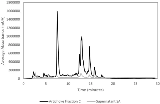
Figure 1.1 - Harvest globe artichokes at left and plant habit of globe artichoke at right. Illustrations removed from Edible Medicinal And Non-Medicinal Plants... 1 Figure 1.2 - Representative compounds present in the artichoke: inulin, lignin and cellulose. .... 4 Figure 1.3 - Representative phenolic compounds in artichoke: cynarin, cynaroside and chlorogenic acid. ... 5 Figure 1.4- Flow diagram of a vacuum filtration system. Illustration removed from Biosolids Tratment Processes. ... 10 Figure 1.5 - Diagram of lyophilization steps of a liquid formulation. Figure 1.5.1. shows the liquid formulation, which is market by ‘A’, in a glass container with the lyophilization closure positioned for the drying process; Figure 1.5.2. shows the frozen ice-product matrix of the formulation, which is market by ‘B’; Figure 1.5.3. represents the primary drying process and the interstitial cake portion is denoted by region market as ‘C’; Figure 1.5.4. illustrated the completion of secondary drying. Figure 1.5.5. demonstrates the final product with the closure in its stoppered position. Illustration adapted from Lyophilization: introduction and basic principles. ... 10 Figure 1.6 - Basal cell layer (B), spinous layer (S), granylar layer (G) and stratum corneum in a hemtoxylin and eosin stains section of normal skin. Illustration from Freinkel et al, 2001. ... 12 Figure 4.1 - Example of chromatogram of artichoke infusion (1mg/mL). ... 40 Figure 4.2 - Scheme of the purification strategies adopted to purify the artichoke extract. ... 41 Figure 4.3 - Quantification of polyphenols in each aliquot collected from the artichoke fraction A during the 24 hours of dialysis. ... 42 Figure 4.4 - Yield of compounds identified for artichoke extract and retentate obtained after dialysis by HPLC-DAD. ... 43 Figure 4.5 - Yield of compounds indentified for retentate and artichoke fraction A, both obtained after dialysis, by HPLC-DAD. ... 43 Figure 4.6 - Yields of compounds identified for the control sample, the retentate, the artichoke fraction A, the artichoke extract and the digested artichoke extract by HPLC-DAD. Control sample is 50% gastric juice and 50% water. Digested artichoke extract chromatogram is represented in a secondary vertical axis. ... 44 Figure 4.7 – Example of chromatogram of artichoke infusion (1mg/mL). ... 46 Figure 4.8 - Scheme of the McCleary method. ... 47 Figure 4.9 - HPLC-DAD analysis of the artichoke fraction C (15 mg/ mL), and the supernatants SA (1,5 mg/ mL) and SB (1,3 mg/ mL). The artichoke fraction C is in a seconday axis (right one). ... 48 Figure 4.10 - HPLC-DAD analysis of the artichoke fraction C (15 mg/ mL) and the supernatant SA (1,5 mg/ mL). ... 49 Figure 4.11 - HPLC-DAD analysis of the SA (1,5 mg/ mL) and SB (1,3 mg/ mL) supernatants. ... 50 Figure 4.12 - HPLC-DAD analysis of the SB supernatant (1,3 mg/ mL) and the pancreatic juice (2,5 mg/ mL). ... 51 Figure 4.13 - HPLC-DAD analysis of the precipitate obtained and the pancreatic juice (2,5 mg/ mL). ... 51 Figure 4.14 - HPLC-DAD analysis of the artichoke fractions B and C. ... 56 Figure 4.15 - HPLC-DAD analysis of the precipitates obtained from both methods, with and without acetic acid. ... 56 Figure 4.16 - Analysis of the artichoke fraction C and its precipitate by HPLC-DAD (1 mg/mL both). ... 58 Figure 4.17 - HPLC-DAD analysis of the artichoke fraction C washed and unwashed. ... 59 Figure 4.18 - Antioxidant activity of artichoke extract. The data are presented as the mean ± SD of at least 3 replicates experiments. ... 60
XIV
Figure 4.19 - Antioxidant activity pattern of Artichoke Fraction A abtained from dialysis. The data are presented as the mean ± SD of at least 3 replicates experiments. ... 61 Figure 4.20 - Antiradicalar activity (%) of the artichoke fraction C. The data are presented as the mean ± SD of at least 3 replicates experiments. ... 61 Figure 4.21 - Antiradicalar activity (%) of the artichoke fraction B. The data are presented as the mean ± SD of at least 3 replicates experiments. ... 62 Figure 4.22 - Antioxidant activity of ascorbic acid. The data are presented as the mean ± SD of at least 3 replicates experiments. ... 62 Figure 4.23 - Antioxidant activities of artichoke extract, artichoke fractions A, B and C, and ascorbic acid The data are presented as the mean ± SD of at least 3 replicates experiments... 63 Figure 4.24 - Cytotoxicity of artichoke extract (black bar) and artichoke fraction C (square bar) (1 mg/mL). SDS (sodium dodecyl sulfate) (dots bar)) was used as positive control and the medium as negative control (horizontal risks bar). The data are presented as the mean ± SD of 6 replicates experiments. ... 65 Figure 4.25 - ROS production after artichoke extract (black bars) and artichoke fraction C (square bars) (1 mg/mL) exposure to HaCaT cells in RPMI medium for 30 minutes before the addiction
of H2O2 and at the same time. Ascorbic acid as a negative control (risks bars). The data are
presented as the mean ± SD of at least 5 replicates experiments. ... 66 Figure 4.26 - ROS production of artichoke (black bar) and artichoke fraction C (squares bar) (1
mg/mL) in HaCaT cells in RPMI médium in presence of H2O2. Ascorbic acid was used as a
negative control (risks bar). The data are presented as the mean ± SD of at least 10 replicates experiments. ... 67 Figure 4.27 - ROS production of artichoke extract (black bar) and artichoke fraction C (squares bar) (1 mg/mL) in HaCaT cells in RPMI médium when the cells are exposed to UVB radiation for 15 minutes. Ascorbic acid (risks bar) was used as a negative control. The data are presented as the mean ± SD of at least 5 replicates experiments. ... 68 Figure 4.28 - Flow curves of artichoke extract, artichoke fraction C and blank creams representing shear stress as function of shear rate. ... 71 Figure 4.29 - Flow curves of artichoke extract, artichoke fraction C and blank gels representing shear stress as function of shear rate. ... 71 Figure 4.30 – MTT assay measurement of cytotoxicity of topical formulations containing the artichoke extract (black bars) and artichoke fraction C (squares bars) and their blanks (vertical risks bars). The data are presented as the mean ± SD of 6 replicates experiments. ... 74 Figure 4.31 - MTT assay measurement of cytotoxicity of artichoke extract (black bars) and artichoke fraction C (square bars) and their respective topical formulations. The data are presented as the mean ± SD of 6 replicates experiments. ... 75 Figure 4.32 – Propidium iodide assay measurement of cytotoxicity of topical formulations containing the artichoke extract (black bars) and artichoke fraction C (squares bars) and their blanks (vertical risks bars). The data are presented as the mean ± SD of 6 replicates experiments. ... 76 Figure 4.33 - ROS production of artichoke extract gel (white bar with black dots) and artichoke
extract cream (black bar with white dots) in HaCaT cells in RPMI médium in presence of H2O2.
Ascorbic acid was used as a negative control (risks bar). The data are presented as the mean ± SD of at least 5 replicates. ... 78 Figure 4.34 - ROS production of artichoke extract gel (white bar with black dots) and artichoke extract cream (black bar with white dots) in HaCaT cells in RPMI médium when the cells are exposed to UVB radiation for 15 minutes. Ascorbic acid was used as a negative control (risks bar). The data are presented as the mean ± SD of at least 5 replicates. ... 78 Figure 4.35 - Oxidant activity % change during the study after the exposure to artichoke extract gel (white bar with black dots) and artichoke extract cream (black bar with white dots). Mean values of all the subjects (n=10). ... 80
XV
List of Tables
Table 3.1 - Percentage of ingredients in artichoke extract and artichoke fraction C emulsions. 33 Table 3.2 - Percentage of ingredients in artichoke extract and artichoke fraction C hydrogels. . 34 Table 4.1 - Quantification of polyphenols in artichoke fraction A after 3, 6, 22 and 24 hours of dialysis... 42 Table 4.2 – Total content of chlorogenic acid, cynaroside and cynarin in the artichoke extract. 46 Table 4.3 - Total content of total phenols present in the artichoke fraction C and in the supernatants SB and SC. The data are presented as the mean ± SD of at least 3 replicates experiments. ... 49 Table 4.4 - Quantity of carbohydrates, proteins, phenols and soluble and insoluble fibers present in a total of 80 mg of artichoke extract. The data are presented as the mean ± SD of at least 3 replicates experiments. ... 52 Table 4.5 - Concentration of total phenols and tannins in digested artichoke extract before dialysis and in the retentate and artichoke fraction A after dialysis. The data are presented as the mean ± SD of at least 3 replicates experiments. ... 53 Table 4.6 - Concentration of chlorogenic acid, cynarin and luteolin in digested artichoke extract before dialysis and in the retentate and artichoke fraction A after dialysis. ... 54 Table 4.7 - Quantification of total phenols and tannins in the supernatants and precipitates obtained with the mucilage precipitation methods with and without acetic acid. The data are presented as the mean ± SD of at least 3 replicates experiments. ... 57 Table 4.8 - Quantification of chlorogenic acid, cynarin and cynaroside in the artichoke fractions B and C. ... 57 Table 4.9 - Quantification of chlorogenic acid, cynarin and cynaroside in the artichoke fraction C and its precipitate. ... 58 Table 4.10 - Total phenols present in the artichoke fraction C before and after the wahing process. The data are presented as the mean ± SD of at least 3 replicates experiments. ... 59 Table 4.11 - Cytotoxicity of the artichoke extract and artichoke fraction C. The data are presented as the mean ± SD of at least 3 replicates experiments. ... 65 Table 4.12 - Half maximal effective concentration (EC₅₀) of the artichoke fractions B and C obtained from the assays with and without acetic acid. The data are presented as the mean ± SD of at least 3 replicates experiments. ... 61 Table 4.13 - Total content of chlorogenic acid, cynarin and cynaroside in the artichoke extract, artichoke fraction A obtained from dialysis and artichoke fraction C obtained from the mucilage precipitation method with no use of acetic acid, and their antioxidant activity. The data are presented as the mean ± SD of at least 3 replicates experiments. ... 63 Table 4.14 - Content of flavonoids in the artichoke extract and the artichoke fraction C. The data are presented as the mean ± SD of at least 3 replicates experiments. ... 68 Table 4.15 - SPF found for the natural oils/extracts. The data are presented as the mean ± SD of at least 3 replicates experiments. ... 69
Table 4.16 - Apparent viscosity values calculated at 61,18s-1 of all formulations. ... 72
Table 4.17 - Determination of pH values of all formulations prepared at environment temperature. The data are presented as the mean ± SD of at least 3 replicates experiments. ... 73 Table 4.18 - Summary results of the evolution of b* color (AU) during the HRIPT assay. ... 80
XVI
List of Abbreviations
AC – Artichoke Extract Cream AG – Artichoke Extract Gel ANOVA – Analysis of variance BC – Blank Cream
BG – Blank Gel
CC – Artichoke Fraction C Cream CG – Artichoke Fraction C Gel DMSO - Dimethyl sulfoxide
DPPH - 1,1-diphenyl-2-picrylhydrazyl EC50 - Half maximal effective concentration
HPLC-DAD – High-performance liquid chromatography–diode array detector MTT - 3-[4,5-dimethylthiazol-2-yl]-2, 5-diphenyltetrazolium bromide
O/W – oil-in-water
PBS - Phosphate buffer saline PI – Propidium Iodide
ROS - Reactive oxygen species SDS - Sodium Dodecyl Sulfate SPF – Sun Protector Factor
TAMC - Total Aerobic Microbial Count
TYMC - Total Combined Yeasts and Molds Count UV – Ultraviolet
1
1. Chapter: Introduction
1.1. Cynara Cardunculus var. Scolymus
1.1.1.
The Domestication of Artichoke
The artichoke was already known and cultivated at the beginning of the Christian era [1]. The first mention to this specie was made by Theophrastus, the Greek (371-287 BCE), who described artichoke cultivation in Sicily and Southern Italy [2], and in 77 EC the medical benefits of this plant were reported by the Roman Pliny the Elder (ad 23–79, in Naturalis Historia). However, it’s estimated that domestication of artichoke only occurred between 800 and 1 500 CE, possibly in monastery gardens [3]. Records point Filippo Strozzi as the first merchant of this specie, who was responsible for trading artichokes from Sicily to Florence in the beginning of 15th century [4].
1.1.2.
Taxonomy and Botanical Description
Cynara cardunculus var. scolymus, usually known as globe artichoke, belongs to the family Asteraceae, subfamily Carduoideae and genus Cynara. Wiklund used a cladistics method based on morphological characterization to search the ancestry of Cynara crops and the results showed that the cultivated artichoke (C. Cardunculus var. Scolymus), leafy cardoon (C. cardunculus var. Altilis) and wild cardoon (C. Cardunculus var. Sylvestris) are included in a single species: C. Cardunculus L. [5]. A more recent study analyzed the phyletic relationships between Cynara species by using internal transcribed spacer sequences of the ribosomal regions, and the results were nearly in accordance with the phylogeny proposed earlier by Wiklund [6]. In Figure 1.1 are represented harvested artichokes and its habitat.
Figure 1.1 - Harvest globe artichokes at left and plant habit of globe artichoke at right. Illustrations removed from Edible Medicinal And Non-Medicinal Plants.
2
The globe artichoke is a perennial herbaceous plant, up to 80-180 cm tall, characterized by greyish green and deeply lobed leaves up to 75 cm in length. The flower heads are 5-10 cm in diameter and consists of green bracts surrounding violet florets. Both bracts and florets are attached to a receptacle, commonly known as the heart [7].
At first the leaves of the globe artichoke are rosulate, but later they sprout and adopt an ovate-lanceolate shape. These leaves are silvery-grey, pinnatifid and normally large, up to 50 cm long by 20 wide. This specie is characterized by a voluminous ovate capitula, usually known as the head, which is characterized by a violet-blue flower that is placed on the top of the flowering stem, up to 1.5 m. This head is composed of thick, fleshy bracts called phyllaries (involucral bracts), which are broadly ovate and green or tinged with purple or wholly purple, and a fleshy base of the receptacle, also called heart. These are the edible parts of globe artichoke.
1.1.3.
Agroecology
Globe artichoke has a wide adaptive range of 7-30°C, being a cool-season crop. Therefore, it should be planted in areas with day and night temperatures between 20-22°C and 12-14°C, respectively. This specie is very sensible to lower temperatures, and severe of frequent frosts can damage or kill it; but it tolerates well high temperatures (>30°C), though it tends to decrease the quality of artichoke heads. It also requires long periods of sunlight, having a critical photoperiod of 10.5 hours [8].
The transition from vegetative to reproductive phase in seed-planted individuals depends on the interaction between the size of the plant, low temperatures and photoperiod. This type of artichoke has deep-rooted and can grow in several varieties of soils. However, it grows best on deep, fertile and well-drained soils with a pH range of 6-8. It requires a raised-bed culture when drainage of the field isn’t known, since globe artichoke dislikes water-logged soils [8].
1.1.4.
Geographic Distribution
Mediterranean diet is characterized by a high consumption of vegetables, fruits, cereals and olive oil, which provide numerous micronutrients, such as vitamins, minerals, fibers
3
and polyphenols [9]. The artichoke is one of those vegetables, and it is consumed raw, boiled, steamed or fried, and therefore it’s used in numerous recipes [10].
This plant plays an important role in the agriculture and economy of the Mediterranean region, with a production of 199 900 t in Spain, 36 423 t in France, 547 799 t in Italy and 28 600 t in Greece; it is also produced on a large scale in the North Africa – 777 671 t. Global production is approximately 1 793 016 t, making Mediterranean countries belonging to Europe – France, Spain, Italy and Greece – responsible for over 45% of artichoke production, which makes it a source of income that is helping the economy in this region [11].
1.1.5.
Phytochemistry
The head of globe artichoke corresponds to 30-40% of its fresh weight [12] and the receptacle accounts for 33-55% of the head [13]. A raw globe artichoke is manly composed of water (84.94%); but it also contains proteins (3.27%), ash (1.13%), fat (0.15%), carbohydrates (10.51%), including inulin (1.8%), fibers (5.4%) and sugars (0.99%), such as glucose, fructose and sucrose. Minerals (0.66%) and vitamins (0.049%) are as well important components [14], [15].
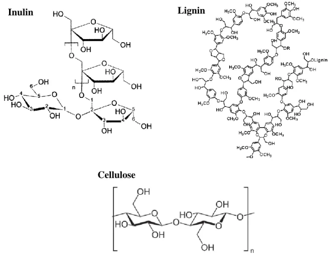
Carbohydrates are polyhydroxy aldehydes or ketones or substances that can originate such compounds through hydrolysis reactions, and they may be divided into three major classes: monosaccharides, oligosaccharides and polysaccharides. Plant polysaccharides can be classified into storage polysaccharide (e.g. inulin; the body can digest), cell-wall polysaccharides (e.g. cellulose; the body can’t digest it) and gums and mucilage. Structural fibers make up the plant cell walls and include lignin, cellulose, hemicelluloses and pectins.
Globe artichoke has high content of carbohydrates, including the storage polysaccharide inulin. Inulin is a fructan-type polysaccharide that consists of (2→1) linked β-d-fructosyl residues (n = 2–60), usually with an (1↔2) α-d-glucose end group (degree of polymerization ranging from 2 to 60 or more). Previous studies report a content between 1.8 and 2.5% of inulin in this plant [16], [17]. Fibers are also one of the most abundant constituents in the artichoke, namely cellulose and lignin. Cellulose is an unbranched glucose water-insoluble polymer with around 3 000 glucose units and it is the most widely
4
distributed component of plant cell walls, and lignin is phenylpropane polymers synthetized from coumatyl, coniferyl and sinapyl alcohols. Artichoke fibers are composed of 75.3 % of cellulose and 4.3% of lignin, being a rich-fiber plant [18]. Chemical structure of some compounds present in artichoke are showed in Figure 1.2.
Phenolic compounds are one of the biggest and widely distributed groups of secondary metabolites in plants. All polyphenols share a common origin, the amino acid phenylalanine, which is deaminated to cinnamic or converted to tyrosine, and then it enters the phenylpropanoid pathway. Therefore, phenolic compounds have as a common characteristic, the presence of at least one aromatic ring hydroxyl-substituted. The polyphenols can be classified into simple phenols, hydroxybenzoic acid and hydroxycinnamic acid derivatives, flavonoids, stilbenes, lignans and hydrolyzable as well as condensed tannins. These compounds are usually bounded to other molecules, commonly sugars and proteins, but they also exist in a small quantity in their free form, possibly due to its toxicity when in free state. They play an important role in plants,
Inulin
Cellulose
Lignin
5
namely in sensorial properties (color, aroma, taste and astringency), structure, pollination, resistance to pests and defense from predators and in grow, germinative and reproduction processes. According to literature, the outer and inner bracts, the receptacle and the leaves of globe artichoke contain polyphenols [10], [19], but its content can be influenced by the harvest time. The phenolic content can increase up to 16 times from winter to Spring harvest, especially in floral stem [20].
Zhu et al. [21] identified four caffeoylquinic acid derivatives, chlorogenic acid, cynarin, 3,5-di-O-caffeoylquinic acid and 4,5-di-O-caffeoylquinic acid, and four flavonoids, luteolin-7-rutinoside, cynaroside, apigenin-7-rutinoside and apigenin-7-O-β-D-glucopyranoside in artichoke leaf extracts, and Falé et al. [22] also identified cynaroside (luteolin-7-O-glucoside) and luteolin-7-O-(6’’malonylglucoside). However, the artichoke extract is mainly composed of caffeoylquinic and dicaffeoylquinic acids, being the most abundant cynarin (1,3-dicaffeoylquinic acid) and chlorogenic acid (3-O-caffeoylquinic acid)) [19], [23]. Chemical structure of some phenolic compounds present in artichoke are showed in Figure 1.3.
Cynarin
Cynaroside
Chlorogenic acid
6
1.1.6.
Therapeutic Properties
Artichoke dried leaves have been used as a medicinal herb in traditional medicine for centuries because of its choleretic and hepatoprotective [24], anticarcinogenic[25], antioxidant [19], [22], [26], antibacterial [21], antifungal [27], anti-inflammatory [28] and antiphotoaging properties [29].
The hepatoprotective activity of artichoke was tested in isolated rat hepatocytes against CCl4 toxicity and the results revealed that cynarin and caffeic acid act in glutamine oxaloacetic transaminase (GOT) and glutamic pyruvic transaminase (GTP), consequently showing hepatoprotective properties, while isochlorogenic acid and cynaroside act only in the GOT activity. Quinic acid and chlorogenic acid didn’t alter the toxicity of CCl4 [30]. Another study showed that the artichoke leaf extract with the highest content in phenolic compounds exerted a more significant effect on bile flow and liver protection, but the results also showed that the administration of cynarin and chlorogenic acid as pure compounds didn’t revealed hepatoprotective or choleretic properties [24]. The investigators suggested that caffeoyl derivates have an important role in therapeutic properties of artichoke, but when administrated in their pure form, they aren’t effective.
Mileo et al. [31] investigated the chemopreventive properties of artichoke edible parts through cytotoxicity assays on several cell lines and the results showed a similar level of toxicity in breast cancer cell lines but not any notable effects, even at high concentrations, on normal breast epithelial cells. This effect is due to the combined activities of several bioactive compounds present in the edible part of artichoke, since a study demonstrated that the artichoke extract has a faster and more pronounced apoptotic activity than the pure chlorogenic acid [32].
Through the measurement of the lag phase prolongation of conjugated diene formation, the decrease in the rate of propagation and a sparing effect on the a-tocopherol within the low density lipoprotein (LDL), artichoke extract showed to have the capacity to retard LDL oxidation in a dose-depend way. Pure luteolin revealed a lipid peroxidation inhibition similar to 20 µg/mL of artichoke extract, and luteolin-7-O-glucosine also showed a dose-dependent inhibition of LDL oxidation but was less effective than luteolin. Studies of cooper-chelating properties of these compounds suggested a potential role in the antioxidant activity of artichoke extract [33]. Artichoke has an EC50 of 123.1 ± 5.7 µg/mL for DPPH scavenging activity and its biochemical activities (acetylcholinesterase
7
inhibition and DPPH scavenging activity) remain similar after gastric and pancreatic digestions [22].
In order to examine the antimicrobial activities of artichoke leaf extract, three fractions of it in different solvents, chloroform, ethyl acetate and n-butanol, were tested by disk assays. The results demonstrated that at least six types of bacteria, including B. subtilis, S. aureus, A. tumefaciens and M. luteus, and five fungi, including C. albicans, S. cereVisiae, S. carlsbergensis, A. niger and P. oxalicum, were susceptible to all artichoke fractions [21]. However, C. lusitaniae and M. mucedo were only susceptible to n-butanol fraction and C. cucumerinum was sensitive only to n-butanol and chloroform fractions. Therefore, the n-butanol fraction was considered the most active to all microorganisms tested. In the same study, eight phenolic compounds were isolated from n-butanol fraction, including chlorogenic acid, cynarin, 3,5-di-O-caffeoylquinic acid, 4,5-di-O-caffeoylquinic acid, luteolin-7-rutinoside, cynaroside, apigenin-7-rutinoside and apigenin-7-O-β-D-glucopyranoside, and the compounds that showed a higher antimicrobial activity were the chlorogenic acid, the cynarin, cynaroside and luteolin-7-rutinoside. In following studies, artichoke leaf extract also showed antifungal activity against 7 foodborne bacterial pathogens, Bacillus subtilis, Staphylococcus aureus, Agrobacterium tumefaciens, Micrococcus luteus, Escherichia coli, Salmonella typhimurium and Pseudomonas aeruginosa, 4 yeasts, Candida albicans, Candida lusitaniae , Saccharomyces cerevisiae and Saccharo myces carlsbergensis , and 4 molds, Aspergillus niger , Penicillium oxalicum , Mucor mucedo and Cladosporium cucumerinum [27].
A group of 10 albino mice were administrated via oral with 10% infusion (dry plant) or 20% (fresh plant) corresponding to 1 or 2 g/kg of several plants, and the analgesic and anti-inflammatory activities were measured by the number of contortions and Evans blue dye diffusion to the peritoneal cavity, respectively [34]. From 10 plants tested, artichoke was one of the two plants that showed powerful anti-inflammatory and analgesic activities [28]. In other study, elevated levels of proinflammatory cytokines in blood serum of patients with chronic viral hepatitis C were normalized after using artichoke preparations during the medical rehabilitation period.
Cynaropicrin is a bioactive sesquiterpene lactone and its content in artichoke extracts ranges between 0.44-1.60 % [35]. The administration of this compound found to be
8
effective against photoaging of the skin through the inhibition of the nuclear factor kappa (NF-κB)-mediated transactivation of basic fibroblast growth factor (bFGF) and the matrix metalloprotease-1 (MMP-1) in a dose-dependent manner [29]. NF- κB is normally retained in the cytoplasm as an inactive complex but it is activated by UV radiation, which induces bFGF and MMP-1 production [36], [37]. Was also confirmed that in an in vivo mouse model, cynaropicrin is able to prevent skin photoaging, which is traduced in a hyperproliferation of melanocytes and keratinocytes [29].
1.1.7.
Artichoke Waste Phytochemicals
The waste produced by the artichoke processing industry, such as leaves, outer bracts and stems, can form up to 80-85% of total biomass of the material, and it is normally used as organic mass, animal feed [38], and fuel [39] and fiber production [40].
In the artichoke waste were reported 45 phenolic compounds, including gallic acid; caffeoylquinic acid 1,2 and 3; dicaffeoylquinic acids 1,2,3,4,5,6 and 7; protocatechuic acid; esculin; chlorogenic acid; p-coumaric acid-O-glucoside isomers 1 and 2; eriodictyolglucuronide; rutin (quercetin-3-O-rutinoside); dicaffeoylquinic acid derivative; hyperoside (quercetin-3-O-galactoside); luteolin-7-O-rutinoside; cynaroside (luteolin-7-O-glucoside); isoquercitrin (quercetin-3-O-glucoside); luteolin-7-O- glucuronide; luteolin-7-O-galactoside; naringenin-O-hexoside; avicularin (quercetin-3-O-arabinoside); isorhoifolin (apigenin-7-O-rutinoside); quercetin-O-pentoside; quercitrin (quercetin-3-O-rhamnoside); luteolin-7-O-neohesperidoside; apigenin-O-glucoside; prunin (naringenin-7-O-glucoside); naringin (naringenin-7-O-neohesperidoside); scolimoside (luteolin-7-O-rhamnoside); apigenin-7-O-glucuronide; quercetin-O-pentoside; phloridzin (phloretin-2-O-glucoside); feruloylquinic acid-O-glucoside; luteolin; quercetin; naringenin; and apigenin chrysoeriol. Among all components, caffeoylquinic and dicaffeoylquinic acids showed to be the main components of artichoke waste [41].
Knowing the therapeutic properties of all artichoke components combined, the actual use of artichoke by-products is questionable and since it has several health benefits, alternatives for its use should be considered. Production of natural nontoxic food additives, neutraceuticals or cosmetics using artichoke as bioactive ingredient are some options for the use or artichoke by-products.
9
1.2. Purification Methods
Extraction is the separation of active ingredients of plant tissues from the inactive or inert compounds through the use of selective solvents in standard extraction techniques. The plant extracts obtained are still a relatively impure liquid, semisolid or powders and include several chemical compounds with different functions and structures, such as polyphenols [42]. The extraction of polyphenols from leaves depends on many factors including pH, solubility, extraction time and temperature [43]. However, the extraction method has effects on the rate, yield and purity of these compounds, so the method choice is crucial.
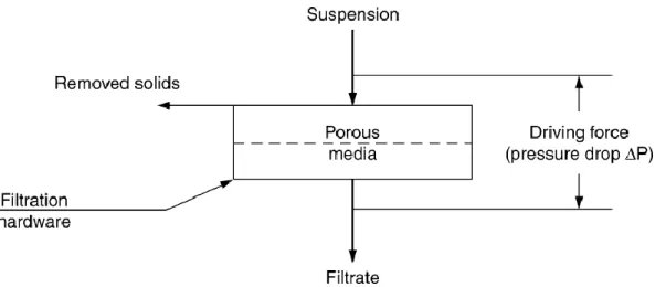
Infusion is a conventional method, in which dried leaves are placed in boiling water for a short period of time. The temperature and extraction time are parameters with impact in the total polyphenol content and consequently in antioxidant activity [42]. Liang et al. [44] investigated the influence of temperature on the extraction efficiency and degradation of polyphenols in green tea. The results showed that at a temperature of 100°C, the green tea catechins suffered alterations on their configuration to their isomers, and at 80°C the catechins epimerization was inhibited. Therefore, the extraction should be executed at a temperature between 80 and 100°C. Another study showed that a prolonged extraction period leads to the degradation of a large fraction of flavonoids present in green tea [45]. The investigators concluded that the conventional procedure of pouring boiling water to the cup with the herbal leaves and let it rest for 10 minutes is the proper way to infuse the herbs. This extraction technique is usually followed by a filtration procedure in order to separate the plant tissues from the solution containing the bioactive compounds. The vacuum filtration is a well-know and simple method that consists in passing the stream through a porous filter, which is capable of retaining the solids. This filtration requires a pressure drop in order for the liquid to flow through the filter, and this pressure can be achieved by creating a vacuum or a centrifugal force, by raising the pressure more than the atmospheric pressure or by designing to make use of gravitational force [46]. The vacuum filtration schematic is represented in Figure 1.4.
10
To finalize the active substances extraction procedure from herbs, a lyophilization of the herb infusion is required to remove its water content, which enhances its stability. To proceed to the lyophilization, the herbal infusion needs to be frozen in order to separate the solvent from the solutes, since the water forms ice crystals and the solutes will be confined in the interstitial region between those crystals. Then the pressure in the system is reduced and heat is applied to the frozen herbal infusion, which will lead to the sublimation of the ice crystals formed previously. A dried mixture, usually named cake, is the result of this process, but it may contain 5-10% (w/w) of water adsorbed onto its surface. In some cases, if the water content in the cake is still too high and consequently its stability isn’t the desired, a second drying is performed through the increasing of temperature and reducing of partial pressure of water vapor in the container [47]. The diagram of a lyophilization is represented in Figure 1.5.
Figure 1.5 - Diagram of lyophilization steps of a liquid formulation. Figure 1.5.1. shows the liquid formulation, which is market by ‘A’, in a glass container with the lyophilization closure positioned for the drying process; Figure 1.5.2. shows the frozen ice-product matrix of the formulation, which is market by ‘B’; Figure 1.5.3. represents the primary drying process and the interstitial cake portion is denoted by region market as ‘C’; Figure 1.5.4. illustrated the completion of secondary drying. Figure 1.5.5. demonstrates the final product with the closure in its stoppered position. Illustration adapted from Lyophilization: introduction and basic principles.
11
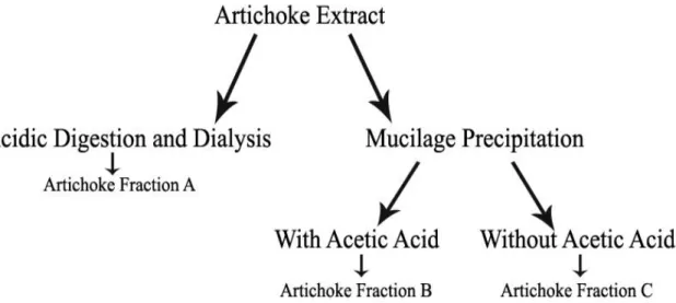
The herbal extracts obtained from the previous method are still impure, so a purification is required in order to enhance its properties, including the antioxidant activity. Two different approaches can be followed in order to purify the herb infusion: gastric digestion followed by dialysis and mucilage precipitation.
Tagliazuchi et al. [48] investigated the bio-accessibility of the major classes of polyphenols from Red Globe grapes through an in vitro model that simulated some chemical (pH, temperature and bile salts) and biological (gastric and pancreatic enzymes) gastro-intestinal conditions. The investigators concluded that the content of bio-accessible polyphenols, flavonoids and anthocyanins increases during gastric digestion, and that the gastric digestion has no effect on the stability of polyphenols. Therefore, it is advantageous to perform a gastric digestion of infusions in order to increase its bio-accessible polyphenols content, which will consequently improve its health benefits. Afterwards, a dialysis is required to separate the polyphenols from other large and complex molecules present in the herbal infusion, and consequently purify it. Dialysis is a separation method, based on the differential diffusion, of solutes with different molecular size through a membrane. This procedure is considered slow but it is efficient in separating small solutes from large molecules, and due to its simplicity, it is a method of choice. In this technique, the solution to be dialyzed is inserted in a dialysis membrane and then it is placed in a container with water for some hours or even days. In this process, a way of stirring the solutions is advisable and for that purpose, a magnetic stirred can be used to gently move continuously the dialysis membrane [49].
Another purification method is the precipitation of mucilage, a polysaccharide mixture that can be found in plant tissues of higher plants, present in herbal infusions. Kim et al. [50] investigated the effects of pectic mucilage removal from cactus cladodes (Opuntia humifusa Raf) and the results showed that along with the mucilage elimination occurs an increase of total polyphenols, and since these compounds contribute to antioxidant activity, it also resulted in an enhanced antioxidant activity.
12
1.3. Oxidative Stress in Skin
The skin covers the entire surface of our bodies and it corresponds approximately to 15% of total adult body weight, being the biggest organ in human body [51]. It consists of differentiated cells and tissues that perform several functions in our body, such as defense against other organisms, maintenance of body temperature and protection from external environment [52]. The protection of external physical, chemical and biological aggressions is due to a complex structure that has different tissues, like epithelial, connective, vascular, muscular and nervous, which are organized in three distinct layers: epidermis, dermis and hypodermis [51]. The skin also has several specialized structures with different functions called appendages (sebaceous, eccrine sweat and apocrine glands, and hair follicles). The epidermis is a tough stratified epithelium composed of cells with different embryonic origins, such as melanocytes, Langerhans cells, Merkel cells and mostly keratinocytes. It exhibits a progressive differentiation (keratinization and cornification) from the basal to the surface, which results in different appearances of cells from one layer to another [52]. Basal cells are attached to an underlying basal lamina in the innermost layer, and above this one is the stratum spinosum composed of prickle cells. Upward is a thin layer consisting of granular cells - stratum granulosum - which form a waterproof barrier, being this one of the most vital functions of the epidermis. This layer is attached to the outermost layer called corneal stratum, which consists of dead cell with no intracellular organelles. This cells rich in keratin are connected by a thin and tough layer of proteins, including cytoplasmic protein involucrin [53]. Epidermis is associated with some appendages like pisosevaceous follicles and sweat glands [51]. Skin layers are represented in Figure 1.6.
Figure 1.6 - Basal cell layer (B), spinous layer (S), granylar layer (G) and stratum corneum in a hemtoxylin and eosin stains section of normal skin. Illustration from Freinkel et al, 2001.
13
Below the epidermis is the dermis, a connective tissue component of the skin that protects the epidermis and provides elasticity and support [51]. The dermis has less cells than the epidermis and it is composed of fibrous molecules, dermal cells, a ground substance, appendances, neurovascular networks and sensory receptors. Its function is to protect the body from physical injuries, to retain water, regulate the temperature and it has receptors of sensory regulation [52]. It has a continuous turnover system that regulates the synthesis and degradation of its protein and it’s composed of the papillary and reticular layers [54]. Papillary layer is the upper layer and it is formed of thin collagen fibrils that form thicker collagen fibers [54], cells (fibroblasts, dermal dendrocytes, mast cells), vessels and nerve endings, being this layer the connection between the dermis and epidermis [51]. Bellow this one is the reticular layer that is composed mostly of type I collagen and fibers, and has only about 15% of dermal collagen which is mostly found in the papillary layer [54]. The deepest part of the appendages, vascular and nerve plexures are retain in this last layer [51]. The deepest layer of the skin is the hypodermis that serves as a reserve of energy, protects the skin from mechanical injuries and has a role in thermoregulation [52]. This layer is mostly composed of large adipocytes with a very weakly osmiophilic cytoplasm, which show pericellular expression of S100 protein and vimentin [51]. The adipocytes are organized in primary and secondary lobules separated by connective tissue septa containing cells, such as fibroblasts, dendrocytes and mast cells [51]. The fat cells are in continuous renovation through the accumulation of lipids on their inside, proliferation of adipocytes and recruitment of undifferentiated cells from mesoderm. This continuous system appears to be regulated by a hormone released from adipocytes called leptin [52]. The hypodermis also accommodates vessels, nerves and the deepest part of sweat glands [51].
In 1955, Denham Harman articulated an innovated biochemistry theory that suggested a correlation between oxygen radicals produced by the cells and the aging process. According to his theory, oxygen radicals such as HO∙ and HO2∙ were generated inside cells, most likely due to the interaction of respiratory enzymes and the molecular oxygen and to the action of catalase on hydrogen peroxide, and they were responsible for damages in DNA and proteins, and consequently for the ageing and cancer [55]. Despite the controversy around this theory back in those days, in the present day it is well proved. Skin is exposed to several harmful agents, such as ultraviolet (UV) and ionizing radiation, air pollution and biologic contaminants, that contribute for the production of reactive
14
oxygen species (ROS) in epidermal tissues. ROS are compounds derived from molecular oxygen, O2, but due to chemical reactions they have extra electrons, which make them unstable. Hydroxyl radicals, HO∙, are a result from homolytic cleavage of hydrogen peroxide, H2O2, and superoxide, O2∙, a result of molecular oxygen reduction. These two radicals and hydrogen peroxide are largely produced by the cells as a result of several biological mechanisms [56].
Among differentiated cells, human keranocytes are the most exposed to harmful agents, including oxidative stress associated with ROS overproduction, and they are further modulated by exposure to UV radiation, chemical compounds, rich-oxygen environment and inflammatory processes. Orciani et al. [57] determined the susceptibility of isolated mesenchymal stem cells from human skin (S-MSCs) to oxidative stress and compared the results with keratinocytes, which are differentiated cells of the same lineage. Cells were exposure to H2O2, a stress oxidative inductor, for 2 hours and the oxidative stress effects were analyzed after 4, 12, 24 and 48 hours of recovery, and the data reported a greater antioxidant defense for the keranocytes, while stem cells, which are surrounded by a protective environment, weren’t able to overcome oxidative stress. However, human keranocytes can still suffer damage at a long-term exposition to harmful agents, like UV radiation [58].
At high levels, ROS can induce damage to cell structures, lipids, nucleic acids and proteins, which can contribute to mutagenesis, carcinogenesis and ageing [59]. Human organism have developed several defensive strategies, like preventative and repair mechanisms, and physical and oxidant defenses [59], but these are not always capable to respond to an ROS overproduction and occasionally some errors may occur. Therefore, the search for new compounds with the capacity of preventing these oxidative stress events is constant.
UV radiation is divided in three distinct types of radiation: UVC (wavelength 200-280), UVB (wavelength 290-315nm) and UVA (315-400nm). The UVB and UVC are mostly filtered by the ozone layer, therefore only 5-10% of highly energetic UVB reaches the skin, being the rest UVA radiation [60]. The UVB radiation is mainly absorbed by the epidermis, while UVA radiation is able to penetrate into the dermis and it also interacts with the stratum corneum and epidermis [61]. There are some chromophores in the skin that can absorb UV radiation, such as melanin, DNA, RNA, proteins, lipids, aromatic
15
amino acids – tyrosine and tryptophan – and urocanic acid [62]. This absorption can lead to several photochemical reactions and secondary interactions with ROS, which can result in inflammation, photoaging, immunosuppression and skin cancers – cutaneous malignant melanoma, basal cell carcinoma and squamous cell carcinoma [61]. UVA and UVB radiation have different wavelength and therefore, they show distinct properties concerning their biological effects on our skin, being UVB radiation more mutagenic and cytotoxic than UVA. UVA radiation interacts with endogenous photosentisizers resulting in ROS production, which may cause damage to DNA, proteins and membranes. On the contrary, UVB radiation is highly absorbed by DNA resulting in its disruption and the production of photoproducts like cyclobutane pyrimidine dimers (CPD) and pyrumidone. A consequence of these photoreactions is the accumulation of defective DNA that can be the trigger to mutations of genes that regulate the tissue homeostasis and genome integrity, for example the p53 tumor suppressor gene [60]. UVB radiation modifies the epidermal morphology, including the thickness increase of the corneal stratum, modification of cell cohesion and mechanical integrity of corneal stratum and disruption of the permeability barrier, resulting in transepidermal water loss, changes in the lipids of corneal stratum and decreased corneal stratum hydration [61].
Polyphenols from plant tissues have been described as powerful antioxidants that can support the skin’s own antioxidant defense against oxidative stress when applied topically [63], [64]. These compounds also have photoprotective properties [65] and sun protection factor (SPF), which ranges between 7 and 29 for stilbenes, flavonoids and hydroxycinnamic acid homologues [66].
1.4. Topical Formulations
Nowadays, plant extracts are being more used in cosmetic formulation, mostly due to the prohibition of the use of animal origin ingredients and to an increase search for ecofriendly and sustainable products. These plant extracts are very different from purified therapeutic agents, since plant extracts are more dilute and usually contain more bioactive ingredients that may be related chemically and therapeutically to the main component responsible for the desired effect [67]. The use of bioactive ingredients or phytochemicals extracted from plant tissues in cosmetics have two major purposes, including body care and as a source of nutrients for a healthy skin [68].
16
In cosmetics is possible to use the entire plant extract, which is mostly applied according with its known therapeutic effects, or selective plant extract, which is used according to the investigations studies on its properties [67]. Usually, these botanical products are rich in vitamins, hydrocolloids, proteins, antioxidants, essential oils and other bioactive compounds [69]. Cosmetic formulations containing bioactive ingredients from a natural source are designed to protect skin against exogenous or endogenous harmful agents and to equilibrate the dermal hydrolipid content, which suffers alterations due to ageing and dermatosis [67].
To develop a good formulation, simplicity is required and the shorter the ingredient list, the better, since it diminishes the possible adverse effects due to a compound present in it and the costs of production. The formulation design has to balance the ingredients used in order to create a product stable and easy to apply, and that maintains its functions during manufacturing, on the shelf and during and after application.
Topical dermatological products can be administered easily and they can be liquid, such as suspensions, solutions and emulsions, or semisolid, including ointments, gels and creams. Active ingredients incorporated in topical formulations can stay on the surface layers of tissues or can penetrate into deeper layers, depending on its physicochemical properties, its action and the formulation approach.
Creams are semisolid emulsions made with two immiscible liquids, water and oil, in which one is considered the dispersion phase and it is dispersed into the other phase, which acts as the dispersion medium, resulting in a stable dispersion. The main ingredients of these formulations are emollients, humectants, surfactants, preservatives, chelating agents, perfumes and others. The equilibrium between water and oily ingredients may produce different formulations in order to suit different purposes (skin types, skin conditions, age of user and living environment). Usually, the main functions of creams are to maintain the moisture balance and keep the skin moist and supple by supplying water, humectants and oils [70]. There are two categories of creams according to the surfactants and oily ingredients used: O/W or W/O emulsions. In O/W emulsions are used hydrophilic surfactants and the oily ingredients can vary widely from non-polar to polar. On the contrary, in W/O emulsions are mostly used lipophilic surfactants and the oily ingredients are mainly non-polar. The W/O type has been used to increase the oily nature and the O/W type when a light feeling is wanted [70].
17
Gels are a type of base with a uniform external appearance that can vary from transparent to semitransparent and they provide a moist feeling. These formulations can be divided into two categories, aqueous or oily gels. Aqueous gels are used as a base material and are known for their water supplying, moisturizing and cooling effects. Oily gels supply oil to the skin and have moisturizing properties as well. The main compounds of gels are polymers and solvents [70]. Polymers are a crucial gel constituent and they can be classified based on source (natural, semi-synthetic and synthetic), structure (linear, branched chain, crosslinked or network polymer), polymerization type (addition or condensation polymers), molecular forces (elastomers, fibers, thermosetting, thermoplastic), chain growth polymerization (free radicals governed) or degradability (biodegradable or non-degradable) [71]. Water soluble polymers can be applied in several industries, like food, paint, textiles, paper, pharmaceuticals, cosmetics, water treatment, among others, and can be divided into two categories, synthetic or natural. Synthetic water soluble polymers can be dissolve, disperse or swell in water and can work as a gelation, thickening or emulsification/stabilization agent, which result in alterations of physical properties of aqueous systems. These include poly(ethylene glycol) (PEG), polyvinyl pyrrolidone (PVP), Polyvinyl alcohol (PVA), Polyacrylamides, Polyacrylic acid (PAA), N-(2-Hydroxypropyl) methacrylamide (HPMA), Divinyl Ether-Maleic Anhydride (DIVEMA), polyoxazoline, Polyphosphates and Polyphosphazenes; and natural water soluble polymers include xanthan gum, pectins, chitosan derivatives, dextran, carrageenan, guar gum, cellulose ethers, hyaluronic acid (HA), albumin and starch or starch based derivatives [71].
The quality, safety and efficacy of a topical formulation is evaluated by physical and chemical parameters such as pH, homogeneity, texture, microbiological control, skin irritation studies, cytotoxicity, stability and biometric parameters, including transepidermal water loss, hydration, strengthening the cutaneous barrier and elasticity, among others.
Under normal physiological condition, skin has an acidic pH between 4.5 and 6.5 that depends on the region. The acidic skin pH contributes to the defense against microbiological or chemical agents, the skin barrier homeostasis and stratum corneum desquamation [72]. Therefore, since skin pH has several important biological functions and its change could compromise the functions of topical formulations, these should present a pH value similar to skin pH.