2014/2015
Raul Miguel de Freitas Lima Neto
Achalasia: Pre-operative tests, post-operative symptoms and
surgical outcomes
Mestrado Integrado em Medicina
Área: Cirurgia Geral
Tipologia: Dissertação
Trabalho efetuado sob a Orientação de:
Professor Doutor José Adelino Lobarinhas Barbosa
Trabalho organizado de acordo com as normas da revista:
Surgery Today
março, 2015
Raul Miguel de Freitas Lima Neto
Achalasia: Pre-operative tests, post-operative symptoms and
surgical outcomes
Dedicatória
Dedico esta tese aos meus pais, à Isabel Sousa, aos meus avós e a todos os familiares e
amigos que me apoiaram de qualquer forma ao longo deste percurso na FMUP, em todo
e qualquer momento. Sozinho seria uma tarefa muito mais difícil. Um profundo
agradecimento a todos eles.
1
Title
: Achalasia: Pre-operative tests, post-operative symptoms and surgical outcomesAuthors:
Name: Raul Miguel de Freitas Lima Neto
Affiliation : Faculdade de Medicina da Universidade do Porto
Address : Alameda Prof. Hernâni Monteiro, 4200 - 319 Porto, PORTUGAL E-mail: raulfmup@hotmail.com
Telephone number:+351 919190022 Fax: N/a
Name: José Adelino Lobarinhas Barbosa
Affiliation: Departamento de Cirurgia Geral, Centro Hospitalar de São João Address: Alameda Prof. Hernâni Monteiro, 4200 - 319 Porto, PORTUGAL E-mail: jalbarbosa@netcabo.pt
Telephone number: +351 913805368 Fax: N/a
Article Type
: Original Article (Clinical original)2
ABSTRACT
Purpose:
The main goal of this study was to search for predictors, of post-operative dysphagia andheartburn, need for medical treatment during follow-up and the occurrence on intra and post-operative morbidity in patients with achalasia.
Methods:
The records of the patients who underwent myotomy for achalasia from 2005 to 2014 werereviewed (n=46). Data regarding pre-operative and post-operative manometry and pH-metry was compiled, along with patients’ symptoms and characteristics..
Results
: Our sample was composed by 25 female patients and 21 male patients. No parameter of theconventional manometry was associated with post-operative dysphagia, heartburn or medical treatment. Pre-operative heartburn and regurgitation were associated with less post-operative dysphagia (p<0.01 and p=0,034, respectively); Pre-operative dilatations were associated with post-operative morbidity (p=0.035) but not with intra-operatory morbidity (p=0.898). Relapse of achalasia was associated with greater usage of PPI during follow-up (p= 0.028).
Conclusions:
Heller myotomy is an effective treatment option for Achalasia. Dilatations should beused carefully since they can lead to an increase in post—operative morbidity. Patients with relapse of achalasia are more likely to need medical treatment after re-intervention while patients with pre-operative regurgitation and heartburn have less post-operative dysphagia.
3
INTRODUCTION
Achalasia is a disorder of oesophageal motility characterized by impaired relaxation of the lower oesophageal sphincter (LES), frequently associated with an increase in the pressure of LES and an absence of peristalsis of the oesophageal body [1]. Complete lower esophageal sphincter (LES) relaxation occurs in approximately 15–30% of patients with achalasia and is not a characteristic of early achalasia [2]. It’s annual incidence is about 1 case per 100000 habitants and the prevalence is around 10 in 10000 [3]. There is no sexual and racial prevalence and, while it is a disease that can appear at any age, there are two peaks of incidence: between 20-40 years old and between 70-80 years old [3, 4].
The main symptom is dysphagia, usually progressive for solids and liquids, although it can also present itself as paradoxal dysphagia. Other common presentation symptoms include thoracic pain, regurgitation, heartburn and weight loss [1, 5].
As of now, the pathogenesis of this disease is unknown, [6] although it is hypothesized that it results from the loss of the ganglionic cells of the myenteric plexus of the esophagus [1]. Other current train of thought deals with a possible viral etiology [7, 8]. Achalasia has been associated with, at various degrees of risk, squamous cell carcinoma. However, there are no recommendations for screening for cancer in patients with Achalasia [3, 9].
Diagnosing Achalasia requires a high index of clinical suspicion. The current gold-standard is the oesophageal manometry [6]. A complete workup usually includes oesophageal manometry, upper gastrointestinal endoscopy and a barium esophagogram [3]. The cardinal feature of this disease is the impaired relaxation of the LES in response to swallowing. Other abnormalities include an increase in the LES pressure and absence of peristalsis in the oesophageal body [10]. Currently, there are two forms of manometry, conventional and the more recent high-resolution manometry (HRM). Conventional manometry has some disadvantages in comparison to HRM manometry, since it is unable to account for the intrabolus pressure, the crural diaphragm relaxation, radial asymmetry of the gastroesophageal junction (GEJ) and deglutive oesophageal shortening [11]. As for the upper gastrointestinal endoscopy, it’s important to exclude a serious differential diagnosis, the Pseudo-Achalasia [6]. Barium esophagogram can also help in the diagnosis, identifying the so called “Bird’s Beak [3, 6]. The advancements brought by HRM allowed the classification of Achalasia into 3 different subtypes, a classification known as the Chicago Classification of Motility Disorders [12]. Type I
4
represents classic achalasia, associated with low intra-oesophageal pressure and minimum levels of oesophageal contractility; Type II is characterized by panoesophageal pressure elevations and an absence of peristalsis [1]; Type III is usually called “spastic achalasia” [5, 10, 13]. HRM proved to be an important evolution in terms of diagnosis and classification of Achalasia [14, 15].
The available treatments are used for symptom relief. Currently, no curative interventions exist [1]. Regarding pharmaceutic interventions, the most common drugs used are calcium channel blockers, but they are ineffective [4, 16]. The other treatment options are injection of botulin toxin, pneumatic dilatation and surgery. Regarding botulin toxin injection, it’s ineffective in the long term, with its’ effects reverting in a period between 6 and 9 months after the intervention [5]. It is currently reserved for elderly patients and high surgical risk patients [4]. Pneumatic dilatation and surgical myotomy are more useful treatment options. Pneumatic dilatation is performed by insufflating a balloon in the distal oesophagus, causing the forced distension of the muscular fibers of the LES. Usually a balloon of 30 mm is used due to a lower perforation rate [17]. It’s possible to achieve similar long-term results to surgery with pneumatic dilatation if multiple dilatations are used. It seems to be a more effective treatment in patients over 40 years old [16, 18]. It is also the most cost effective procedure [4] and has a lower risk of gastroesophageal reflux disease [17]. Surgery is the gold-standard treatment and the most frequently used surgical procedure is Heller’s myotomy, performed laparoscopically (section of the muscular fibers of the LES). The laparoscopic approach results in shorter hospitalization times [19]. To prevent gastroesophageal reflux a partial fundoplicature must also be performed in conjunction with the myotomy [3, 16]. There seems to be no difference between the two different partial fundoplicatures, Dor or Toupet [20]. For the myotomy, most surgeons will perform a section of 6-8 cm of the LES fibres in the oesophagus and prolong it 1-2 cm into the stomach [21]. Surgery has great success rate in the long term [22], even though it is not completely free of complications and does not warrant the absence of relapse. Choosing between pneumatic dilatation or surgery has been a topic of recent discussion. A recent European RCT could not determine which approach was better, finding no statistical difference between them [17]. The symptom which shows greater improvement with surgery is regurgitation, in contrast with heartburn [20]. In general, post-surgical complications are scarce, with one studying relating incidence of complications in less than 4% of patients [23]. The most frequent adverse effect of the surgical intervention is gastroesophageal reflux. Another complication is the perforation of the oesophagus,
5
usually reported in around 5-10% of myotomies. A robotically assisted myotomy has been shown to lower the rate of perforation to 0% [24].
A new technique has been developed, known as POEM (per-oral endoscopy myotomy). In this technique the LES is sectioned through a tunnel created in the esophagus submucosa. [22]. The results using this technique have been promising, with significant decreases of the LES pressure [25]. One of the main criticisms of this approach is the lack of an anti-reflux procedure. In fact a study showed that around 46% patients had significative reflux just 6 months after the intervention [26].
The main goal of this study was to search for a relation between the parameters evaluated in the conventional manometry and the relapse of dysphagia post-operatively, the presence of post-operative heartburn and the usage of protein-pump inhibitors during follow-up. A secondary objective was to provide a report of the patients’ symptoms (pre-op and post-op) and characteristics’ and whether any of these factors could predict the development of post-operative dysphagia, heartburn and the usage of PPI drugs as well as the occurrence of intra-operative and post-operative morbidity.
6
METHODS
Subjects and study protocol:
We performed a retrospective observational study of all patients, admitted in the Upper Gastrointestinal Unit of Centro Hospitalar de São João, who underwent surgery for Achalasia, be it primary or relapse, between 1st of January 2005 and 14th May 2014. The sampleconsisted of 46 patients, 25 female and 21 male. Patients younger than 18 years were excluded. To fulfil our aims, patient data was collected using the informatized systems of the hospital, and by consulting the manometry reports archived on paper. Patient data was compiled in an electronic database.
To evaluate the relation between manometric parameters and the post-operative symptoms, surgical complications, relapse of dysphagia and need for medical treatment, we reviewed all available manometric records, both pre-operatively and post-operatively. A sub-population of 20 patients, which had complete manometric data for both the pre-operative and post-operative periods, was created to evaluate the relation between pre-operative manometric parameters and post-operative outcomes. The variables used were: age at diagnosis; gender; diagnosis (primary achalasia or relapse); number of comorbidities; number of previous surgical procedures; pre-operative dilatations; type of surgery (heller-dor or totalization of myotomy); approach (laparoscopy or laparotomy); intra-operative morbidity; hospitalization length; post-operative morbidity; proton pump inhibitors (PPI) treatment during follow-up (starting in the first post-operative consultation or later) . Comorbidities were evaluated using Charlson Comorbidity Index [27] and morbidity was evaluated using the Clavien-Dindo Classification [28].
The symptoms recorded were: aspiration; siallorhea; halitosis; weight loss; chest Pain; bloating; heartburn; dysphagia; nocturnal cough; eructation; regurgitation; vomiting; nausea. Manometric variables evaluated were LES resting pressure; LES basal pressure; esophageal peristalsis; percentage of LES relaxation and LES functional length for both pre-operative and post-operative examinations.
Ambulatory 24-hour Esophageal pH Monitoring: This examination was performed using a pH
Orion II® (single crystal antimony multi-use pH catheter ) catheter equipped with 2 electrodes. Catheterswere introduced through transnasal approach and the sensors were placed at 15 cm and 5 cm from the GEJ (pH channel 1 and 2 respectively), whose location had been previously determined by manometry. We instructed patients to perform their regular routines, and to record their meal times, the time of sleep and whenever they experienced symptoms. After an average of 24 hours, patients returned to the hospital
7
to remove the sensors and check the information recorded. Data analysis using MMS® software was
performed, including the calculation of the DeMeester score.
Manometry:
Patients were submitted to the test in the supine position, after a fasting period of 12 hours. The test was performed using a manometric tube with 8 sensors, 4 placed in the distal end and 4 along the tube, 5 cm apart. This tube was inserted transnasally until the EGJ and then removed, cm by cm, by stationary pull-through so as to measure LES basal (normal: 10-25 mmHg) and residual pressures. LES relaxation was analysed using wet swallows of water (5mL). Relaxation was considered complete when LES pressure decreased to levels of gastric baseline pressure. Peristalsis was evaluated with a series of ten wet swallows.Surgical Procedure: The surgery was performed either by laparoscopy or laparotomy. The patient was
placed in a modified lithotomy position. In the laparoscopic procedure the procedure began with the creation of a pneumoperitoneum. Afterwards five trocars were inserted. The liver was retracted. After the opening of the crura, the oesophagus was dissected into the mediastinum. Next the short gastric vessels were mobilized so as to facilitate the partial fundoplicature. The EGJ was identified and the myotomy was then performed using an L-shaped hook electrocautery device or an UltraCision ® device, cutting the longitudinal muscle fibers first. The circular muscle fibers were then exposed and sectioned. The myotomy was performed with a length of 6-8 cm in the esophagus and prolonged distally 1-2 cm to the stomach. After finishing the myotomy, a Dor fundoplicature was performed. As for the laparotomy approach, the procedure was similar, except that it was performed with a supraumbilical median incision.
Statistical analysis: Statistical analysis was performed using IBM SPSS22 Statistics ®. Student t test
was used to analyse independent samples when comparing two groups and the variables were quantitative. To assert whether a variable had a normal distribution or not, the Levene and the Kolmogorov-Smirnov tests were used. If normality could not be asserted, the proper non-parametric test (Mann-Whitney test) was used. When studying categorical variables, crosstabs were created and χ2-test or Fisher’s exact test were applied, according to the situation. To evaluate whether pre-operative factors affected the risk of post-operative outcomes, we performed logistic regression. Tests results were considered statistical significant for a level of p<0.05.8
RESULTS
A total of 46 patients were operated in our centre from 2005 to 2014. Mean age at diagnosis was 48.02 ± 17.79 years old (range: 18-81 years old). 45.7% of patients were male and 54.3% were female. 40 patients had been diagnosed for the first time with Achalasia and 6 patients had relapse of the disease. Male patients had a lower age at diagnosis (46.10 years) when compared to female patients (49.64 years) without statistical difference (p=0.516).
In our sample, 52.2% of patients had no comorbidities, 10.9% had 1, 17.4% had 2 comorbidities and 19.6 had more than 2. Applying the Charlson Comorbidity Index, 4 patients had grade I complications, 3 had grade II, 2 had grade III and only one had grade IV comorbidities. It’s also important to point out that one patient had multiple sclerosis and another had Down syndrome.
The majority of the patients in our study had no prior surgical procedures (76.1%); 17.4% had 1; 4.3% had 2 surgical procedures and only 2.2% had been subjected to 3 surgeries before.
In terms of pneumatic dilatations, 14 patients (30. 4%) had dilatations prior to surgery. No information was found regarding the specific number of dilatations of 2 patients. Mean number of dilatations was 2.25 ± 1.288.
In terms of the surgical technique used, 4 patients underwent myotomy totalization and the remaining patients were subjected to Heller-Dor surgery. No Toupet fundoplicature was performed. A cholecystectomy was performed in one patient in the same operative time, while 3 others underwent hiatal hernia correction in the same operative period.
Most operations were performed laparoscopically, with only 10.9% of interventions performed through laparotomy. Only one patient was previously operated using the POEM technique. This patient relapsed and was operated laparoscopically, having complaints of heartburn and light dysphagia for solids after the surgery but no need for PPI treatment.
The mean hospitalisation length was 6.10 days ± 10.67; the minimal period of stay was 2 days, while the maximum was 76 days (patient with Down syndrome, which developed nosocomial pneumonia and respiratory insufficiency).
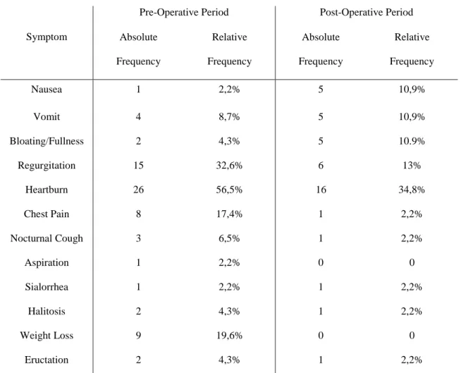
A complete report on all the patients’ symptoms and complaints pre-operatively and post-operatively, can be seen in table 1.Virtually all patients in the pre-operative period had dysphagia. Information regarding the type of dysphagia was difficult to collect using the medical reports, with information about 22 patients missing. Despite that, the data available showed that 2.2% had dysphagia just for liquids, 8.7% had
9
isolated dysphagia for solids, 1 patient (2.2%) had paradoxal dysphagia and 18 patients had dysphagia for liquids and solids. In our sample, 21.8% of patients had dysphagia post-operatively. 5 patients had dysphagia for solids, 4 had dysphagia for liquids and solids and 1 had dysphagia for liquids only.
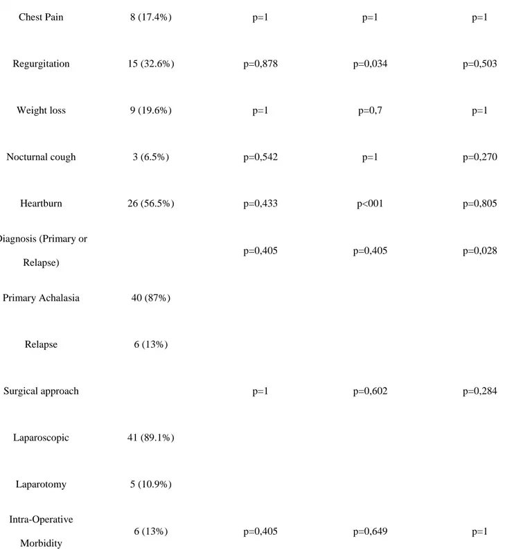
No statistically significant associations were found between post-operative heartburn and pre-operative symptoms or patients’ characteristics (Table 2).
A total of 23 patients performed a 24-hour pH-metry after the surgery. In our sample, 17.4% of patients had acid pathological reflux detected in the examination. Reflux was only detected in channel pH2, which was located at 5 cm from the GEJ. 75% of the patients who performed the examination showed no symptoms. 12.5% of patients had a negative symptom index while the remaining 12.5% had a positive symptom index. The association between post-operative complaints of heartburn and post-operative reflux demonstrated in 24-hour pH-metry was non-significant (p=0.281).
No associations were found between operative data and post-operative dysphagia, except for pre-operative regurgitation and pre-pre-operative heartburn (Table 2). Women had more dysphagia post-operatively (40% vs 37.5) but there was no statistical difference (p=0.538).
Patients who had pre-operative complaints of heartburn were less likely to have dysphagia post-operatively (25% vs 75%, p<0.01). Pre-operative heartburn and age at diagnosis were not associated (p=0.254). Patients that had pre-operative regurgitation had less dysphagia when compared with patients who did not have regurgitation prior to the surgery (13.2% vs 45.2% respectively, p=0.034). In our study, patients who had pre-operative regurgitation had a higher age at diagnosis (56.53 vs 43.90; p= 0.024). The number of pre-operative co-morbidities was higher in patients with post-operative dysphagia (1.63 vs 0.86) but this difference was not significant (p=0.074). Patients who had post-operative dysphagia also had a higher age at diagnosis (50.94 vs 46.47 years old) but it wasn’t statistically significant (p=0334). In our population, 39.1% of patients started therapy with proton-pump inhibitors during follow-up. There were no differences between the patients who needed PPI treatment and those who didn’t when it comes to the number of comorbidities (p=0.449), age (p=0.350), the number of surgical procedures (p=0.683) and the number of dilatations (p=0.148). There was an association between the type of diagnosis (primary achalasia or relapse) and the need for PPI treatment in the post-operative period (32.5% vs 83.3% respectively, p= 0.028).
No other statistically significant associations regarding post-operative PPI treatment were found (Table 2).
10
There was no association between the patients who had pre-operative dilatations and post-operative heartburn (p=0.189), post-operative dysphagia (p=0.739), need for medical treatment (p=0.332) or reflux in the post-operative pH-metry (p= 0.273).
The rate of intra-operative complications was 13%. Iatrogenic perforation of the oesophagus happened in 4 patients, and 2 other had intra-operatory aspiration of vomit. One of these patients also suffered a spleen laceration. Regarding post-operative morbidity, 8.7% of patients had complications. Surgical wound dehiscence (Grade I of the Clavien-Dindo classification) was present in one patient, while another had surgical wound infection (Grade II). One patient experienced left superior member palsy (Grade I) and finally one had nosocomial pneumonia (Grade IV). There was no mortality in our sample, except for the patient with Down syndrome, who died due to complications of the disease.
Concerning intra-operative morbidity, no predictors were found (Table 3). As for post-operative morbidity, patients who had a higher number of pre-operative dilatations had more morbidity after the surgery (p=0.035).
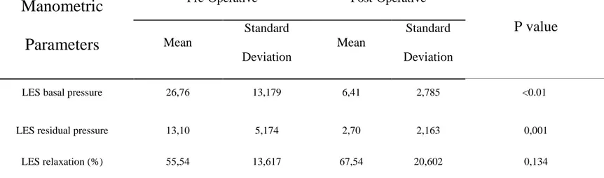
We compared the manometric profile of patients before and after the surgery (Table 4). A significant decrease of LES basal and residual pressures was verified. Average LES functional length was 3.4 cm ±1 cm. Before the surgery, 17 patients had positive intraoesophageal pressure and 7 had negative pressure. Post-operatively, only one patient had positive intraoesophageal pressure, while the remaining 24 had negative intraeosophageal resting pressure. The pre-operative and post-operatives manometric profiles did not differ with gender.
We performed logistic regression to see if any pre-operative aspect of the patients’ manometric profile could have an impact in post-operative dysphagia, post-operative heartburn and the need for medical treatment. There were no statistically significant differences (Table 5).
11
DISCUSSION
Achalasia has no curative treatment. All available treatments are palliative and have the main purpose of assuring the passage of liquids and solids through the gastroesophageal junction, therefore alleviating the symptomatology, preventing food stasis in the oesophagus and ultimately allowing the patient to eat. Furthermore, our knowledge of the disease, in pathophysiological terms, is still suboptimal. This leads to difficulty in identifying predictors of favourable outcomes for all treatments, including surgery.
There have been previous reports regarding predictors of surgical success. One study reported that patients with a higher pre-operative score of dysphagia had better surgical outcomes [23], while other reported that a duration of symptoms longer than 10 years predicted more post-operative dysphagia [29]. This was also supported by Krishnamohan P et al., whose study also found that a sigmoid oesophagus shape in pre-operative tests was also a predictor of surgical success [30].
In this study we provide a complete report of the patients’ complaints, both pre-operatively and post-operatively. The most commons symptoms before surgery, besides dysphagia, were heartburn and regurgitation. Heartburn remained the most common symptom even after surgery (34.8%). This is probably due to the fact that pre-operative and post-operative heartburn have different pathophysiological origins. This rate of heartburn is similar to other reports in the literature [31]. Despite this, only 17.4% of patients had had acid pathological reflux detected in the pH-metry post-operatively. This is in accordance to previous literature results [32] and can be explained by the low correlation between a patient complaining of heartburn and reflux and the demonstration of said reflux in the pH-metry.
One of our main goals was to try and find which pre-operative characteristics could predict better outcomes after the surgery, namely less dysphagia, less reflux, less intra-operative and post-operative complications and a lower usage of PPI medication.
Regarding post-operative dysphagia, we found two associations. First, patients which had pre-operative regurgitation had less post-operative dysphagia when compared with patients who did not have operative regurgitation (13.3% vs 45.2%, respectively, p=0.034). Interestingly, patients with pre-operative regurgitation were older, at the time of diagnosis, than patients without regurgitation (56.53 vs 43.90; p= 0.024). This was the only symptom which showed an association with age. It is a well-known fact that older patients, particularly over 60, have better outcomes after surgery, including less post-operative dysphagia [33]. As such, we think that this predictor effect of regurgitation of a better outcome was due to the fact that, in our sample, patients who complained of said symptom were older. Secondly,
12
another interesting finding was that patients who had pre-operative complaints of heartburn were less likely to have dysphagia post-operatively (p<0.01). As in the case of regurgitation, patients who experienced pre-operative heartburn were older than patients who didn’t (51.04 years old vs 44.10 years old), even though it was not statistically significant (p=0.254). It’s possible that the explanation for this association is the same that we propose for the regurgitation and dysphagia association, and that our sample simply did not have the statistical power to demonstrate it.
The type of diagnosis, the type of surgical approach, the number of co-morbidities did not prove to be predictors of post-operative dysphagia. Although women had more dysphagia post-operatively (40% vs 37.5%) there was no significance (p=0.538). Contrary to what has been reported about the patient’s age as a predictor of surgical outcomes [31], no association was found in our sample, even though patients who had post-operative dysphagia also had a higher age at diagnosis (50.94 vs 46.47 years old; p= 0.334). In terms of heartburn, we did not find any factors that could predict a higher incidence of this symptom post-operatively. This seems to be the case with other similar studies [29]. 39.1% of our patients started proton-pump inhibitor medication during follow-up. Although we could not find any predictors for post-op heartburn, we did discover that the diagnosis can predict the need for PPI medication. Patients who had relapse of Achalasia were more likely to need PPI treatment (83.3% of patients with relapse needed PPI vs only 32.5% of patients with primary Achalasia; p= 0.028). No similar reports were found in the literature. A possible explanation may result from the fact that patients who relapse must endure a second myotomy, which further weakens the physiological anti-reflux mechanism, predisposing to more severe heartburn and a greater need for medication.
LHM plus Dor fundoplicature proved to be an excellent treatment option for Achalasia. Our rate of post-operative dysphagia rounded 21.8%, which is in accordance to the rates reported in the literature [4]. After the surgery there was a significant decrease in LES basal pressure (26.76 vs 6.41; p<0.01), as well as LES Residual pressure (13.10 vs 2,70, p<0.01). LES relaxation increased after the surgery (from 55.54% to 67,54%) even though this was not a statistically significant increase ( p=0.134). Other studies have showed a non-significant increase as well [32].
As for the pre-operative manometric parameters, we did not find any association between these characteristics and post-operative dysphagia, need for medical treatment and post-operative heartburn (Table 4).
13
We also evaluated intra-operative and post-operative morbidity. Our rate of complications, 13% and 7%, respectively, is within the normal range reported in previous works [23, 30, 31].
When trying to assert possible predictors of morbidity, we found no factors that could predict intra-operative morbidity. However, we did find an association between pre-intra-operative pneumatic dilatations and post-operative morbidity (p=0.035). Dilatations are one of the most effective treatments available for Achalasia [4]. Nonetheless, one must take into account that previous dilatations can make the surgical technique more challenging and can perhaps lead to more complications for patients. Multiple reports in the literature have associated the existence of previous treatments and poorer surgical outcomes, this effect being greater with botulin toxin injection [5, 34, 35]. Smith et al. state that, in patients with previous dilatations, the submucosal dissection plane is obliterated [36]. Nevertheless, other articles found no association between pre-operative dilatations and surgical outcomes [29, 31].
Taking this information into consideration, we think that pneumatic dilatations should be used carefully and that there should be a strict decision-making process on which patient does dilatation before surgery. One recent development that might help in this decision is the Chicago Classification, since it has been shown that patients with type II achalasia as defined by this classification respond better to any form of treatment and this subtype is a predictor of therapeutical success [10]. On the other hand, type I and type III of the Chicago Classification tend to predict a poorer outcome [5].
The limitations of this study are the retrospective approach and the small sample size. Due to our sample size, a risk for a Type II error exists. Another limitation was the lack of complete data regarding the manometric reports of all patients.
In conclusion, our study reinforces the efficacy of LHM as the current gold-standard of treatment for Achalasia while introducing the notion that there should be an extra care in terms of reflux for patients with relapse of the disease. It also shows that pneumatic dilatations, while curiously a safe option in terms of intra-operative morbidity, can lead to a higher incidence of post-operative morbidity and, as such, should be used with great care. It is our opinion that prospective studies with long follow-up are needed to uncover new predictors of surgery success or failure, specially taking into account the differences between subtypes of Achalasia elucidated by the new Chicago Classification.
14
REFERENCES
1. Tolone S, Limongelli P, Del Genio G, Brusciano L, Russo A, Cipriano L, et al. Recent trends in endoscopic management of achalasia. World journal of gastrointestinal endoscopy. 2014;6(9):407-14. 2. Felix VN, DeVault K, Penagini R, Elvevi A, Swanstrom L, Wassenaar E, et al. Causes and treatments of achalasia, and primary disorders of the esophageal body. Annals of the New York Academy of Sciences. 2013;1300:236-49.
3. Katada N, Sakuramoto S, Yamashita K, Shibata T, Moriya H, Kikuchi S, et al. Recent trends in the management of achalasia. Annals of thoracic and cardiovascular surgery : official journal of the Association of Thoracic and Cardiovascular Surgeons of Asia. 2012;18(5):420-8.
4. Chuah SK, Chiu CH, Tai WC, Lee JH, Lu HI, Changchien CS, et al. Current status in the treatment options for esophageal achalasia. World journal of gastroenterology : WJG. 2013;19(33):5421-9.
5. Eckardt AJ, Eckardt VF. Treatment and surveillance strategies in achalasia: an update. Nat Rev Gastroenterol Hepatol. 2011;8(6):311-19.
6. O'Neill OM, Johnston BT, Coleman HG. Achalasia: a review of clinical diagnosis, epidemiology, treatment and outcomes. World journal of gastroenterology : WJG. 2013;19(35):5806-12. 7. Lau KW, McCaughey C, Coyle PV, Murray LJ, Johnston BT. Enhanced reactivity of peripheral blood immune cells to HSV-1 in primary achalasia. Scandinavian journal of gastroenterology. 2010;45(7-8):806-13.
8. Ghoshal UC, Daschakraborty SB, Singh R. Pathogenesis of achalasia cardia. World journal of gastroenterology : WJG. 2012;18(24):3050-7.
9. Ravi K, Geno DM, Katzka DA. Esophageal cancer screening in achalasia: is there a consensus? Diseases of the esophagus : official journal of the International Society for Diseases of the Esophagus / ISDE. 2014.
10. Pandolfino JE, Kwiatek MA, Nealis T, Bulsiewicz W, Post J, Kahrilas PJ. Achalasia: a new clinically relevant classification by high-resolution manometry. Gastroenterology. 2008;135(5):1526-33. 11. Carlson DA, Pandolfino JE. The Chicago criteria for esophageal motility disorders: what has changed in the past 5 years? Current opinion in gastroenterology. 2012;28(4):395-402.
12. Roman S, Gyawali CP, Xiao Y, Pandolfino JE, Kahrilas PJ. The Chicago Classification of motility disorders: an update. Gastrointestinal endoscopy clinics of North America. 2014;24(4):545-61. 13. Bredenoord AJ, Fox M, Kahrilas PJ, Pandolfino JE, Schwizer W, Smout AJ. Chicago classification criteria of esophageal motility disorders defined in high resolution esophageal pressure topography. Neurogastroenterology and motility : the official journal of the European Gastrointestinal Motility Society. 2012;24 Suppl 1:57-65.
14. Greene CL, Chang EJ, Oh DS, Worrell SG, Hagen JA, DeMeester SR. High resolution manometry sub-classification of Achalasia: does it really matter? : Does Achalasia sub-classification matter? Surgical endoscopy. 2014.
15. Xiao Y, Kahrilas PJ, Kwasny MJ, Roman S, Lin Z, Nicodeme F, et al. High-resolution manometry correlates of ineffective esophageal motility. The American journal of gastroenterology. 2012;107(11):1647-54.
16. Friedel D, Modayil R, Iqbal S, Grendell JH, Stavropoulos SN. Per-oral endoscopic myotomy for achalasia: An American perspective. World journal of gastrointestinal endoscopy. 2013;5(9):420-7. 17. Wu JC. Pneumatic Dilation versus Laparoscopic Heller's Myotomy for Idiopathic Achalasia (N Engl J Med 2011;364:1807-1816). Journal of neurogastroenterology and motility. 2011;17(3):324-6. 18. Eckardt VF, Aignherr C, Bernhard G. Predictors of outcome in patients with achalasia treated by pneumatic dilation. Gastroenterology. 1992;103(6):1732-8.
19. Ramacciato G, Mercantini P, Amodio PM, Corigliano N, Barreca M, Stipa F, et al. The laparoscopic approach with antireflux surgery is superior to the thoracoscopic approach for the treatment of esophageal achalasia. Experience of a single surgical unit. Surgical endoscopy. 2002;16(10):1431-7. 20. Arain MA, Peters JH, Tamhankar AP, Portale G, Almogy G, DeMeester SR, et al. Preoperative lower esophageal sphincter pressure affects outcome of laparoscopic esophageal myotomy for achalasia. Journal of gastrointestinal surgery : official journal of the Society for Surgery of the Alimentary Tract. 2004;8(3):328-34.
21. Teitelbaum EN, Soper NJ, Pandolfino JE, Kahrilas PJ, Boris L, Nicodeme F, et al. An extended proximal esophageal myotomy is necessary to normalize EGJ distensibility during Heller myotomy for achalasia, but not POEM. Surgical endoscopy. 2014;28(10):2840-7.
22. Yang D, Wagh MS. Peroral Endoscopic Myotomy for the Treatment of Achalasia: An Analysis. Diagnostic and therapeutic endoscopy. 2013;2013:389596.
15
23. Parise P, Santi S, Solito B, Pallabazzer G, Rossi M. Laparoscopic Heller myotomy plus Dor fundoplication in 137 achalasic patients: results on symptoms relief and successful outcome predictors. Updates in surgery. 2011;63(1):11-5.
24. Shaligram A, Unnirevi J, Simorov A, Kothari VM, Oleynikov D. How does the robot affect outcomes? A retrospective review of open, laparoscopic, and robotic Heller myotomy for achalasia. Surgical endoscopy. 2012;26(4):1047-50.
25. Friedel D, Modayil R, Stavropoulos SN. Per-oral endoscopic myotomy: Major advance in achalasia treatment and in endoscopic surgery. World journal of gastroenterology : WJG. 2014;20(47):17746-55.
26. Swanstrom LL, Kurian A, Dunst CM, Sharata A, Bhayani N, Rieder E. Long-term outcomes of an endoscopic myotomy for achalasia: the POEM procedure. Annals of surgery. 2012;256(4):659-67. 27. D'Hoore W, Sicotte C, Tilquin C. Risk adjustment in outcome assessment: the Charlson comorbidity index. Methods of information in medicine. 1993;32(5):382-7.
28. Clavien PA, Barkun J, de Oliveira ML, Vauthey JN, Dindo D, Schulick RD, et al. The Clavien-Dindo classification of surgical complications: five-year experience. Annals of surgery. 2009;250(2):187-96.
29. Carter JT, Nguyen D, Roll GR, Ma SW, Way LW. Predictors of long-term outcome after laparoscopic esophagomyotomy and Dor fundoplication for achalasia. Archives of surgery (Chicago, Ill : 1960). 2011;146(9):1024-8.
30. Schuchert MJ, Luketich JD, Landreneau RJ, Kilic A, Gooding WE, Alvelo-Rivera M, et al. Minimally-invasive esophagomyotomy in 200 consecutive patients: factors influencing postoperative outcomes. The Annals of thoracic surgery. 2008;85(5):1729-34.
31. Krishnamohan P, Allen MS, Shen KR, Wigle DA, Nichols FC, 3rd, Cassivi SD, et al. Long-term outcome after laparoscopic myotomy for achalasia. The Journal of thoracic and cardiovascular surgery. 2014;147(2):730-6; Discussion 36-7.
32. Ortiz A, de Haro LF, Parrilla P, Lage A, Perez D, Munitiz V, et al. Very long-term objective evaluation of heller myotomy plus posterior partial fundoplication in patients with achalasia of the cardia. Annals of surgery. 2008;247(2):258-64.
33. Roll GR, Ma S, Gasper WJ, Patti M, Way LW, Carter J. Excellent outcomes of laparoscopic esophagomyotomy for achalasia in patients older than 60 years of age. Surgical endoscopy. 2010;24(10):2562-6.
34. Portale G, Costantini M, Rizzetto C, Guirroli E, Ceolin M, Salvador R, et al. Long-term outcome of laparoscopic Heller-Dor surgery for esophageal achalasia: possible detrimental role of previous endoscopic treatment. Journal of gastrointestinal surgery : official journal of the Society for Surgery of the Alimentary Tract. 2005;9(9):1332-9.
35. Finley CJ, Kondra J, Clifton J, Yee J, Finley R. Factors associated with postoperative symptoms after laparoscopic Heller myotomy. The Annals of thoracic surgery. 2010;89(2):392-6.
36. Smith CD, Stival A, Howell DL, Swafford V. Endoscopic therapy for achalasia before Heller myotomy results in worse outcomes than heller myotomy alone. Annals of surgery. 2006;243(5):579-84; discussion 84-6.
16
Agradecimentos
Gostaria de agradecer a todos os elementos do Departamento de Cirurgia Geral do Centro Hospitalar de São João, em particular ao Professor Doutor José Barbosa, pelo apoio disponibilizado e pela atenção dada à realização deste projecto, bem como pela correcta orientação durante todos os momentos.
Gostaria também de agradecer à Doutora Manuela Baptista, por responder sempre às minhas questões e por me permitir consultar os processos dos doentes. Um agradecimento também para a auxiliar Sara Taveiras, pelo auxílio na procura de informação nos relatórios dos doentes.
17
18
ANNEX I- Tables
Symptom
Pre-Operative Period Post-Operative Period Absolute Frequency Relative Frequency Absolute Frequency Relative Frequency Nausea 1 2,2% 5 10,9% Vomit 4 8,7% 5 10,9% Bloating/Fullness 2 4,3% 5 10.9% Regurgitation 15 32,6% 6 13% Heartburn 26 56,5% 16 34,8% Chest Pain 8 17,4% 1 2,2% Nocturnal Cough 3 6,5% 1 2,2% Aspiration 1 2,2% 0 0 Sialorrhea 1 2,2% 1 2,2% Halitosis 2 4,3% 1 2,2% Weight Loss 9 19,6% 0 0 Eructation 2 4,3% 1 2,2%
19
Pre-operative symptoms/Patient data N (%) Post-op Dysphagia Post-Op HeartburnPost-Op PPI Treatment
Chest Pain 8 (17.4%) p=1 p=1 p=1 Regurgitation 15 (32.6%) p=0,878 p=0,034 p=0,503 Weight loss 9 (19.6%) p=1 p=0,7 p=1 Nocturnal cough 3 (6.5%) p=0,542 p=1 p=0,270 Heartburn 26 (56.5%) p=0,433 p<001 p=0,805 Diagnosis (Primary or Relapse) p=0,405 p=0,405 p=0,028 Primary Achalasia 40 (87%) Relapse 6 (13%) Surgical approach p=1 p=0,602 p=0,284 Laparoscopic 41 (89.1%) Laparotomy 5 (10.9%) Intra-Operative Morbidity 6 (13%) p=0,405 p=0,649 p=1
20
Patient Data
Mean
Intra-Operative
Morbidity (p value)
Post-Operative
Morbidity (p value)
Age 48.02 0.794 0.575 Number of dilatations 0.61 0.898 0.035 Number of surgeries 0.33 0.778 0,781 Number of comorbidities 1.11 0.997 0,28121
Manometric
Parameters
Pre-Operative
Post-Operative
P value
Mean
Standard
Deviation
Mean
Standard
Deviation
LES basal pressure 26,76 13,179 6,41 2,785 <0.01
LES residual pressure 13,10 5,174 2,70 2,163 0,001
LES relaxation (%) 55,54 13,617 67,54 20,602 0,134
22
Pre-operative Manometric
parameters
Post-operative Heartburn (p
value)
Post-Operative Dysphagia (p
value)
PPI Treatment
(p value)
LES basal pressure
0.906
0.421
0.395
LES residual pressure
0.728
0.233
0.265
LES relaxation pressure
0.866
0.471
0.387
LES functional length
0.326
0.358
0.851
ANNEX 2 – Surgery Today magazine rules
Surgery Today
Instructions for Authors
The editors of Surgery Today abide by the recommendations formulated by the International Committee of Medical Journal Editors (ICMJE) (http://www.icmje.org/).
PREREQUISITES FOR PUBLICATION
A Certification Form, included in each issue and available from the ScholarOne Manuscripts site, must be submitted to the journal’s editorial office via ScholarOne Manuscripts. A scanned file (PDF, TIFF, or JPEG) of the original signed Certification Form should be uploaded at the same time you submit your manuscript via ScholarOne Manuscripts.
IMPORTANT: Upon receipt of a Certification Form, manuscripts are officially recognized as submissions.
Please note: No changes concerning authorship are allowed once the manuscript is accepted for publication. Secondary publication of material published in other journals or online should be prepared and submitted in accordance with the ICMJE Recommendations at: http://www.icmje.org/ This journal is committed to upholding the integrity of the scientific record. As a member of the Committee on Publication Ethics (COPE) the journal will follow the COPE guidelines on how to deal with potential acts of misconduct. http://publicationethics.org/
ETHICAL STANDARDS
Manuscripts submitted for publication must contain a statement to the effect that all human and animal studies have been approved by the appropriate ethics committee and have therefore been performed in accordance with the ethical standards laid down in the 1964 Declaration of Helsinki and its later amendments. It should also be stated clearly in the text that all persons gave their informed consent prior to their inclusion in the study. Details that might disclose the identity of the subjects under study should be omitted. The editors reserve the right to reject manuscripts that do not comply with the abovementioned requirements. The author will be held responsible for false statements or failure to fulfill these requirements.
Clinical Trials Registration: The journal requires all clinical trials that prospectively assign human subjects to medical interventions, comparison groups, or control groups for the purpose of examining the potential health effects of such interventions, to be registered in one of several free, publicly accessible, non‐profit electronically searchable databases such as the one administered by the National Library of Medicine (NLM), which is located at http://www.clinicaltrials.gov.
Submitted manuscripts must include the unique registration number in the abstract as evidence of registration. For details regarding the required minimal registration data set, please go to the International Committee of Medical Journal Editors (ICMJE) site at http://www.icmje.org/#clin_trials. The journal accepts registration in the following registries:
‐ http://www.clinicaltrials.gov/ (Clinical Trials)
‐ http://actr.org.au (Australian Clinical Trials Registry) ‐ http://isrctn.org (ISRCTN Register)
‐ http://www.trialregister.nl/trialreg/index.asp (Netherlands Trial Register) ‐ http://www.umin.ac.jp/ctr (UMIN Clinical Trials Registry)
Potential Conflict of Interest:
Authors must indicate whether or not they have a financial relationship with the organization that sponsored the research. They should also state that they have full control of all primary data and that they agree to allow the journal to review their data if requested. Therefore the manuscript must be accompanied by the “Conflict of Interest Disclosure Statement”. This form can be obtained from http://www.springer.com/journal/595/ In a manuscript submitted to Surgery Today, all disclosures should be inserted by the corresponding author in the “Conflict of Interest Statement” before the reference list, as shown in the following example.
Conflict of Interest Statement
A (author name) serves as a consultant to Z (entity name); B’s spouse is chairman of Y; C received a research grant from X; D received lecture fees from V; E holds a patent on U; F has been reimbursed by T for attending several conferences; G received honoraria for writing promotional material for S; H has no conflict of interest.
MANUSCRIPT SUBMISSION
Submission of a manuscript implies: that the work described has not been published before;that it is not under consideration for publication anywhere else; that its publication has been approved by all
coauthors, if any, as well as by the responsible authorities – tacitly or explicitly – at the institute where the work has been carried out. The publisher will not be held legally responsible should there be any claims for compensation.
Permissions: Authors wishing to include figures, tables, or text passages that have already been published elsewhere are required to obtain permission from the copyright owner(s) for both the print and online format and to include evidence that such permission has been granted when submitting their papers. Any material received without such evidence will be assumed to originate from the authors.
Online Submission: Authors should submit their manuscripts to Surgery Today online. Electronic submission substantially reduces the editorial processing and reviewing times and shortens overall publication times. Please log in directly at: http://mc.manuscriptcentral.com/st and upload your manuscript following the instructions given on the screen. Please use the Help option to see the most recently updated system requirements. If you are unable to submit your manuscript via ScholarOne Manuscripts, please contact the editorial office: Japan Surgical Society, 8th floor, World Trade Center Bldg., 241 Hamamatsucho,Minatoku,Tokyo 1056108,Japan. email:info@jssoc.or.jp
MANUSCRIPT PREPARATION
Incomplete or improperly prepared manuscripts will be returned to authors without review.
Text Formatting: Manuscripts should be submitted in Word. Save your file in docx format (Word 2007 or higher) or doc format (older Word versions). Manuscripts must be double spaced with wide margins throughout. Use a normal, plain font (e.g., 10point Times Roman) for text. To number the pages, use the automatic page numbering function. Do not use field functions. Use tab stops or other commands for indents, not the space bar. Use the table function, not spreadsheets, to make tables. Abstract, text, acknowledgments, references, tables, legends, and figures should begin on separate pages and follow in that order. Define abbreviations at first appearance and use them consistently thereafter, and avoid their use in the title and abstract. Use generic names of drugs.
Headings: Please use no more than three levels of displayed headings.
Footnotes: Footnotes can be used to give additional information, which may include the citation of a reference included in the reference list. They should not consist solely of a reference citation, and they should never include the bibliographic details of a reference. They should also not contain any figures or tables. Footnotes to the text are numbered consecutively; Those to tables should be indicated by superscript lowercase letters (or asterisks for significance values and other statistical data). Footnotes to the title or the authors of the article are not given reference symbols. Always use footnotes instead of endnotes.
Acknowledgments: Acknowledgments of people, grants, funds, etc. should be placed in a separate section before the reference list. The names of funding organizations should be written in full. Title page: A separate title page should be provided, and it should include:
The full name(s) of the author(s) A brief, specific, and informative title
The affiliation(s) and address(es) of the author(s)
The email address, telephone, and fax numbers of the corresponding author
The article type: Review Article, Original Article (Clinical Original or Experimental Original), Short Communication, How to Do It, or Letter to the Editor.
3–5 key words or phrases for indexing
Review Article and Original Article: The abstract should not exceed 200 words, and should be arranged under the following subheadings for original articles: Purpose; Methods; Results; and Conclusion(s). The text of original articles should be divided into the following sections: Introduction, Methods, Results, and Discussion.
Short Communication: To be presented without subdivision into sections such as Methods, Results, etc., the manuscript should not exceed 9 typed pages with no more than 2 figures (or tables), including an abstract of a maximum 150 words and 3 key words for indexing.
How to Do It: This section includes short articles on methods or techniques recommendable for practical surgery. Articles should not exceed 9 manuscript pages with 4 figures (or tables), including an abstract of a maximum of 150 words and 3 key words for indexing.
Letters to the Editor: A letter must not exceed 500 words including references. The Editorial Board has the right to accept or reject any letter. It is understood that, if accepted, letters may be edited so long as the writer’s views are not misrepresented.
References: Number references consecutively in the order cited in the text, not alphabetically. Identify references in text, tables, and legends by Arabic numerals in square brackets on the line. Examples:
Ames et al. [1] reported…Negotiation research spans many disciplines [3]. This result was later contradicted by Becker and Seligman [5]. This effect has been widely studied [1–3, 7].
The list of references should only include works that are cited in the text and that have been published or accepted for publication. Accuracy of reference data is the author’s responsibility.
Personal communications and unpublished data should be cited in parentheses in the text. If such a citation is from someone other than the authors, a letter should be submitted in which the direct quotation is given with the author’s signature. Provide inclusive page numbers for all references. In citation of articles list the first six authors only, and add “et al” if there are seven or more authors. Journal titles should be abbreviated according to Index Medicus. For papers written in Japanese, follow the style of example 2. If such a paper has an English abstract, see example 3. For papers cited only by DOI, see example 4.
Examples:
1.Mulford DK, Dawson AE. Atypia in fineneedle aspiration cytology of nonpalpable and palpable mammographically detected breast lesions. Acta Cytol. 1994; 38:9–17.
2.Nakajima T. Tabular analysis of 10 000 cases of gastric cancer in the Cancer Institute Hospital (in Japanese). Gan to Kagakuryoho (Jpn J Cancer Chemother). 1994; 21:1813–97.
3.Imada T, Takehana T, Rino Y, Suzuki M, Takahashi M, Chin C, et al. Indications for
pyloruspreserving gastrectomy for early gastric cancer (in Japanese with English abstract). Nihon Syokakigeka Gakkaizasshi (Jpn J Gastroenterol Surg). 1995; 28:2248–55.
4.Mitchell AJ, Vaze A, Rao S. Clinical diagnosis of depression in primary care: a metaanalysis. Lancet. 2009; doi: 10.1016/S01406736(09)608795.
5.Watanabe H, Jass JR, Sobin LH. Histopathological typing of oesophageal and gastric tumours, 2nd ed. Berlin Heidelberg New York: Springer; 1990. p. 23.
6.Wyatt JI. Helicobacter pylori, duodenitis and duodenal ulceration. In: Rathbone BJ, Heatley RV, editors. Helicobacter pylori and gastroduodenal disease. 2nd ed. Oxford: Blackwell; 1992. p. 140–9.
7.Doe J. Title of subordinate document. In: The dictionary of substances and their effects. Royal Society of Chemistry. 1999. http://www.rsc.org/dose/title of subordinate document. Accessed 15 Jan 1999.
Figures: All figures should be cited in the text and numbered consecutively throughout. The height and thickness of letters and numbers in illustrations must be legible when the figures are reduced. Figure parts should be identified by lowercase roman letters (a, b, etc.). If illustrations are supplied with uppercase labeling, lowercase letters will still be used in the figure legends and citations. Figure legends should be typed on a separate page. If a figure has been published previously, acknowledge its source and submit written permission of author and publisher. The previously published source should also be included in the list of references.
Color illustrations will be accepted. Color illustrations will always be published in color in the online version. In print, however, the authors will be expected to make a contribution toward the printing costs (¥152,000 per article ). For more information about preparing illustrations, please refer to the artwork guidelines available at the end of this document.
Tables: Tables should always be cited in text in consecutive numerical order. Each table should be given a number using Arabic numerals and a brief informative title, and should appear on a separate page. Explain in footnotes all abbreviations used. Identify any previously published material by giving the original source in the form of a reference at the end of the table footnotes. Footnotes to tables should be indicated by superscript lowercase letters (or asterisks for significance values and other statistical data) and included beneath the table body.
Electronic Supplementary Material: Electronic supplementary material will be published in the online version only. It may consist of Information that cannot be printed: animations, video clips, sound recordings Information that is more convenient in electronic form: sequences, spectral data, etc. Large original data, e.g. additional tables, illustrations, etc. If supplying any supplementary material, the text must make specific mention of the material as a citation, similar to that of figures and tables. Refer to the supplementary files as “Online Resource”, e.g., "... as shown in the animation (Online Resource 3)", “... additional data are given in Online Resource 4”. Name the files consecutively, e.g. “ESM_3.mpg”, “ESM_4.pdf”. For each supplementary material, please supply a concise caption describing the content of the file.
Electronic supplementary material will be published as received from the author without any conversion, editing, or reformatting. For more information about preparing electronic supplementary material, please refer to the electronic supplementary material guidelines at the end of this
document.
ACKNOWLEDGMENTS AND FUNDING INFORMATION
Acknowledgments of people, grants, funds, etc. should be placed in a separate section before the reference list. The names of funding organizations should be written in full. In addition, please provide the funding information in a separate step of the submission process in the peer review system. Funder names should preferably be selected from the standardized list you will see during submission. If the funding institution you need is not listed, it can be entered as free text. Funding information will be published as searchable metadata for the accepted article, whereas
acknowledgements are published within the paper.
Upon acceptance of your article you will receive a link to the special Author Query Application at Springer’s web page where you can sign the Copyright Transfer Statement online. Once the Author
Query Application has been completed, your article will be processed and you will receive the
proofs. You will also receive a separate email for ordering offprints and printing of figures in color. OpenChoice: In addition to the normal publication process (whereby an article is submitted to the journal and access to that article is granted to customers who have purchased a subscription), Springer provides an alternative publishing option: Springer OpenChoice. A Springer OpenChoice article receives all the benefits of a regular subscription based article, but in addition is made available publicly through Springer’s online platform SpringerLink. Springer OpenChoice [http://springer.com/openchoice]
Copyright Transfer: Authors will be asked to transfer copyright of their articles to Springer Japan. This will ensure the widest possible protection and dissemination of information under
copyright laws. OpenChoice articles do not require transfer of copyright as the copyright remains with the author. In opting for open access, they agree to the Springer OpenChoice License.
Fees and Offprints: There is no publication charge for articles. Offprints can be ordered by the corresponding author.
Color Illustrations: Online publication of color illustrations is free of charge. For color in the print version, authors will be expected to make a contribution toward the extra costs (¥152,000 per article). Proofreading: The purpose of the proof is to check for typesetting or conversion errors and the completeness and accuracy of the text, tables and figures. Substantial changes in content, e.g., new results, corrected values, title and authorship, are not allowed without the approval of the editor responsible. In such a case, please contact the editorial office before returning the proofs to the publisher. After online publication, further changes can only be made in the form of an Erratum, which will be hyperlinked to the article. The editors reserve the right to make minor revisions in manuscripts accepted for publication, in the interest of clarity, consistency, and readability. However, they cannot accept responsibility for opinions expressed by contributors.
English script and rewrite are under the supervision of Amanda Tompson, Sydney, Australia, and Brian T. Quinn, Fukuoka, Japan.
Online First: The article will be published online after receipt of the corrected proofs. This is the official first publication citable with the DOI. After release of the printed version, the paper can also be cited by issue and page numbers.
ARTWORK GUIDELINES
Electronic Figure Submission:
Supply all figures electronically. For vector graphics, the preferred format is EPS; for halftones, please use TIFF format. MS Office files are also acceptable. Vector graphics containing fonts must have the fonts embedded in the files. Name your figure files with "Fig" and the figure number, e.g., Fig1.eps. Line Art: Definition: black and white graphic with no shading. Do not use faint lines and/or lettering and check that all lines and lettering within the figures are legible at final size.
All lines should be at least 0.1 mm (0.3 pt) wide. Scanned line drawings and line drawings in bitmap format should have a minimum resolution of 1200 dpi.
Halftone Art:
Definition: photographs, drawings, or paintings with fine shading, etc. If any magnification is used in the photographs, indicate this by using scale bars within the figures themselves. Halftones should have a minimum resolution of 300 dpi.
Combination Art:
Definition: a combination of halftone and line art, e.g., halftones containing line drawing, extensive lettering, color diagrams, etc. Combination artwork should have a minimum resolution of 600 dpi. Color Art:
Color art is free of charge for online publication. If black and white will be shown in the print version, make sure that the main information will still be visible. Many colors are not distinguishable from one another when converted to black and white. A simple way to check this is to make a xerographic copy to see if the necessary distinctions between the different colors are still apparent. If the figures will be printed in black and white, do not refer to color in the captions. Color illustrations should be submitted as RGB (8 bits per channel).
Figure Lettering:
To add lettering, it is best to use Helvetica or Arial (sans serif fonts). Keep lettering consistently sized throughout your finalsized artwork, usually about 2–3 mm (8–12 pt). Variance of type size within an illustration should be minimal, e.g., do not use 8pt type on an axis and 20pt type for the axis label. Avoid effects such as shading, outline letters, etc. Do not include titles or captions into your illustrations.
Figure Numbering:
All figures are to be numbered using Arabic numerals. Figures should always be cited in the text in consecutive numerical order. Figure parts should be denoted by lowercase letters (a, b, c, etc.). If an appendix appears in your article/chapter and it contains one or more figures, continue the
consecutive numbering of the main text. Do not number the appendix figures, "A1, A2, A3, etc." Figures in online appendices (Electronic supplementary Material) should, however, be numbered separately.
Figure Captions:
Each figure should have a concise caption describing accurately what the figure depicts. Include the captions in the text file of the manuscript, not in the figure file. Figure captions begin with the term Fig. in bold type, followed by the figure number, also in bold type. No punctuation is to be included after the number, nor is any punctuation to be placed at the end of the caption. Identify all elements found in the figure in the figure caption; and use boxes, circles, etc., as coordinate points in graphs. Identify previously published material by giving the original source in the form of a reference citation at the end of the figure caption.
Figure Placement and Size:
When preparing your figures, size figures to fit in the column width. Figures should be 39 mm, 84 mm, 129 mm, or 174 mm wide and not higher than 234 mm. The publisher reserves the right to reduce or enlarge figures.
Permissions:
If you include figures that have already been published elsewhere, you must obtain permission from the copyright owner(s) for both the print and online format. Please be aware that some publishers do not grant electronic rights for free and that Springer will not be able to refund any costs that may have occurred to receive these permissions. In such cases, material from other sources should be used.
Accessibility:
In order to give people of all abilities and disabilities access to the content of your figures, please make sure that all figures have descriptive captions (blind users could then use a texttospeech software or a texttoBraille hardware). Patterns are used instead or in addition to colors for conveying information (colorblind users would then be able to distinguish the visual elements) Any figure lettering has a contrast ratio of at least 4.5:1.
ELECTRONIC SUPPLEMENTARY MATERIAL GUIDELINES
Submission:
Supply all supplementary material in standard file formats. To accommodate user downloads, please keep in mind that largersized files may require very long download times and that some users may experience other problems during downloading.
Audio, Video, and Animations: Always use MPEG1 (.mpg) format. Text and Presentations:
Submit your material in PDF format; .doc or .ppt files are not suitable for longterm viability. A collection of figures may also be combined in a PDF file.
Spreadsheets:
Spreadsheets should be converted to PDF if no interaction with the data is intended.
If the readers should be encouraged to make their own calculations, spreadsheets should be submitted as .xls files (MS Excel).
Collecting Multiple Files:
It is possible to collect multiple files in a .zip or .gz file. Accessibility:
In order to give people of all abilities and disabilities access to the content of your supplementary files, please make sure that the manuscript contains a descriptive caption for each supplementary material
Video files do not contain anything that flashes more than three times per second (so that users prone to seizures caused by such effects are not put at risk)