Universidade de Lisboa
Faculdade de Ciências
Departamento de Biologia Vegetal
The impact of the phytopathogen
Pectobacterium carotovorum
in
Drosophila melanogaster development
Filipe José Dias Vieira
Dissertação
MESTRADO EM MICROBIOLOGIA APLICADA
2014
Universidade de Lisboa
Faculdade de Ciências
Departamento de Biologia Vegetal
The impact of the phytopathogen
Pectobacterium carotovorum
in
Drosophila melanogaster development
Filipe José Dias Vieira
Dissertação
MESTRADO EM MICROBIOLOGIA APLICADA
Dissertação orientada pela Doutora Karina de Bivar Xavier e pelo Professor Doutor
Francisco Dionísio
The impact of the phytopathogen
Pectobacterium carotovorum in
Drosophila melanogaster development
Filipe José Dias Vieira
2014
This thesis was fully performed at Instituto Gulbenkian de Ciência (Oeiras, Portugal) under the direct supervision of Dr. Karina de Bivar Xavier in collaboration with Dr. Luis Teixeira in the scope of the Master in Applied Microbiology of the Faculty of Sciences of the University of Lisbon.
Prof. Dr. Francisco Dionísio was the internal designated supervisor in the scope of the Master in Applied Microbiology of the Faculty of Sciences of the University of Lisbon.
Acknowledgements
First of all I would like to thank to my supervisor Karina Xavier. Thank you for the guidance and support. Thank you for believing in me and in my potential. But most of all, thank you for hearing my opinion and for letting me explore my own ideas.
I would like to thank to Francisco Dionísio, my internal supervisor, for giving me the opportunity to work at is laboratory. It was my first scientific experience and I have learned a lot.
I also would like to thank to Luis Teixeira and the Host-Microorganism Interactions group for the support, critical thoughts and new ideas, and for helping me setting up all my fly assays.
It would be impossible to forget my lab mates:
To Rita, thank you for showing me every single tool available in the lab. For teaching me how to work with Graphpad, Vector NTI, Filemaker, Flowing software, Excel and even Word sometimes. Thank you for sharing 3X1 Telepizza at the weekends during 14 hours FACS experiments, for singing with me every day, and for correcting my thesis. But most of all, thank you for being just right there, at your desk.
To Pol, for accompanying my first steps in the Erwinia world, for teaching me how to construct primers and to work with flies in the beginning. Also, for the scientific discussions and support. To Ana Rita, for the scientific discussions and many tips, the support, encouragement and for enjoying the Buraka moments on Fridays with me.
To Jess, for the kindness and encouragement. For all the “How are you this morning?” and for correcting my terrible English. Sorry if I killed your language too many times…
To Özhan, for being so much fun to hang out with. For the beers and hours of friendly talk. For your amazing dramas and for showing me the gym. Peace Rastaman!
To Joana Amaro, for laughing every single time with my jokes, even when they were terrible. For telling me always the size of the antibiotic cassettes and for making the best agarose ever!
To Joana Dias, for being such good and kind person. For making me laugh and most of all for being capable of laugh about her, something that I really appreciate.
It would be impossible to thank every single thing you have done for me in this few words.
A special thanks to João Batista. The proof that you can find a friend in your work place. Thank you for listening to my complaints and for sharing beers with me. Thank you for helping me with my midnight experiments, for all your ideas, for taking my plates and count my larvae at weekends. This thesis would not be possible without you, thanks for everything.
E agora à minha família e amigos:
À minha mãe, por aturar o meu mau feitio, não é nada fácil. Por encorajar e apoiar as minhas decisões mesmo sem perceber totalmente aquilo que faço. Pelo apoio, carinho, amor incondicional e por me ter dado o espaço necessário para concluir esta tese.
Ao meu pai, pelo apoio, carinho e discussões constantes. Por me encorajar e não deixar desistir. Mas sobretudo, por me ter ensinado que fazer melhor todos os dias é mais importante que tentar ser o melhor.
Aos meus tios Jaquelina e Mário. À minha tia por aos 18 anos me ter ajudado a encontrar o caminho a seguir. Ao meu tio por estar sempre disponível para me ajudar, sobretudo com os meus problemas informáticos. Aos dois pelo carinho, apoio e conversas constantes.
À minha Avó Bernardina e Avô Pitota, pelo carinho e apoio incondicional. Ao meu avô pelas conversas sobre a actualidade e por ouvir os meus desabafos. À minha avó pela comida fantástica, por me elogiar sempre, por sorrir cada vez que ouvia o meu assobio e porque não ouvir “Bijou-Bijou” deixa saudades.
Aos meus padrinhos Rosário e Zé Manel, por me conseguirem fazer rir sempre. Por serem dados e verdadeiramente bons. Porque qualquer recordação que tenha vossa é sinónimo de felicidade no seu estado mais puro.
À minha irmã, Rita e Paula, pelo apoio, encorajamento e pelos bons momentos que passamos juntos.
À Ana por me ensinar coisas novas todos os dias, pelo carinho e amor. Por aturar os meus períodos de silêncio. Por todos os pequenos momentos que passamos juntos. Significa muito, tu significas muito.
Aos meus amigos: Frodo, Maie, Mário, Fábio, Xana, Dani, Tozé, Miau, Clara, Cláudia, Sara Costa, Sara Mendes, Inês, Catarina P., Catarina Ruiva, Renato e Gonçalo. Obrigado a vocês todos pelos bons momentos, pelas cervejas e boa disposição, pelos jantares de natal, aniversário ou simplesmente jantares. Pelas saídas nocturnas, concertos, convívios e por fazerem de mim uma pessoa melhor. Cada um de vocês marcou-me de uma forma especial.
Por último, agradecer às duas pessoas a quem dedico por inteiro esta tese:
À minha Avó Graciete, por ser a pessoa mais forte que conheço. Por nunca baixar os braços e porque com ela, andar é sempre para a frente. À tua maneira ensinaste-me a não desistir. Somos demasiados parecidos e por isso discutimos tanto.
Ao meu Avô Adelino, por ser o meu melhor amigo, por ter visto (e decorado) comigo todos os episódios do Dragonball. Por me fazer rir quando só me apetece chorar, por aquele sorriso que enche o coração e por ser a mão nas costas que me empurra para a frente. Por ter estado ali para mim, sempre. Mas sobretudo, por me ter tratado como uma criança quando todos queriam que fosse adulto e por me tratar como um adulto quando todos queriam que fosse criança. Estou-te eternamente grato.
O meu caminho começou agora mas com vocês chego onde quiser. Muito obrigado a todos!
"Don't bother just to be better than your contemporaries or predecessors. Try to be better than yourself."
Abstract
Cell-to-cell communication or quorum sensing is an important bacterial process of intercellular communication that enables bacteria to determine their numbers by producing, secreting and detecting the accumulation of signalling molecules, called autoinducers. This process allows bacterial populations to synchronize gene expression and coordinate, at the group level, complex behaviours such as antibiotic production, biofilm formation or virulence.
Pectobacterium carotovorum are Gram-negative bacteria which inhabit the soil and
infect vegetables, causing large economic problems. Production of virulence factors in this specie is tightly regulated by two quorum sensing mechanisms: ExpI/ExpR system which uses the N-acyl homoserine lactone signal; and the GacSA/Rsm system in which the signal is unknown. Ecc15 is a strain of P. carotovorum that is able to persist in the gut, when oral ingested, by Drosophila melanogaster larvae. This persistence ability was attributed to the acquisition of a gene denominated Erwinia virulence factor (evf). Although the host immune response to this bacterium was extensively studied, the effect at the host development level upon bacterial infection was never addressed. Additionally, the genetic network controlling evf expression and the Evf specific mechanism of action in the gut remains poorly understood.
In this work we studied the effect of the exposure to Ecc15 in the host development by following the time to pupation of larvae exposed to bacteria. We also investigated the Evf specific mechanism of action in the context of the host-microbe interaction by testing the effect of bacterial free supernatants and purified Evf protein. Moreover, we investigated the genetic network regulating evf transcription by monitoring the expression of the promoter regions of the known and putative regulators of evf (hor and evr, respectively). Finally we studied the role of quorum sensing and evf possible regulators in this specific interaction by exposing larvae to mutants in these genetic systems.
In conclusion we observed that larvae exposed to wild-type Ecc15 have a development delay of approximately one day and that this effect is mediated. We concluded the evf-effect is caused by interruption of feeding behaviour and that the product of the evf gene is not extracellularly toxic. In addition, we optimized a method to construct mutants in Ecc15 that allowed us to elucidate part of the genetic network involved in regulation of evf. Lastly we showed the role of the quorum sensing in the evf-mediated effect of Ecc15 in Drosophila development in vivo
Keywords: quorum sensing; evf; Drosophila development; microbe-host interactions; bacterial
Resumo
Na maior parte dos casos, em ambientes naturais, os microrganismos fazem parte de uma comunidade grande e diversa. Assim como os Humanos desenvolveram a linguagem em resposta à necessidade de comunicação entre indivíduos, os cientistas descobriram que as bactérias também são capazes de comunicar umas com as outras através de sinais químicos. A comunicação célula-a-célula, ou quorum sensing, é um importante processo bacteriano que permite às bactérias determinar a sua densidade populacional através da produção, secreção e deteção de moléculas sinalizadoras chamadas auto-indutores. Este processo permite que as populações bacterianas sincronizem a expressão de genes e coordenem complexos comportamentos de grupo, como a produção de antibióticos, formação de biofilmes ou virulência.
Pectobacterium spp. são bactérias Gram-negativas que habitam o solo e causam a
doença da podridão mole em legumes. Estes agentes fitopatogénicos infetam diversos vegetais tais como batatas, cenouras ou aipos, causando importantes perdas económicas e de produção agrícola. A virulência desta bactéria está associada à produção de enzimas que degradam a parede celular. Estas enzimas são produzidas em elevadas quantidades e secretadas para o meio extracelular, resultando na maceração dos tecidos vegetais. Apesar do potencial virulento destas enzimas, a sua produção implica grandes custos energéticos para as bactérias. Portanto, de forma a serem efetivas, a sua produção é altamente regulada por dois sistemas de quorom sensing: o sistema ExpI/ExpR que usa as homoserinas lactonas aciladas como moléculas sinalizadoras e o sistema GacSA/Rsm cujo sinal é ainda desconhecido.
O sistema ExpI/ExpR é um sistema de quorum sensing do tipo LuxI/LuxR, composto por uma sintetase de homoserinas lactonas ExpI e por dois recetores ExpR1 e ExpR2. As homoserinas lactonas têm a particularidade de se difundir livremente através da membrana e a sua concentração depende da densidade populacional. A baixas densidades celulares, quando a concentração de homoserinas lactonas é baixa, os recetores ExpR1 and ExpR2 ligam-se à região promotora da proteína reguladora RsmA estimulando a sua expressão. Esta proteína liga-se ao RNA de vários alvos, incluindo o das enzimas que degradam os componentes da parede celular vegetal, promovendo a sua degradação e reprimindo a virulência. Num cenário de altas densidades celulares, quando a concentração de homoserinas lactonas aumenta, estas ligam-se aos recetores, levando à restrição da interação DNA-proteína, que resulta na diminuição da expressão de rsmA e assim promovendo a expressão dos genes associados à virulência.
O sistema GacSA/Rsm é um sistema canónico de dois componentes. É composto pela cinase sensora GacS e pelo regulador de resposta GacA. Apesar do sinal que despoleta a ativação deste sistema ser desconhecido, sabe-se que a ativação é dependente da densidade celular. Num cenário de alta densidade celular, a cinase GacS fosforila o regulador de resposta GacA promovendo a transcrição do RNA não codificante rsmB. Este sRNA liga-se à proteína RsmA inativando-a, o que resulta na ativação da expressão de genes relacionados com virulência.
Assim, o sistema ExpI/ExpR e o sistema GacSA/Rsm coordenam a produção de fatores de virulência, como as enzimas que degradam componentes da parede celular vegetal, através da regulação dos níveis de RsmA e rsmB.
Ecc15 é uma estirpe da espécie fitopatogénica Pectobacterium carotovorum que tem a particularidade de persistir no aparelho intestinal de Drosophila melanogaster quando ingerida oralmente. Esta capacidade foi atribuída à aquisição de um único gene denominado evf (erwinia virulence factor). Juntamente com este gene, dois reguladores foram identificados:
hor uma proteína da família dos reguladores SlyA, e evr (erwinia virulence regulator) cujo papel
na expressão de evf nunca foi investigado.
Foi também descrito que a persistência da estirpe Ecc15 no intestino das larvas de
Drosophila é um fenómeno transitório, em que a bactéria é eventualmente eliminada pelo
sistema imunitário da larva. No entanto, numa fase crítica do desenvolvimento dos insetos, como é a fase larvar, uma infeção provoca o deslocamento dos recursos nutricionais de acumulação de reservas para ativação do sistema imunitário, o que pode ter consequências ao nível do desenvolvimento do hospedeiro. De facto, apesar de a resposta imunitária à infeção transitória causada pela estirpe Ecc15 ter sido intensamente estudada, a consequência desta para o desenvolvimento do hospedeiro nunca foi avaliada.
Neste trabalho, propusemo-nos a estudar a rede genética que regula a transcrição do gene evf e o papel do quorum sensing na interação microrganismo-hospedeiro, com base na hipótese de que a expressão de evf é regulada por quorum sensing usando os componentes envolvidos na regulação da produção de factores de virulência associados à degradação das plantas. Para além disso, investigámos o efeito da exposição bacteriana no desenvolvimento do hospedeiro e o mecanismo de acção específico do Evf ao nível desta interação
De modo a investigar o efeito da exposição à bactéria Ecc15 ao nível do hospedeiro, monitorizámos o desenvolvimento das larvas ao longo do tempo, em específico a passagem para o estádio de pupa, após infeção com Ecc15. Os resultados mostraram que larvas infetadas com a estirpe wild type (wt) de Ecc15 tinham um atraso no desenvolvimento, com apenas 50%
(alimentadas só com comida de mosca) com mais de 90% a atingirem o estado de pupa no mesmo período de tempo. Por outro lado, larvas infetadas com uma bactéria mutante no gene
evf não mostraram qualquer atraso no desenvolvimento (93% tinham pupado ao dia 2)
quando comparado com as larvas infetadas com a bactéria wt. Estes resultados revelam que a infeção transiente da bactéria Ecc15 provoca um atraso no desenvolvimento do hospedeiro e que este atraso é dependente do gene evf.
Tendo em conta que a paragem de ingestão de comida é uma resposta comum durante uma infeção e que este comportamento pode causar problemas ao nível do desenvolvimento, monitorizámos a quantidade de comida ingerida pelas larvas após exposição à bactéria. As larvas foram infectas com a respetiva bactéria e postas em tubos contendo comida de mosca corada. A quantidade de comida ingerida foi inferida, quantificando a porção de pigmento azul ingerido 3 e 22 horas após infeção. Os resultados mostraram que 3 horas após o tratamento as larvas infetadas com a bactéria wt tinham ingerido pouca ou nenhuma comida quando comparado com as larvas infectas com o mutante evf ou com o controlo. Passadas 22 horas após infeção, pudemos observar pigmento nas larvas infetadas com a bactéria wt indicando que as larvas tinham retomado o consumo de comida. Estes resultados sugerem que a infeção que provoca o comportamento anormal das larvas foi eliminada, permitindo-lhes recomeçar a ingestão de comida de modo a prosseguir o desenvolvimento.
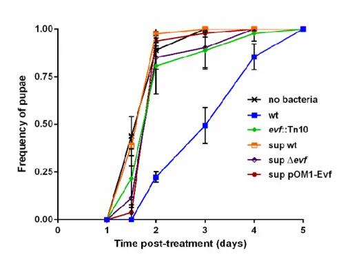
Apesar de ter sido anteriormente postulado que o gene evf altera a fisiologia normal do hospedeiro cessando os movimentos peristálticos normais do intestino, o mecanismo específico pelo qual evf produz este efeito permanece desconhecido. Para perceber se o mecanismo de acção da proteína Evf ao nível do hospedeiro estaria relacionado com toxicidade extracelular, testámos sobrenadantes de uma cultura overnight e proteína Evf purificada no desenvolvimento das larvas. Foram analisados sobrenadantes de culturas wt, mutante evf, e Ecc15 wt a sobreexpressar o gene evf. Os resultados revelaram que nenhum dos sobrenadantes teve efeito ao nível do desenvolvimento larvar, com todas as larvas a comportaram-se de forma similar às larvas controlo. Suportando os dados dos sobrenadantes, a aplicação da proteína purificada também não teve um efeito relevante no desenvolvimento do hospedeiro. Estes resultados indicam que se a proteína Evf é uma toxina não é activa no meio extracelular, sendo provavelmente injetada no hospedeiro.
Para estudar a rede genética envolvida na regulação da expressão do gene evf construímos, pela primeira vez, mutantes na estirpe Ecc15. Para isso usámos uma adaptação do método de recombinação descrito por Wanner e Datsenko. Escolhemos, como sendo os genes relevantes para compreensão da rede regulatória do evf, os genes expI e gacA como parte integrante da regulação por quorum sensing, e os genes hor e evr por serem conhecidos
como reguladores de virulência. Sendo Ecc15 uma estirpe patogénica de plantas, os mutantes foram caracterizados para a expressão de fatores de virulência associados à degradação de componentes vegetais. Para isso usámos um protocolo para medir a maceração de batatas. Ambos os mutantes nos genes de quorum sensing (expI e gacA) apresentaram níveis de maceração muito baixos quando comparado com a bactéria wt, tal como os mutantes nos genes hor e evr. Curiosamente, o mutante evf apresentou níveis de maceração semelhantes ao wt. Estes resultados revelaram a importância do quorum sensing na expressão de genes associados à virulência em infeções de vegetais nesta estirpe bacteriana. Para além disso, mostraram que os genes hor e evr desempenham um papel na produção destes fatores ou em genes associados à sobrevivência na batata. Contrastando com a infeção das larvas, os resultados mostraram que o gene evf não tem qualquer papel na expressão dos fatores de virulência envolvidos na degradação das plantas, o que suporta a hipótese de aquisição génica especifica para a interação com a mosca da fruta.
De forma a avaliar se a expressão do gene evf seria também regulada por quorum
sensing fizemos fusões transcricionais com GFP (Green Fluorescent Protein) do gene conhecido
como seu regulador (hor) e do regulador putativo (evr) e avaliámos a sua expressão ao longo do tempo. Para isso usámos citometria de fluxo e inferimos a expressão destes genes medindo a quantidade de GFP por célula em cada um dos mutantes. Os nossos resultados revelaram que num mutante expI a expressão génica (mais evidente no gene hor) está diminuída. Esta pode ser complementada adicionando homoserinas lactonas de forma exógena, o que é característico de regulação por quorum sensing. No entanto, os resultados também mostraram que o perfil de expressão de ambos os genes é diferente do perfil padrão de um gene regulado por quorum sensing. A expressão destes genes atinge o seu pico a meio da fase exponencial (4 horas de crescimento), contrastando com o observado normalmente em genes regulados por
quorum sensing onde os picos de expressão ocorrem na fase estacionária. Os nossos
resultados mostram que o quorum sensing tem um efeito na expressão destes genes in vitro, uma vez que a sua expressão é alterada na presença de homoserinas lactonas. No entanto, contrasta com a típica regulação por quorum sensing, já que na fase estacionária a expressão é reprimida por um mecanismo desconhecido.
Por último, avaliámos o papel do quorum sensing e dos reguladores do gene evf no contexto do desenvolvimento in vivo. Para isso infetámos as larvas com os respetivos mutantes usando o protocolo descrito anteriormente. Em relação aos mutantes em genes de
quorum sensing, os resultados mostraram que larvas infetadas com o mutante expI não
apresentaram um fenótipo parcial, com um atraso no desenvolvimento (53% de pupas ao dia 2) significativamente diferente do controlo mas também do wt. Quanto aos mutantes nos genes reguladores do evf, larvas infetadas com o mutante hor não apresentaram qualquer atraso no desenvolvimento. Contrastando com este resultado, larvas infetadas com o mutante
evr exibiram um fenótipo semelhante ao wt, com apenas 42% das larvas a atingirem o estádio
de pupa ao dia 2. Isto revela que provavelmente este gene não tem um papel importante na expressão do gene evf. Coletivamente, estes resultados mostram que os genes expI e hor têm um papel importante no efeito causado por evf, assim como gene gacA parece ter uma função
in vivo. No que toca ao gene evr os resultados indicam que não parece ter um papel relevante
no efeito mediado por evf.
Em suma, este trabalho elucidou o efeito da infeção transiente da bactéria Ecc15 no desenvolvimento da larva de mosca da fruta. Mostrámos que o efeito é causado por interrupção do comportamento alimentar normal e que o produto do gene evf não é toxico, pelo menos ao nível extracelular. Otimizámos ainda um método de construção de mutantes por recombinação homóloga em Ecc15 que nos permitiu elucidar parcialmente a rede genética reguladora da expressão do gene evf. Por fim mostrámos ainda o papel do quorum sensing in
vivo, embora não tenha ficado completamente esclarecido se o efeito é direto, afetando o
gene evf ou indireto, comprometendo a capacidade da bactéria permanecer e sobreviver no interior do hospedeiro.
Palavras-chave: quorum sensing; evf; Desenvolvimento de Drosophila; Interacções
Index
1. Introduction ... 1
1.1 Quorum sensing: the language of bacteria ... 1
1.2 The QS system of Pectobacterium carotovorum ... 3
1.3 P. carotovorum and Drosophila - a host-microbe interaction ... 5
2. Materials and methods ... 11
2.1 Bacterial strains and growth conditions ... 11
2.2 Genetic and molecular techniques ... 12
2.3 Construction of Ecc15 mutants by homologous recombination ... 13
2.4 Construction of the promoter gfp reporter fusions ... 13
2.5 Time-course gfp analysis of the promoter gfp expression ... 14
2.6 P. carotovorum virulence assay in potatoes ... 14
2.7 Drosophila oral bacterial infection and development assay ... 14
2.8 Oral infection with Supernatants ... 15
2.9 Food dye quantification ... 15
2.10 Statistical analysis ... 16
3. Results ... 17
3.1 Ecc15 infection causes a development delay in D. melanogaster ... 17
3.2 Ecc15 changes the feeding behaviour of D. melanogaster larvae ... 18
3.3 Evf function at the host level ... 20
3.4 Network controlling Evf production ... 22
3.5 Phenotypes of Ecc15 mutants in plant virulence ... 24
3.6 QS regulation of evf expression ... 26
3.6 Effect of QS and Evf regulator mutants in the development of Drosophila larvae ... 31
4. Discussion ... 34
5. Bibliography ... 39
1.
Introduction
1.1 Quorum sensing: the language of bacteria
Quorum sensing (QS) is a process of intercellular communication that enables bacteria to determine their own numbers by producing, secreting and detecting the accumulation of signalling molecules1,2, also called autoinducers. These molecules accumulate in the extracellular environment as bacteria multiply and when a threshold concentration is reached, the signal is detected and changes at the level of gene expression of the population occur2. This way, bacteria are able to coordinate gene expression and synchronise complex group behaviours across the population, such as antibiotic production, biofilm formation, expression of virulence factors, sporulation or competence3.
The first QS system described was the Lux system4,5 of Vibrio fisheri and is present in a large number of Gram-negative bacteria. This marine bacterium uses this system to coordinately activate the expression of the lux operon, which leads to the production of light also known as bioluminescence. The Lux system is composed of an acyl-homoserine lactone (AHLs) synthase, LuxI, and a cytoplasmic receptor, LuxR. At high cell densities the threshold concentration of AHLs is reached and LuxR binds to the signal molecules. This causes a conformational change in LuxR which will act as a transcription regulator inducing the expression of the lux operon. This operon includes the genes responsible for expressing luciferase which is responsible for producing bioluminescence. This process is essential for the symbiotic interaction of V. fisheri with its natural host, Euprymna scolopes, the bobtail squid6. Bacteria colonizing the squid light organ produce light that is used by the squid to better escape from its predators. In exchange, the squid provides a nutrient rich environment for the bacteria. It was shown, using V. fischeri mutants, that light production and the ability to colonize are tightly related7. V. fischeri mutants affected in the genes responsible for light production or QS components display impaired colonization of the light organ. The enzyme luciferase catalyses a chemical reaction in which one of the unstable intermediates emits light8. It was shown that mutants in the synthesis of luciferase are impaired in the ability to persist in the light organ. Likewise, QS mutants affected in the signal synthase, LuxI, or in its receptor, LuxR, are also unable to produce light and similarly to what was observed for the luciferase mutants, QS mutants are also unable to establish a symbiotic relationship with the squid.
Gram-negative bacteria are not the only organisms regulating behaviours in a density-dependent manner. Gram-positive bacteria use a similar system where small peptides are used
as signals instead of AHLs. These peptides are secreted to the extracellular media where they can be detected by a two-component system comprising a sensor kinase and a response regulator9,10. The resulting signalling and changes in gene expression leads to changes in group behaviours. For example the production of virulence factors in Staphylococcus aureus is regulated by the Agr QS system, a typical Gram-positive QS mechanism9.
Although the two mechanisms presented here are species-specific, there is also a third, the autoinducer-2 (AI-2) system, which has been proposed to act as an interspecies cell-to-cell communication system. The AI-2 signal is produced by the LuxS synthase and can be recognized by both Gram-positive and Gram-negative bacteria11. It has been shown to control a variety of group behaviours, such as biofilm formation (Bacillus cereus), motility (Helicobacter pylori) or bioluminescence (Vibrio harveyi)12.
Though QS has been studied for five decades, proper in vivo models to study its function in host-microbe interactions are still limited. Although it has been shown that QS signals and regulators are essential for activating functions that are necessary for microbe-host interactions (bioluminescence), the benefit of regulating those in a density-dependent manner has not been formally demonstrated. Indeed, it has been postulated that QS dependent regulation might have a role in regulating complex host-microbe interactions such as transmission to new hosts and gut colonization but experimental models to test this hypothesis are missing.
One group of bacteria that relies on QS to precisely synchronise the production of their virulence factors is the Pectobacterium spp. These bacteria are Gram-negative
Enterobacteriaceae which inhabit the soil and cause soft rot disease. These plant pathogens
can infect several vegetables, such as potatoes, carrots or celery, causing important agricultural and economic losses. The virulence of these bacteria is associated with the production of plant cell wall-degrading enzymes (PCWDEs)13,14. High levels of these important enzymes are secreted into the extracellular milieu, resulting in general plant tissue maceration. Known PCWDEs include proteases, cellulases and pectinases, which convert cell wall components into nutrients that can be used by bacteria. Pectinases are the most relevant to disease development. These include pectin lyases, pectate lyases and polygalacturonases, all of which break down pectin, an essential component that maintains the structure of the plant cell wall by creating a matrix that involves the cellulose-hemicellulose network. Despite the virulence potential of PCWDEs, their production comprises great energy costs to bacteria and in order to be effective they are activated only when the optimum conditions are met. Therefore, the production of these enzymes in Pectobacterium spp. is tightly regulated by QS.
The phytopathogenic Enterobacteriaceae Pectobacterium carotovorum (formerly
Erwinia carotovora) strain Ecc15, has been shown by Bruno Lemaitre and colleagues as being
capable of persisting in the gut of Drosophila melanogaster and might be using this insect as an transmission vector. Given the tractability of this model system both from the microbe and the host side we decided to investigate the role of QS in this specific interaction. The
Pectobacterium-Drosophila interaction allows us to study the importance of QS in bacterial
infections in vivo and ultimately explore the possible role of QS in transmission between hosts.
1.2 The QS system of Pectobacterium carotovorum
P. carotovorum strain Ecc15 was first isolated in 1978 from an infected potato field in
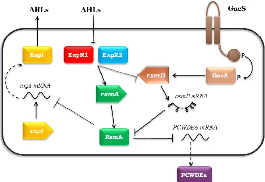
France15. Despite its economical relevance, no studies were performed to understand its signalling networks regulating virulence, though the closely related strain Ecc71 has been studied in detail16–18 and the regulation of virulence in these two strains was assumed to be identical. Moreover, other Pectobacterium spp. like P. wasabiae or P. atroseptica have been extensively studied and their signalling networks shown to be very similar to P. carotovorum. The production of PCWDEs in Pectobacterium spp. is regulated by two QS systems: an AHLs system17,19 and the GacSA/Rsm system16 (Figure 1). The AHL system, a LuxI/LuxR type QS system, is the most studied communication system in Pectobacterium. It is composed by ExpI (LuxI homolog) that synthesizes AHLs, which diffuse across the membrane to the extracellular environment, and by two receptors, ExpR1 and ExpR218,20 (LuxR homologs). At low cell densities (low concentrations of AHLs) these receptors bind to the promoter region of the RNA-binding protein, RsmA21, leading to an increase of its expression22. RsmA binds to several targets, such as the mRNAs of PCWDEs, promoting their degradation and repressing virulence. Interestingly, the ExpR1 and ExpR2 regulators can only bind DNA in absence of AHLs, contrasting to what is observed in the majority of the LuxI/LuxR type QS systems23. At high cell densities, AHLs are recognized by ExpR1 and ExpR2, restricting binding to the rsmA promoter, thus stopping activation of rsmA transcription, which results in the relief of RsmA-mediated repression of virulence and increasing expression of virulence genes like PCWDEs. As in the LuxI/LuxR systems, at high cell densities, high concentration of AHLs also regulates activation of virulence.
Figure 1 –Quorum Sensing network for the regulation of plant virulence in Pectobacterium spp. At low cell densities (low levels of AHLs), ExpR1 and ExpR2 bind to rsmA promoter region inducing its transcription. RsmA binds to the PCWDEs mRNA promoting its degradation and consequently repressing virulence. At high cell densities the AHLs are at high concentration and bind to the two receptors, which no longer promote the expression of rsmA leading to derepression of virulence. Additionally, the GacSA/Rsm system is activated and promotes the expression of rsmB which binds to the existent RsmA proteins blocking their action and thus also promoting expression of the virulence factors.
The GacSA/Rsm is the second system which controls the expression of PCWDEs16. This canonical two component system works together with the ExpI/ExpR system to control the expression of PCWDEs. The GacSA/Rsm is homologous to the GacSA/Rsm two component system from Pseudomonas aeruginosa and the BarA/UvrY/Csr system from Escherichia coli24. It is composed by a sensor kinase GacS and a response regulator GacA. Although the sensor is activated by an unidentified signal, it is known that the activation is regulated in a density dependent manner. At high cell density, upon activation, GacS phosphorylates the response regulator GacA, promoting the transcription of the small non-coding RNA rsmB25. This sRNA binds RsmA and acts as a competitor with RsmA targets. RsmA sequestration by rsmB results in inactivation of RsmA and derepression of virulence26. Recently, it has been shown that rsmB expression is not only controlled by the GacS/GacA system but also by the ExpI/ExpR system (unpublished data from our laboratory). At low cell densities, rsmB expression is inhibited and
rsmA transcription is promoted by ExpR1 and ExpR2, upregulating the RsmA levels and
consequent repression of virulence. Together, the ExpI/ExpR and the GacSA/Rsm systems control the production of PCWDE’s by regulating RsmA. At high cell density, AHLs promote inhibition of rsmA transcription while rsmB acts via a post-translational mechanism to inhibit RsmA by sequestration.
The integration of these two cell-to-cell communication systems tightly controls the expression of these virulence factors, which are essential for the success of bacteria in their natural environment. Presumably, by using QS systems, bacteria can ensure that virulence factors are only expressed when they are effective, highlighting the relevance of these systems in the bacterial world.
1.3 P. carotovorum and Drosophila - a host-microbe interaction
How Pectobacterium spp. are transmitted between their plant hosts has been a question intriguing scientists since 1980. The first obvious thought was that, being a phytopathogen, it survives in the soil and infects injured plants. Though, studies performed in the last century showed a rapid decline of the population size in the soil27, and even though the presence of plant material in the soils (e.g. the remains of the harvesting) enhanced the resistance of these bacteria to the soil conditions, promoting survival27, it was concluded that in general Pectobacterium spp. are present in the soil in amounts below detection levels but can expand their numbers when exposed to plant materials. However, invasion of new sites and the routes of transmission to new hosts are still not understood. In 1981 it was proposed by J.W.Kloepper, J.W.Brewer and M.D.Harrison that insects from the Drosophila family could act as vectors of Pectobacterium spp28 as occurs with many other pathogens, including those of human. They suggested that transmission could take place by three non-specific interactions: carriage on the body, regurgitation and defecation. Although these are valid hypothesis, most likely these interactions are not passive, as previously assumed. In fact, nowadays, most researchers think dissemination of pathogens by vectors involves specific interactions between the host and the bacteria29. Supporting this hypothesis, it has been shown that insects have sensitive mechanisms to recognize microbes and mediate interactions with these organisms, relying upon multiple innate immune responses that are shared with higher organisms30.
Drosophila melanogaster, commonly known as fruit fly, is a model organism to study
immunity and responses to pathogens31,32. Due to the similarities between the innate immune system of D. melanogaster and that of mammals, this insect is considered to be a good model to study host-microbe interactions33. In addition to the presence of the innate immune system, other Drosophila characteristics such as the short life cycle, the availability of genetic tools and easy maintenance in the laboratory make this insect an excellent model to study bacterial-host interactions and identify mechanisms to fight infections. D. melanogaster is highly resistant to microbes; living in an environment rich in fungi, bacteria and viruses, this insect has developed efficient ways to kill pathogens34. The lack of adaptive immune system does not seem to be a
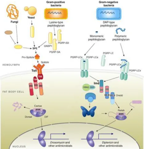
problem in the insect world and they depend on their innate immunity to fight against pathogens35. The Imd and the Toll cascades are the two main regulatory pathways controlling immune responses in flies30. The Imd pathway acts upon recognition of Gram-negative bacteria whilst the Toll pathway responds to both Gram-positive and fungi recognition (Figure 2). Peptidoglycan recognition proteins (PGRPs) that recognize different forms of peptidoglycan (PGN) are present in both cell membranes and hemolymph of flies. PGN from Gram-positive and -negative bacteria differs mostly in two features: composition of the peptide chain and structure of the PGN membrane. At the third position of the peptide chain Gram-negative bacteria have a meso-diaminopimelic acid (DAP) molecule instead of the lysine residue (Lys) that exists in Gram-positive. On the other hand, Gram-negative have a single layer of PGN hidden in the periplasmic space, whereas the PGN of Gram-positive bacteria is multilayered and exposed at the bacterial surface. The Toll pathway is activated by Lys-type PGN and induces the synthesis of several peptides, such as drosomycin. DAP-type PGN activates the Imd pathway, inducing the production of different AMPs such as diptericin36. This production of AMPs can be systemic as well as locally associated with a tissue. Usually, in local immune responses, the first line of defence is the production of reactive oxygen species (ROS) while the production of AMPs comprises a second line of defence. Thus, in the context of an oral infection, to survive in the gut, the invading pathogens have to initially resist to ROS- and then to AMPs-mediated death34.
Although the immune response is critical for survival, its action against foreign microorganisms is energetically costly37–39. In order to successfully eliminate pathogens the host has to produce a battery of antimicrobial peptides, shifting energy utilization to activation of Toll or Imd pathways37, instead of growth and substrates storage. In Drosophila, the fat body is the central organ responsible for synthesis and secretion of antimicrobial peptides40 as well as primary storage of energy sources. During an infection/inflammation, lipids metabolism is altered, leading to inhibition of storage and activation of hydrolysis of triglycerides in order to provide enough energy for immune reactions39,41.
Figure 2 – Schematic representation of Toll and Imd pathway activation in Drosophila spp. Production of antimicrobial peptides occurs upon recognition of potential pathogenic organisms. The Toll pathway (top left) is largely activated by fungi and Gram-positive bacteria whereas Imd pathway (top right) is activated by Gram-negative bacteria. Both pathways possess membrane receptors which sense bacterial components such as the peptidoglycan. The Toll receptor is activated by a cleaved form of the protein Spatzle processed by recognition molecules (PGRP-SA, PGRP-SD or GNBP1) involved in identification of Gram-positive bacteria and fungi cellular components. This mature form of Spatzle binds to Toll42 as dimer leading to series of signalling cascades concluding in cactus degradation43. This event leads to the release and reallocation of the Rel transcription factors Dif and Dorsal from the cytoplasm to the cell nucleus44. The allocation promotes the expression of AMPs such as Drosomycin. The Imd receptor PGRP-LC directly binds to DAP-type peptidoglycan. This interaction promotes the recruitment of the caspase Dredd45. This caspase associates with a phosphorylated form of Relish and cleaves it46,47 realising the Rel domain. This domain is translocated to nucleus and promotes the expression of AMPs such as Diptericin. (Adapted from 30).
The majority of insects have development checkpoints that need to be reached in order to continue the development process. The body size and growth rate are two essential parameters tightly controlled in insects and higher organisms through the development process48,49. Body size is regulated by genetic, physiological and environmental factors such as temperature, population density and nutrition49. It was found that insulin signalling, and in particular Drosophila insulin-like peptides (DILPs) are crucial for normal growth, acting as mediators between nutrition and cell growth50. DILPs activate Drosophila insulin receptors (dlnR)51,52 which trigger a series of signalling cascades, leading to production and accumulation of the cell signal phosphatidylinositol 3,4,5-triphosphate (PIP3) which promotes accumulation
of nutrients and cell growth53. The fat body responds to nutrients availability and produces an unknown growth factor that regulates DILPs/dlnR signalling controlling the growth rate54.
During development there are several size assessment events that determine if a larva is ready to enter metamorphosis or whether more growth is necessary. For example, in order to enter pupation, last stage larvae need to surpass both minimal viable weight (MVW) and critical weight (CW). MVW is defined as the minimal fat body mass needed to survive through metamorphosis49. CW is the development switch in which no more growth is necessary to commit to metamorphosis55. In Drosophila, contrary to other insects, these two development checkpoints appear to occur nearly at the same time, around 70 hours after egg laying56, and thus were considered the same for a long time. However, experiments done in 1938 by Beadle and co-authors reported that larvae starved before 70 hours of development were unable to continue metamorphosis whereas larvae starved after that period entered metamorphosis but originated smaller adults57. It was also observed that larvae starved before reaching CW had a delay in their development for a period equivalent to the starvation period57. When CW is reached, an endocrine cascade for metamorphosis begins in which the titers of juvenile hormone (JH) decrease and lead to the release of the prothoracicotropic hormone (PTTH). PTTH acts on the prothoratic gland stimulating the synthesis of the hormone ecdysone. In the absence of JH, ecdysone induces metamorphosis and temporary suspension of feeding.
In a scenario of bacterial infection, a common physiological response is feed ceasing, larvae enter in starvation, and allocate resources to the activation of the immune system. This behaviour produces changes at the endocrine level which can lead to development delays37,58,59.
Recently, two strains of P. carotovorum, Ecc15 and Ecc1488, were shown to trigger an immune response in D. melanogaster by oral ingestion, causing a non-lethal transient infection15. Presumably, this immune response is due to the ability of those bacteria to persist in the gut of D. melanogaster larvae. Importantly, a single gene present in these two P.
carotovorum strains, was shown to be essential and sufficient to this phenotype60. This gene was named evf, standing for Erwinia virulence factor. Additionally, another gene, hor, was identified as an evf regulator60. The gene hor encodes a transcriptional regulator from the SlyA family and besides regulating evf it also shown to regulate production of antibiotics and production of extracellular enzymes in other bacteria61. evf seems to be strain restricted, it was found only in the Ecc15 and Ecc1488 P. carotovorum strains, while hor is present in many P.
carotovorum and also in human pathogens that live in close interaction with insects, such as Yersinia or Serratia spp.61. Thus the described interaction of strains Ecc15 and Ecc1488 with D.
persistence. An event of single acquisition dramatically changing bacterial life style is not new: acquisition of the gene ymt (Yersinia murine toxin)62 allowed Yersinia pestis to colonize and proliferate in the gut of fleas turning this bacterium in one of the worst plagues in human history.
Although it was demonstrated that evf improves bacterial survival rate in the
Ecc15-Drosophila model, the mechanism behind this phenomenon is not clear. In 2007, Muniz et al.
showed that Ecc15 evf mutants were unable to persist in the gut of the imd deficient larvae. Thus it does not seem that evf influences directly the Imd-dependent immune response. They also hypothesized that Evf could be acting extracellularly since co-infections with Ecc15 wt and
evf mutant allowed the mutant to persist in the gut. However, they could not detect Evf
extracellularly and showed that Evf was present in the cytoplasm. Lastly, by removing the gut from the larvae body, evf mutants were able to persist in the same titers as the wt bacteria. They concluded by proposing that Evf might be affecting the normal gut physiology by antagonizing the peristaltic movements and thus avoiding natural bacterial elimination. Although this still remains a plausible hypothesis, no mechanism has been proposed for how Evf could have such effect. Additionally, the regulatory pathway controlling Evf production still remains to be elucidated. It was shown that Hor controls the expression of evf60 and, in other
Pectobacterium species, hor is regulated by QS via RsmA63. However, it was never evaluated if
evf regulation by hor is independent or dependent of other putative regulators such as evr.
This gene was described in NCBI gene database as a putative regulator of evf (http://www.ncbi.nlm.nih.gov/nuccore/AY167733.1) but no paper was published reporting its function in the regulation cascade.
Given the known role of QS in regulating hor in other bacterial species and its importance in regulating virulence in this strain, we hypothesize that in Ecc15 Evf production is regulated by QS by the same genetic pathway as the PCWDEs (Figure 3). Therefore, we proposed to study the regulation network of evf production.
Figure 3 – Propose model for regulation of Evf production via QS. At low cell densities, ExpR1 and ExpR2 bind to rsmA promoting its transcription. RsmA binds to the hor mRNA blocking its expression. If
hor regulates evf transcription then this would result on a decrease of evf transcription at low cell
densities. At high cell densities AHLs bind to ExpR receptors, and these no longer can activate transcription of rsmA. RsmA protein levels decrease and Hor protein levels increase, then two possible signalling pathways can occur. Hor directly acts on evf promoting its expression, or Hor promotes the expression of a second regulator, evr, which consequently regulates transcription of evf.
The rarity of natural bacterial pathogens in Drosophila 29,64 makes the relationship between Pectobacterium and this insect a very interesting case to study. Being non-lethal, this interaction allows us to study both the host and the microbial side on a dynamic system and determine the influence of bacterial cell to cell communication systems in this host-microbe interaction.
In this work we have used the P. carotovorum-Drosophila model to study the role of QS in this interaction in vivo. We have studied the effect of Ecc15 on host physiology and identified a new phenotype for Evf which provides new tools to study its function. We also investigated the regulation of evf expression by QS and by its known and putative regulators. Ultimately, we studied the function of Evf in the context of this host-microbe interaction and the specific mechanism through which this protein changes the normal gut physiology of the host.
2. Materials and methods
2.1 Bacterial strains and growth conditions
All strains, plasmids and primers used in this study are listed in Table 1 and Table 2, respectively. P. carotovorum strains used are derived from wild-type (wt) strain Ecc1515 and were routinely grown at 30oC with aeration in Luria-Bertani (LB) broth. Antibiotics were used at the following concentrations, except otherwise specified: ampicillin (amp) 25 mgL-1; kanamycin (kan) 50 mgL-1; spectinomycin (spec) 50 mgL-1; chloramphenicol (cm) 10 mgL-1. Optical density (OD600 nm) was determined by measuring absorbance at 600 nm in a spectrophotometer (Spectronic, Thermo Scientific).
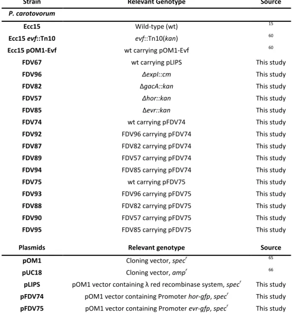
Table 1 – Strains and plasmids used in this study.
Strain Relevant Genotype Source
P. carotovorum
Ecc15 Wild-type (wt) 15
Ecc15 evf::Tn10 evf::Tn10(kan) 60
Ecc15 pOM1-Evf wt carrying pOM1-Evf 60
FDV67 wt carrying pLIPS This study
FDV96 ∆expI::cm This study
FDV82 ∆gacA::kan This study
FDV57 ∆hor::kan This study
FDV85 ∆evr::kan This study
FDV74 wt carrying pFDV74 This study
FDV92 FDV96 carrying pFDV74 This study
FDV87 FDV82 carrying pFDV74 This study
FDV89 FDV57 carrying pFDV74 This study
FDV94 FDV85 carrying pFDV74 This study
FDV75 wt carrying pFDV75 This study
FDV93 FDV96 carrying pFDV75 This study
FDV88 FDV82 carrying pFDV75 This study
FDV90 FDV57 carrying pFDV75 This study
FDV95 FDV85 carrying pFDV75 This study
Plasmids Relevant genotype Source
pOM1 Cloning vector, specr 65
pUC18 Cloning vector, ampr 66
pLIPS pOM1 vector containing λ red recombinase system, specr This study pFDV74 pOM1 vector containing Promoter hor-gfp, specr This study pFDV75 pOM1 vector containing Promoter evr-gfp, specr This study
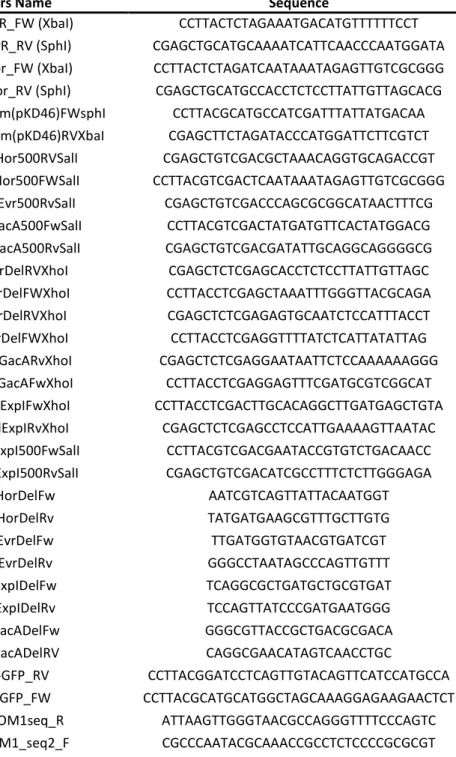
Table 2 – Primers used in this study to construct the GFP reporter fusions, engineer the homologous recombination fragments and for confirmation of deletion mutants.
Primers Name Sequence
1003-PevR_FW (XbaI) CCTTACTCTAGAAATGACATGTTTTTTCCT 1004-PevR_RV (SphI) CGAGCTGCATGCAAAATCATTCAACCCAATGGATA 1005-Phor_FW (XbaI) CCTTACTCTAGATCAATAAATAGAGTTGTCGCGGG 1006-Phor_RV (SphI) CGAGCTGCATGCCACCTCTCCTTATTGTTAGCACG 1108-Redsystem(pKD46)FWsphI CCTTACGCATGCCATCGATTTATTATGACAA 1109-Redsystem(pKD46)RVXbaI CGAGCTTCTAGATACCCATGGATTCTTCGTCT 1127-500Hor500RVSalI CGAGCTGTCGACGCTAAACAGGTGCAGACCGT 1128-500Hor500FWSalI CCTTACGTCGACTCAATAAATAGAGTTGTCGCGGG 1129-500Evr500RvSalI CGAGCTGTCGACCCAGCGCGGCATAACTTTCG 1130-500gacA500FwSalI CCTTACGTCGACTATGATGTTCACTATGGACG 1131-500gacA500RvSalI CGAGCTGTCGACGATATTGCAGGCAGGGGCG 1087-HorDelRVXhoI CGAGCTCTCGAGCACCTCTCCTTATTGTTAGC 1088-HorDelFWXhoI CCTTACCTCGAGCTAAATTTGGGTTACGCAGA 1090-EvrDelRVXhoI CGAGCTCTCGAGAGTGCAATCTCCATTTACCT 1091-EvrDelFWXhoI CCTTACCTCGAGGTTTTATCTCATTATATTAG 1132-DelGacARvXhoI CGAGCTCTCGAGGAATAATTCTCCAAAAAAGGG 1133-DelGacAFwXhoI CCTTACCTCGAGGAGTTTCGATGCGTCGGCAT 1134-DelExpIFwXhoI CCTTACCTCGACTTGCACAGGCTTGATGAGCTGTA 1135-DelExpIRvXhoI CGAGCTCTCGAGCCTCCATTGAAAAGTTAATAC 1136-500ExpI500FwSalI CCTTACGTCGACGAATACCGTGTCTGACAACC 1137-500ExpI500RvSalI CGAGCTGTCGACATCGCCTTTCTCTTGGGAGA 1186-HorDelFw AATCGTCAGTTATTACAATGGT 1187-HorDelRv TATGATGAAGCGTTTGCTTGTG 1188-EvrDelFw TTGATGGTGTAACGTGATCGT 1189-EvrDelRv GGGCCTAATAGCCCAGTTGTTT 1190-ExpIDelFw TCAGGCGCTGATGCTGCGTGAT 1191-ExpIDelRv TCCAGTTATCCCGATGAATGGG 1192-GacADelFw GGGCGTTACCGCTGACGCGACA 1193-GacADelRV CAGGCGAACATAGTCAACCTGC 0665-GFP_RV CCTTACGGATCCTCAGTTGTACAGTTCATCCATGCCA 0576-GFP_FW CCTTACGCATGCATGGCTAGCAAAGGAGAAGAACTCT 0531_pOM1seq_R ATTAAGTTGGGTAACGCCAGGGTTTTCCCAGTC 0752-pOM1_seq2_F CGCCCAATACGCAAACCGCCTCTCCCCGCGCGT
2.2 Genetic and molecular techniques
Unless otherwise mentioned, all PCR reactions were performed using Dream Taq Polymerase (Fermentas). Digestions were done using Fast Digest Enzymes (Fermentas) according to manufactures instructions and ligations performed with T4 DNA ligase (New England Biolabs)(See appendices). Electrocompetent cells were prepared using the glycerol method (See appendices) and a 2.2V shock was applied to favour DNA entry in the competent
2.3 Construction of Ecc15 mutants by homologous recombination
The plasmid pLIPS was constructed by introducing the arabinose inducible λ Red recombinase system67 into the pOM1 vector. The λ Red recombinase system (composed by the genes araC, gam, bet, exo and af60A total of 3421 bp) was amplified by PCR from the vector pKD4668 using the Bio-x-act proof reading enzyme and the primers P1108- λRedsystem(pKD46)FW (SphI) and P1109-λRedsystem(pKD46)RV (XbaI). The fragment was digested with SphI/XbaI and ligated to the SphI/XbaI digested pOM1.
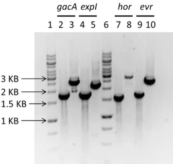
Mutants were constructed by chromosomal gene replacement with an antibiotic marker using the λ Red recombinase system. The DNA region of the gene to be replaced, including approximately 500 bp upstream and 500 bp downstream of the gene, was amplified with Bio-x-act and cloned into pUC18. The 500bp-expI-500bp, 500bp-gacA-500bp, 500bp-hor-500bp and 500bp-hor-500bp-evr-500bp-hor-500bp fragments were cloned into pUC18 using SalI. These constructs, containing the gene to be deleted and its flanking regions, were divergently amplified by PCR using the primers containing XhoI (1087-1135) from table 2 to introduce a XhoI restriction site in the 5’ and 3’ regions of the gene to be deleted. The antibiotic resistance genes cm or kan were amplified with primers containing the XhoI site, from pKD368 and pKD468 respectively, digested with XhoI and ligated to the XhoI digested PCR fragments. The final constructs contained the antibiotic marker flanked by the upstream and downstream regions of the gene to be replaced. The 500bp-antibiotic-500bp fragment was amplified by PCR using Bio-x-act and before electroporation a clean-up was performed using a PCR purification kit (Quiagen). The fragment was eluted in 50 µl of miliQ water and approximately 3000 ng of DNA was electroporated in 50 µl of Ecc15 electrocompetent cells expressing the λ Red recombinase system from pLIPS, to allow recombination. Recombinants were selected in LB plates with the respective antibiotic (kan 50 or cm 10). In order to lose the plasmid pLIPS after recombination, chosen recombinants were streaked in LB plates. The plates were then incubated at 30ºC or 37ºC (to induce temperature stress and increase the probability to lose the plasmid) and random colonies streaked in LB spec plates to check for plasmid loss.
2.4 Construction of the promoter gfp reporter fusions
The pFDV74 and pFDV75 vectors were constructed by introducing the promoter region of the genes hor and evr into pOM1 vector65.
hor
andevr
promoter regions located 500 bp upstream of the genes starting codon were amplified by PCR from genomic DNA using Bio-x-act proof reading enzyme and primers P1005-Phor_FW (XbaI) and P1006-Phor_RV (SphI) forBoth fragments were digested with XbaI/SphI and ligated to the XbaI/SphI digested pOM1. A 792 bp gfp promoterless fragment was amplified from pCMW1 and cloned into pOM1 adjacent to the promoter region fragment using the primers 0576-GFP_FW (SphI) and 0665-GFP_RV (BamHI).
2.5 Time-course gfp analysis of the promoter gfp expression
P. carotovorum strains containing the reporter plasmids pFDV74 and pFDV75 were
grown overnight in LB+Spec and inoculated into fresh medium at a starting OD600nm of 0.05. At the indicated time points, aliquots were collected to evaluate growth and to assess hor or
evr expression. For the analysis of hor or evr expression, aliquots of the cultures were
diluted 1:100 into Phosphate Buffered Solution (PBS) and Phor-GFP or Pevr-GFP expression
was measured by flow cytometry (LSR Fortessa, BD) and analysed with Flowing Software v2.5.0. A minimum of 10,000 cells were acquired per sample and hor or evr expression was recorded as the median of GFP expression. All assays are reported as the mean of the median GFP expression from at least three biological replicates and error bars represent the standard deviation.
2.6 P. carotovorum virulence assay in potatoes
Virulence was analysed using a modified protocol to assess maceration of potato tubers69. Potatoes were washed and surface sterilized by soaking for 10 min in 10% bleach followed by 10 min in 70% EtOH. Overnight cultures were washed twice and diluted to an OD600nm of 0.05 in PBS and 30 μL were inoculated in the previously punctured potatoes. Potato tubers were incubated at 28ºC in a humid environment and at 48 h of incubation, potatoes were sliced and macerated tissue was collected and weighed. All assays are reported as the mean of the maceration from at least five biological replicates and error bars represent the standard error of the mean.
2.7 Drosophila oral bacterial infection and development assay
Transgenic Oregonr larvae carrying a gfp gene tagged to diptericin promoter region70 in both arms of the third chromosome were used in all assays. Egg laying was performed in cages containing 40 adult flies at a ratio of 3 females to 1 male. The flies were incubated for 4 to 6 hours at 25ºC in the presence of standard corn meal fly medium. After this period, the eggs were removed and incubated at 25ºC for 70 hours to obtain L3-stage larvae.
For the bacterial infections, approximately 30 third-instar larvae were placed in a 2 ml Eppendorf containing 200 µl of concentrated bacteria pellet (OD600 = 200) from an overnight culture and 400 µl of standard corn meal fly medium. Larva, bacteria and food were
thoroughly mixed in the Eppendorf. The Eppendorf was then closed with a foam plug and incubated at room temperature for 30 min. The mix was then transferred to a 25 ml plastic tube containing 7.5 ml of standard corn-meal fly medium and incubated at 28ºC. The development of the larvae post-infection, comprising the larva to pupa and pupa to fly transition, was then followed for 8 days. The frequency of larvae that proceeded to the pupae stage was assessed by counting the number of pupae present inside the plastic tube. Similarly, the frequency of eclosed flies was determined based on the number of flies that emerged from the pupae. Unless otherwise mentioned, all assays are reported as the mean of frequency from at least five biological replicates and error bars represent the standard error of the mean.
2.8 Oral infection with Supernatants
For each strain, 100 ml of an overnight culture were centrifuged at 4000 RPM for 20 min at room temperature, in an Eppendorf centrifuge model 5810 in order to pellet the bacteria. The supernatant was transferred to a new recipient and bacterial cells were discarded. Supernatants were then filtered through a 2 µm diameter pore filter (Pall Corporation) to remove remaining bacteria or cellular debris. 200 µl of the supernatant were then used to infect larvae following the same protocol described above in the Drosophila oral
bacterial infection and development assay section.
2.9 Food dye quantification
To evaluate the amount of food ingested by the larvae post-treatment, pigment quantification in larvae fed with blue dyed fly medium was performed. Larvae were treated according to the protocol described previously for the infection with some changes. After the 30 min infection period larvae, bacterial pellet and food were transferred to a new plastic tube containing standard corn meal fly medium to which was added 30,5 µl of a blue food dye (Rayner’s). At the indicated time points, larvae were collected from the tubes using a 15% sucrose solution and washed in a petri dish containing PBS 1x. 30 larvae were randomly collected to an Eppendorf and homogenized with a blender in 200 µl of 80% methanol. The suspension was then centrifuged for 10 min to deposit larvae residue. 100 µl of the mixture methanol /blue dye were recovered and measured at 625 nm using the multiskanGO (Thermo scientific). All assays are reported as the mean of absorbance (625 nm) from at least five biological replicates and error bars represent the standard error of the mean.
2.10 Statistical analysis
The data presented here were analyzed using Graphpad Prism6 software and program R version 3.0.2. The Mann-Whitney test was performed to evaluate significance and P-values
were adjusted using the Holm-Bonferroni correction for multiple comparisons. An adjusted P-value <0.05 was used as the cut-off for statistical significance. ns Not significant; *P-value
3. Results
3.1 Ecc15 infection causes a development delay in D. melanogaster
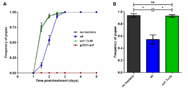
It was recently shown that single acquisition of the gene evf provided P. carotovorum strain Ecc15 the ability to persist in the gut of D. melanogaster60,71. This persistence triggered the activation of the immune system, resulting in a state of transient infection. Though the host immune response against P. carotovorum was investigated, the impact of the evf-dependent infection in host development was never assessed. In order to understand the effect of this infection in host development we monitored the development of larvae over time, by counting the numbers of larvae which progressed to the pupation stage after infection with Ecc15 (Figure 4). L3 stage larvae were fed with a mixture of standard corn meal fly food and a bacterial pellet or, as control, only with standard fly food.
Figure 4 – Effect of Ecc15 on D. melanogaster larvae development. Larvae pupation frequency was evaluated by counting the number of larvae (30±5), reaching pupae stage after infection with Ecc15 (wt) (blue), evf mutant (green) bacteria and Ecc15 pOM1-evf (strain overexpressing evf) (red) in each independent replicate (n=5). As control larvae were fed with standard fly food without bacteria (dark grey). (A) Time-course frequency of pupation after infection. (B) Represents the average pupation frequency of each treatment at day 2. Error bars represent the standard error of the mean, * stands for Pvalue<0.05 andns for Not significant.
The results showed that the majority of the control larvae reached the pupae stage during the first two days after infection (Figure 4A, dark grey crosses). In opposition, larvae fed with wt Ecc15 displayed a development impairment of approximately one day when compared to the control (Figure 4A, blue squares and dark grey crosses), only reaching 50% of pupae in two days (Figure 4B, blue bar and dark grey bar). Importantly, larvae fed with an evf mutant strain of P. carotovorum had no delay in development (Figure 4B, green bar) displaying a temporal
dynamic similar to the control larvae not exposed to bacteria (Figure 4A, green diamonds and dark grey crosses). Moreover, larvae infected with Ecc15 overexpressing evf died without reaching the pupa stage (Figure 4A, red inverted triangles).
These results demonstrated that infection of D. melanogaster with Ecc15 had a significant impact in larvae development. Moreover, the observed development delay could be specifically attributed to evf, since this effect was absent in infections with mutants lacking this gene and led to larvae lethality when it was overexpressed. However, the mechanisms behind this phenotype are not clear, highlighting the importance of studying the effect of evf in the context of infection and development of larvae.
3.2 Ecc15 changes the feeding behaviour of D. melanogaster larvae
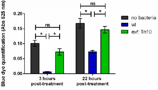
The control of body size is essential for the development of both insects and mammals. Several studies performed in insects, and in particular in D. melanogaster, showed that this parameter is regulated by molecular, environmental and physiological mechanisms, such as critical weight48,49. The critical weight is the development switch where no more growth is needed to enter the pupation stage55. Several environmental factors such as temperature, larval density or nutritional quality are important to the regulation of body size and consequently have an impact on the critical weight. Our previous results showed that larvae fed with wt Ecc15 had a development delay, which could be related with a problem in attaining the critical weight necessary to commit to pupae stage. Since both temperature and population size were well controlled in our experiments, a likely cause to this delay was a nutritional problem. Supporting this hypothesis, we noticed that larvae infected with wt bacteria were thinner than both control larvae and larvae infected with an evf mutant, suggesting an altered nutrition in wt-infected larvae. Therefore, in order to test our hypothesis, we monitored the feeding behaviour of infected larvae, by staining the standard fly food with a blue dye (Rayman’s) and quantifying the amount of blue pigment internalized by the larvae at the indicated time points (Figure 5). Strikingly, 3 hours after treatment, larvae infected with wt bacteria (left blue bar) displayed a reduced amount of internalized pigment, suggesting that they were indeed consuming little or no food. This contrasts to what is observed for the control or evf-infected larvae (left dark grey and green bars, respectively), where the amount of pigment detected indicates food ingestion.
At 22 hours post-treatment we could observe the presence of the pigment in wt-infected larvae (right blue bar), which shows that by this time they had resumed food consumption. This observation suggests that with time the infection with wt bacteria preventing food uptake was eliminated, allowing the larvae to resume food consumption until
reaching the critical weight development checkpoint needed to enter the pupation stage. Interestingly, even after 22 hours post infection we could still observe a difference in the amount of food ingested by the wt-infected larvae, showing the impact of the transient P.
carotovorum infection for the larvae development (Figure 5, right blue bar with green and dark
grey bars).
Figure 5 – Estimation of food uptake by D. melanogaster larvae after infection with Ecc15. Food uptake was inferred by quantifying the amount of blue pigment (Abs 625 nm) internalized by non-infected control larvae (dark grey bars), Ecc15 wt-non-infected (blue bars) or evf::Tn10-non-infected (green bars). For each treatment a pool of 30 larvae was collected (n=5). Error bars represent the standard error of the mean, * stands for Pvalue<0.05 andns to Not significant.
Our results showed a transient nutritional impairment in D. melanogaster larvae mediated by evf. Bacteria lacking this gene were unable to disrupt the normal larval feeding behaviour, contrary to what was observed in the first hours in wt-infected larvae. This in turn suggests that Evf has a temporary toxic effect on larvae, impacting the progression of the life cycle of the fly.