ROLE OF PROTEIN-PROTEIN
INTERACTIONS IN METABOLISM:
GENETICS, STRUCTURE, FUNCTION
EDITED BY : Amit V. Pandey, Colin J. Henderson, Yuji Ishii,
Michel Kranendonk, Wayne L. Backes and Ulrich M. Zanger
PUBLISHED IN : Frontiers in Pharmacology
Frontiers Copyright Statement
© Copyright 2007-2018 Frontiers Media SA. All rights reserved. All content included on this site, such as text, graphics, logos, button icons, images, video/audio clips, downloads, data compilations and software, is the property of or is licensed to Frontiers Media SA (“Frontiers”) or its licensees and/or subcontractors. The copyright in the text of individual articles is the property of their respective authors, subject to a license granted to Frontiers. The compilation of articles constituting
this e-book, wherever published, as well as the compilation of all other content on this site, is the exclusive property of Frontiers. For the conditions for downloading and copying of e-books from Frontiers’ website, please see the Terms for Website Use. If purchasing Frontiers e-books from other websites or sources, the conditions of the website concerned apply. Images and graphics not forming part
of user-contributed materials may not be downloaded or copied without permission. Individual articles may be downloaded
and reproduced in accordance with the principles of the CC-BY licence subject to any copyright or other notices. They may not be re-sold as an e-book. As author or other contributor you grant a CC-BY licence to others to reproduce your articles, including any graphics and third-party materials supplied by you, in accordance with the Conditions for Website Use and subject to any copyright notices which you include in connection with your articles and materials. All copyright, and all rights therein,
are protected by national and international copyright laws. The above represents a summary only. For the full conditions see the Conditions for Authors and the Conditions for Website Use.
ISSN 1664-8714 ISBN 978-2-88945-385-6 DOI 10.3389/978-2-88945-385-6
About Frontiers
Frontiers is more than just an open-access publisher of scholarly articles: it is a pioneering approach to the world of academia, radically improving the way scholarly research is managed. The grand vision of Frontiers is a world where all people have an equal opportunity to seek, share and generate knowledge. Frontiers provides immediate and permanent online open access to all its publications, but this alone is not enough to realize our grand goals.
Frontiers Journal Series
The Frontiers Journal Series is a multi-tier and interdisciplinary set of open-access, online journals, promising a paradigm shift from the current review, selection and dissemination processes in academic publishing. All Frontiers journals are driven by researchers for researchers; therefore, they constitute a service to the scholarly community. At the same time, the Frontiers Journal Series operates on a revolutionary invention, the tiered publishing system, initially addressing specific communities of scholars, and gradually climbing up to broader public understanding, thus serving the interests of the lay society, too.
Dedication to Quality
Each Frontiers article is a landmark of the highest quality, thanks to genuinely collaborative interactions between authors and review editors, who include some of the world’s best academicians. Research must be certified by peers before entering a stream of knowledge that may eventually reach the public - and shape society; therefore, Frontiers only applies the most rigorous and unbiased reviews.
Frontiers revolutionizes research publishing by freely delivering the most outstanding research, evaluated with no bias from both the academic and social point of view. By applying the most advanced information technologies, Frontiers is catapulting scholarly publishing into a new generation.
What are Frontiers Research Topics?
Frontiers Research Topics are very popular trademarks of the Frontiers Journals Series: they are collections of at least ten articles, all centered on a particular subject. With their unique mix of varied contributions from Original Research to Review Articles, Frontiers Research Topics unify the most influential researchers, the latest key findings and historical advances in a hot research area! Find out more on how to host your own Frontiers Research Topic or contribute to one as an author by contacting the Frontiers Editorial Office: [email protected]
METABOLISM: GENETICS, STRUCTURE, FUNCTION
The POR located into the endoplasmic reticulum, interacts with the cytochrome P450s and other redox partners for metabolic reactions. Taken from: Pandey AV and Sproll P (2014) Pharmacogenomics of human P450 oxidoreductase. Front. Pharmacol. 5:103. doi: 10.3389/fphar.2014.00103.
Cover image: Structural models of P450 oxidoreductase (POR), Cytochrome P450s, adrenodoxin, UDP-Glucuronosyltransferase 2B7, PPAR gamma, cholesterol and androstenedione, highlighting the role of protein-protein interactions in metabolism. Cover image created by Amit V. Pandey.
Topic Editors:
Amit V. Pandey, University Children’s Hospital Bern, Switzerland and University of Bern,
Switzerland
Colin J. Henderson, University of Dundee, United Kingdom Yuji Ishii, Kyushu University, Japan
Michel Kranendonk, Universidade Nova de Lisboa, Portugal
Wayne L. Backes, Louisiana State University Health Sciences Center – New Orleans, United States Ulrich M. Zanger, Dr. Margarete Fischer-Bosch-Institute of Clinical Pharmacology, Germany
Genetic variations may change the structure and function of individual proteins as well as affect their interactions with other proteins and thereby impact metabolic processes dependent on protein-protein interactions. For example, cytochrome P450 proteins, which metabolize a vast array of drugs, steroids and other xenobiotics, are dependent on interactions with redox and allosteric partner proteins for their localization, stability, (catalytic) function and metabolic diversity (reactions). Genetic variations may impact such interactions by changing the splicing and/or amino acid sequence which in turn may impact protein topology, localization, post translational modifications and three dimensional structure. More generally, research on single gene defects and their role in disease, as well as recent large scale sequencing studies suggest that a large number of genetic variations may contribute to disease not only by affecting gene function or expression but also by modulating complex protein interaction networks.
The aim of this research topic is to bring together researchers working in the area of drug, steroid and xenobiotic metabolism who are studying protein-protein interactions, to describe their recent advances in the field. We are aiming for a comprehensive analysis of the subject from different approaches including genetics, proteomics, transcriptomics, structural biology, biochemistry and pharmacology. Of particular interest are papers dealing with translational research describing the role of novel genetic variations altering protein-protein interaction. Authors may submit original articles, reviews and opinion or hypothesis papers dealing with the role of protein-protein interactions in health and disease.
Potential topics include, but are not limited to:
• Role of protein-protein interactions in xenobiotic metabolism by cytochrome P450s and other drug metabolism enzymes.
• Role of classical and novel interaction partners for cytochrome P450-dependent metab-olism which may include interactions with redox partners, interactions with other P450 enzymes to form P450 dimers/multimers, P450-UGT interactions and proteins involved in posttranslational modification of P450s.
• Effect of genetic variations (mutations and polymorphisms) on metabolism affected by protein-protein interactions.
• Structural implications of mutations and polymorphisms on protein-protein interactions. • Functional characterization of protein-protein interactions.
• Analysis of protein-protein interaction networks in health and disease.
• Regulatory mechanisms governing metabolic processes based on protein-protein interactions.
• Experimental approaches for identification of new protein-protein interactions including changes caused by mutations and polymorphisms.
Citation: Pandey, A. V., Henderson, C. J., Ishii, Y., Kranendonk, M., Backes, W. L., Zanger, U. M.,
eds. (2018). Role of Protein-Protein Interactions in Metabolism: Genetics, Structure, Function. Lausanne: Frontiers Media. doi: 10.3389/978-2-88945-385-6
Table of Contents
06 Editorial: Role of Protein-Protein Interactions in Metabolism: Genetics, Structure, Function
Amit V. Pandey, Colin J. Henderson, Yuji Ishii, Michel Kranendonk, Wayne L. Backes and Ulrich M. Zanger
UGT Interactions and Regulation of Metabolism
09 Structure and Protein–Protein Interactions of Human UDP-Glucuronosyltransferases
Ryoichi Fujiwara, Tsuyoshi Yokoi and Miki Nakajima
24 Introduction of an N-Glycosylation Site into UDP-Glucuronosyltransferase 2B3 Alters Its Sensitivity to Cytochrome P450 3A1-Dependent Modulation
Tatsuro Nakamura, Naho Yamaguchi, Yuu Miyauchi, Tomoki Takeda, Yasushi Yamazoe, Kiyoshi Nagata, Peter I. Mackenzie, Hideyuki Yamada and Yuji Ishii
33 Endogenous Protein Interactome of Human UDP-Glucuronosyltransferases Exposed by Untargeted Proteomics
Michèle Rouleau, Yannick Audet-Delage, Sylvie Desjardins, Mélanie Rouleau, Camille Girard-Bock and Chantal Guillemette
Nuclear Receptors and Gene Regulation
46 Pregnane X Receptor (PXR)-Mediated Gene Repression and Cross-Talk of PXR with Other Nuclear Receptors via Coactivator Interactions
Petr Pavek
62 Membrane Associated Progesterone Receptors: Promiscuous Proteins with Pleiotropic Functions – Focus on Interactions with Cytochromes P450
Chang S. Ryu, Kathrin Klein and Ulrich M. Zanger
Protein Interactions in Genetics and Pharmacology
70 Altered CYP19A1 and CYP3A4 Activities Due to Mutations A115V, T142A, Q153R and P284L in the Human P450 Oxidoreductase
Sameer S. Udhane, Shaheena Parween, Norio Kagawa and Amit V. Pandey
81 Evaluation of Selected CYP51A1 Polymorphisms in View of Interactions with Substrate and Redox Partner
Tadeja Režen, Iza Ogris, Marko Sever, Franci Merzel, Simona Golic Grdadolnik and Damjana Rozman
Structural Biology of Protein Interactions
92 Advances in the Understanding of Protein-Protein Interactions in Drug Metabolizing Enzymes through the Use of Biophysical Techniques
105 Physical Studies of P450–P450 Interactions: Predicting Quaternary Structures of P450 Complexes in Membranes from Their X-ray Crystal Structures
James R. Reed and Wayne L. Backes
Biochemistry, Biophysics and Biotechnological Implications
125 The Hinge Segment of Human NADPH-Cytochrome P450 Reductase in Conformational Switching: The Critical Role of Ionic Strength
Diana Campelo, Thomas Lautier, Philippe Urban, Francisco Esteves, Sophie Bozonnet, Gilles Truan and Michel Kranendonk
138 Human Cytochrome P450 3A4 as a Biocatalyst: Effects of the Engineered Linker in Modulation of Coupling Efficiency in 3A4-BMR Chimeras
Danilo Degregorio, Serena D’Avino, Silvia Castrignanò, Giovanna Di Nardo, Sheila J. Sadeghi, Gianluca Catucci and Gianfranco Gilardi
doi: 10.3389/fphar.2017.00881
Edited and reviewed by: Marcelo Rizzatti Luizon, Universidade Federal de Minas Gerais, Brazil
*Correspondence: Amit V. Pandey [email protected]
Specialty section: This article was submitted to Pharmacogenetics and Pharmacogenomics, a section of the journal Frontiers in Pharmacology
Received: 06 November 2017 Accepted: 14 November 2017 Published: 27 November 2017
Citation: Pandey AV, Henderson CJ, Ishii Y, Kranendonk M, Backes WL and Zanger UM (2017) Editorial: Role of Protein-Protein Interactions in Metabolism: Genetics, Structure, Function. Front. Pharmacol. 8:881. doi: 10.3389/fphar.2017.00881
Editorial: Role of Protein-Protein
Interactions in Metabolism: Genetics,
Structure, Function
Amit V. Pandey1, 2*, Colin J. Henderson3, Yuji Ishii4, Michel Kranendonk5, Wayne L. Backes6and Ulrich M. Zanger7, 8
1Pediatric Endocrinology, Diabetology and Metabolism, University Children’s Hospital Bern, Bern, Switzerland,2Department of Biomedical Research, University of Bern, Bern, Switzerland,3Division of Cancer Research, Jacqui Wood Cancer Centre, School of Medicine, University of Dundee, Dundee, United Kingdom,4Laboratory of Molecular Life Sciences, Graduate School of Pharmaceutical Sciences, Kyushu University, Fukuoka, Japan,5Center for Toxicogenomics and Human Health (ToxOmics), Genetics, Oncology and Human Toxicology, Universidade Nova de Lisboa, Lisbon, Portugal,6Department of Pharmacology and Experimental Therapeutics, Louisiana State University Health Sciences Center New Orleans, New Orleans, LA, United States,7Department of Molecular and Cell Biology, Dr. Margarete Fischer-Bosch-Institute of Clinical Pharmacology, Stuttgart, Germany,8Eberhard Karls University of Tübingen, Tübingen, Germany
Keywords: cytochrome P450, POR, UGT, PXR, drug metabolism
Editorial on the Research Topic
Role of Protein-Protein Interactions in Metabolism: Genetics, Structure, Function
This editorial describes the articles published under our research topic “Role of protein-protein interactions in metabolism: Genetics, structure, function.” Our aim was to bring together researchers working on drug, steroid, and xenobiotic metabolism with interest in protein-protein interaction for presenting their latest findings and share their opinions on recent advances in the field. Recent advances in genetics (Meyer, 2004) and structural biology have greatly enhanced our understanding of molecular details of diversity and differences behind control of metabolic processes. The topic attracted a wide range of manuscripts using genetics, proteomics, biochemical, and structural biological approaches in study of protein-protein interactions.
In six original articles, four reviews, and one mini-review, leading experts in the field described different approaches and use of advanced technologies in the study of protein-protein interactions related to metabolic processes.
In a review of human UDP-glucuronosyltransfares (UGTs) Fujiwara et al. discussed the current understanding of the structure and function of UGTs in relation to protein-protein interactions and oligomerization and summarized their own as well as other related studies on interactions of UGTs with other proteins (Fujiwara et al., 2010).Nakamura et al.described the modification of UGT2B3 by creation of an N-glycosylation site to alter its sensitivity toward CYP3A1. It has been known for some time that CYP3A4 can change the activities of UGTs in isoform specific manner (Ishii et al., 2014).Rouleau et al.used a proteomics approach to study the protein-protein interactions of human UGT1As with other proteins including UGTs, transporters, and dehydrogenases. Role of UGTs in gene regulation as well as metabolic regulation by interactions with other proteins has become an emerging area of interest (Audet-Delage et al., 2017).
Pavekprovided a review of pregnane X receptor (PXR) interactions with other nuclear receptors and its role in gene regulation (Rulcova et al., 2010).Ryu et al.have reviewed the interactions of cytochrome P450 proteins (Omura, 2010; Zanger and Schwab, 2013) with the membrane associated progesterone receptors (MAPR). Many MAPRs share similarities to cytochrome b5 and therefore are evolutionary adapted for interactions with cytochrome P450s (Xie et al., 2011).
Pandey et al. Role of Protein-Protein Interactions in Metabolism
Udhane et al. explored the role of genetic variations in human NADPH cytochrome P450 oxidoreductase (POR) found in apparently normal human population, in the metabolism of drugs and steroid hormones. Human POR (Pandey and Flück, 2013) is a diflavin reductase containing both the flavin mononucleotide (FMN) and flavin adenine dinucleotide (FAD) co-factors in separate domains that are linked by a hinge segment and interacts with cytochrome P450 proteins (Zanger and Schwab, 2013) and other redox partners (Pandey and Flück, 2013; Riddick et al., 2013). Using CYP19A1 (aromatase) (Pandey et al., 2007) for steroid metabolism and CYP3A4 for drug metabolism (Flück et al., 2010),Udhane et al. found variable effects of POR genetic variants in the FMN binding and hinge regions of POR on the activities of CYP11A1 and CYP3A4.Režen et al.studied the polymorphisms of CYP51A1 (Lewinska et al., 2013) for impact on interactions with POR. Using computational modelsRežen et al.
predicted that CYP51A1 variants R277L and D152G have lower binding affinity for POR.
A thorough review of the biophysical techniques used in study of Cytochrome P450 proteins as well their interactions with other proteins involved in xenobiotic metabolism was provided by Lampe. By examining the X-ray crystal structures of P450 enzymes, Reed and Backes were able to identify potential contact points for the formation of P450-P450 complexes when interacting in membranes. This information allows for the predictions of how P450 system proteins are organized in the endoplasmic reticulum as well as the functional consequences of these interactions (Davydov et al., 2013).
Campelo et al.performed a study on the salt-induced changes of the dynamics properties of human POR (also described as CPR, CYPOR) by changing specific amino acids of the hinge segment which were postulated to play a critical role in electron transfer to its redox partners. Striking changes in the salt-profile of cytochrome c reduction by POR were observed with several of mutations created byCampelo et al. These results demonstrated that both electrostatics and flexibility of the hinge segment in POR are critical. Knowledge on the molecular mechanism of POR’s gated electron transfer is of importance for the understanding the crucial role POR’s for the activity of its redox-partners. Such knowledge may shed light on the impact of specific human polymorphic variants of POR (Sim et al., 2009) in specific pathologies but may also find biotechnological applications, such as P450 mediated metabolite production (Bernhardt and Urlacher, 2014).Degregorio et al.applied a chimeric approach for finding the optimal redox conditions to support cytochrome
P450s reactions. A chimeric protein consisting of the reductase domain of bacterium Bacillus megaterium BM3 and a modified CYP3A4 was created to achieve a P450 containing its own reductase domain for a stable and efficient electron transfer during catalytic reactions. By using different linkers in between reductase and P450,Degregorio et al.could achieve 2 to 3-fold maximum velocity and coupling efficiency compared to use of separate P450 and redox partner proteins (Munro et al., 1996).
In conclusion, this research topic illustrated the up-to-date status of the field by leading scientists and provided a current state of the art on the importance of protein-protein interactions in metabolism and their role in a range of human diseases as well as biotechnological applications of the findings obtained from basic studies. We hope that the information gained from publication of this research topic will stimulate research on the role of protein-protein interactions in metabolism and facilitate further advances in the field.
AUTHOR CONTRIBUTIONS
All authors listed have made a substantial, direct and intellectual contribution to the work, and approved it for publication.
FUNDING
AP was funded by grants from the Swiss National Science Foundation (31003A-134926), Bern University Research Foundation and Department of Clinical Biomedical Research, University of Bern, Switzerland. YI was supported by Grants-in-Aid for Scientific Research (B)[#25293039] from the Japanese Society for the Promotion of Science. MK was funded by a joint ANR/FCT program; France: ANR-13-ISV5-0001 (DODYCOEL), and Portuguese national funds, through the Fundação para a Ciência e a Tecnologia (Project FCT-ANR/BEX-BCM/0002/2013). WB was supported by the National Institutes of Health (USA) Grants ES004344 and ES013648 from NIEHS, USPHS. UZ was supported by grants from the Robert Bosch Foundation, Stuttgart, Germany.
ACKNOWLEDGMENTS
The Editors acknowledge valuable contributions from all the authors and thank the Review Editors and external Reviewers who provided their critical reviews and expertise. We appreciated the professional support from the Frontiers in Pharmacology editorial office and production team for their help during the publication process.
REFERENCES
Audet-Delage, Y., Rouleau, M., Rouleau, M., Roberge, J., Miard, S., Picard, F., et al. (2017). Cross-talk between alternatively spliced ugt1a isoforms and colon cancer cell metabolism. Mol. Pharmacol. 91, 167–177. doi: 10.1124/mol.116.106161
Bernhardt, R., and Urlacher, V.B. (2014). Cytochromes P450 as promising catalysts for biotechnological application: chances and limitations. Appl. Microbiol. Biotechnol. 98, 6185–6203. doi: 10.1007/s00253-014-5767-7
Davydov, D. R., Davydova, N. Y., Sineva, E. V., Kufareva, I., and Halpert, J. R. (2013). Pivotal role of P450–P450 interactions in CYP3A4 allostery: the case of α-naphthoflavone. Biochem. J. 453, 219–230. doi: 10.1042/bj201 30398
Flück, C.E., Mullis, P.E., and Pandey, A.V. (2010). Reduction in hepatic drug metabolizing CYP3A4 activities caused by P450 oxidoreductase mutations identified in patients with disordered steroid metabolism. Biochem. Biophys. Res. Commun. 401, 149–153. doi: 10.1016/j.bbrc.2010. 09.035
Fujiwara, R., Nakajima, M., Oda, S., Yamanaka, H., Ikushiro, S., Sakaki, T., et al. (2010). Interactions between human UDP-glucuronosyltransferase (UGT) 2B7 and UGT1A enzymes. J. Pharm. Sci. 99, 442–454. doi: 10.1002/jps. 21830
Ishii, Y., Koba, H., Kinoshita, K., Oizaki, T., Iwamoto, Y., Takeda, S., et al. (2014). Alteration of the function of the UDP-glucuronosyltransferase 1A subfamily by cytochrome P450 3A4: different susceptibility for UGT isoforms and UGT1A1/7 variants. Drug Metabol. Dispos. 42, 229–238. doi: 10.1124/dmd.113.054833
Lewinska, M., Zelenko, U., Merzel, F., Golic Grdadolnik, S., Murray, J.C., and Rozman, D. (2013). Polymorphisms of CYP51A1 from cholesterol synthesis: associations with birth weight and maternal lipid levels and impact on CYP51 protein structure. PLoS ONE 8:e82554. doi: 10.1371/journal.pone.0082554 Meyer, U.A. (2004). Pharmacogenetics – five decades of therapeutic lessons from
genetic diversity. Nat. Rev. Genet. 5:669. doi: 10.1038/nrg1428
Munro, A. W., Daff, S., Coggins, J. R., Lindsay, J. G., and Chapman, S. K. (1996). Probing electron transfer in flavocytochrome P-450 BM3 and its component domains. Eur. J. Biochem. 239, 403–409. doi: 10.1111/j.1432-1033.1996.0403u.x Omura, T. (2010). Structural diversity of cytochrome P450 enzyme system. J.
Biochem. 147, 297–306. doi: 10.1093/jb/mvq001
Pandey, A. V., and Flück, C.E. (2013). NADPH P450 oxidoreductase: structure, function, and pathology of diseases. Pharmacol. Ther. 138, 229–254. doi: 10.1016/j.pharmthera.2013.01.01
Pandey, A. V., Kempná, P., Hofer, G., Mullis, P. E., and Flück, C. E. (2007). Modulation of human CYP19A1 activity by mutant NADPH P450 oxidoreductase. Mol. Endocrinol. 21, 2579–2595. doi: 10.1210/me.2007-0245 Riddick, D. S., Ding, X., Wolf, C. R., Porter, T. D., Pandey, A. V., Zhang,
Q. Y., et al. (2013). NADPH-cytochrome P450 oxidoreductase: roles inphysiology, pharmacology, and toxicology. Drug Metab. Dispos. 41, 12–23.
doi: 10.1124/dmd.112.048991
Rulcova, A., Prokopova, I., Krausova, L., Bitman, M., Vrzal, R., Dvorak, Z., et al. (2010). Stereoselective interactions of warfarin enantiomers with the pregnane X nuclear receptor in gene regulation of major drug-metabolizing cytochrome P450 enzymes. J. Thromb. Haemost. 8, 2708–2717. doi: 10.1111/j.1538-7836.2010.04036.x
Sim, S. C., Miller, W. L., Zhong, X. B., Arlt, W., Ogata, T., Ding, X., et al. (2009). Nomenclature for alleles of the cytochrome P450 oxidoreductase gene. Pharmacogenet. Genomics 19, 565–566. doi: 10.1097/FPC.0b013e3283 2af5b7
Xie, Y., Bruce, A., He, L., Wei, F., Tao, L., and Tang, D. (2011). CYB5D2 enhances HeLa cells survival of etoposide-induced cytotoxicity. Biochem. Cell Biol. 89, 341–350. doi: 10.1139/o11-004
Zanger, U. M., and Schwab, M. (2013). Cytochrome P450 enzymes in drug metabolism: regulation of gene expression, enzyme activities, and impact of genetic variation. Pharmacol. Ther. 138, 103–141. doi: 10.1016/j.pharmthera.2012.12.007
Conflict of Interest Statement: The authors declare that the research was conducted in the absence of any commercial or financial relationships that could be construed as a potential conflict of interest.
Copyright © 2017 Pandey, Henderson, Ishii, Kranendonk, Backes and Zanger. This is an open-access article distributed under the terms of the Creative Commons Attribution License (CC BY). The use, distribution or reproduction in other forums is permitted, provided the original author(s) or licensor are credited and that the original publication in this journal is cited, in accordance with accepted academic practice. No use, distribution or reproduction is permitted which does not comply with these terms.
fphar-07-00388 October 20, 2016 Time: 17:9 # 1 REVIEW published: 24 October 2016 doi: 10.3389/fphar.2016.00388 Edited by: Yuji Ishii, Kyushu University, Japan
Reviewed by: Karl Walter Bock, University of Tübingen, Germany Ben Lewis, Flinders University, Australia
*Correspondence: Ryoichi Fujiwara [email protected]
Specialty section: This article was submitted to Pharmacogenetics and Pharmacogenomics, a section of the journal Frontiers in Pharmacology
Received: 15 August 2016 Accepted: 05 October 2016 Published: 24 October 2016
Citation: Fujiwara R, Yokoi T and Nakajima M (2016) Structure and Protein–Protein Interactions of Human UDP-Glucuronosyltransferases. Front. Pharmacol. 7:388. doi: 10.3389/fphar.2016.00388
Structure and Protein–Protein
Interactions of Human
UDP-Glucuronosyltransferases
Ryoichi Fujiwara1*, Tsuyoshi Yokoi2and Miki Nakajima31Department of Pharmaceutics, School of Pharmacy, Kitasato University, Tokyo, Japan,2Department of Drug Safety Sciences, Division of Clinical Pharmacology, Nagoya University Graduate School of Medicine, Nagoya, Japan,3Drug Metabolism and Toxicology, Faculty of Pharmaceutical Sciences, Kanazawa University, Kanazawa, Japan
Mammalian UDP-glucuronosyltransferases (UGTs) catalyze the transfer of glucuronic acid from UDP-glucuronic acid to various xenobiotics and endobiotics. Since UGTs comprise rate-limiting enzymes for metabolism of various compounds, co-administration of UGT-inhibiting drugs and genetic deficiency of UGT genes can cause an increased blood concentration of these compounds. During the last few decades, extensive efforts have been made to advance the understanding of gene structure, function, substrate specificity, and inhibition/induction properties of UGTs. However, molecular mechanisms and physiological importance of the oligomerization and protein– protein interactions of UGTs are still largely unknown. While three-dimensional structures of human UGTs can be useful to reveal the details of oligomerization and protein– protein interactions of UGTs, little is known about the protein structures of human UGTs due to the difficulty in solving crystal structures of membrane-bound proteins. Meanwhile, soluble forms of plant and bacterial UGTs as well as a partial domain of human UGT2B7 have been crystallized and enabled us to predict three-dimensional structures of human UGTs using a modeling technique. The homology-modeled structures of human UGTs do not only provide the detailed information about substrate binding or substrate specificity in human UGTs, but also contribute with unique knowledge on oligomerization and protein–protein interactions of UGTs. Furthermore, various in vitro approaches indicate that UGT-mediated glucuronidation is involved in cell death, apoptosis, and oxidative stress as well. In the present review article, recent understandings of UGT protein structures as well as physiological importance of the oligomerization and protein–protein interactions of human UGTs are discussed.
Keywords: UDP-glucuronosyltransferase (UGT), protein–protein interactions, glucuronidation, glucuronides, drug-metabolizing enzymes
INTRODUCTION
Humans are exposed on a daily basis to xenobiotics that may potentially be toxic or pharmacologically active. Xenobiotics are often hydrophobic and therefore may accumulate in the body. To facilitate the excretion of xenobiotics, detoxifying enzymes metabolize them mostly in the liver to increase their hydrophilicity. Since such xenobiotic-metabolizing enzymes play an important role in the metabolism of clinically used drugs, they are also called drug-metabolizing enzymes. Phase I drug-drug-metabolizing enzymes, such as cytochrome P450s (CYPs) and
Fujiwara et al. Structure and Protein–Protein Interactions of Human UGTs
esterases, catalyze oxidation, reduction, and hydrolysis of xenobiotics (Satoh and Hosokawa, 1998; Nebert and Russell, 2002). The formed metabolites, as well as parental compounds, are further metabolized by phase II drug-metabolizing enzymes, such as UDP-glucuronosyltransferases (UGTs), sulfotransferases, and glutathioneS-transferases (Jancova et al., 2010;Miners et al., 2010). Among these, UGTs have been reported with the highest contribution to drug metabolism (Williams et al., 2004).
UDP-glucuronosyltransferases-mediated glucuronidation can be a rate-limiting step in the clearance of endogenous and exogenous substances. Therefore, inhibition of UGTs by co-administered drugs or genetic deficiency in the UGT gene can increase blood concentrations of their substrates in vivo, whereas induction of UGT genes would result in a decrease of blood concentrations of their substrates (Lankisch et al., 2009; Hirashima et al., 2016). Three-dimensional crystal structures are useful to facilitate the understanding of protein structures that determine substrate- and/or inhibitor-binding. Due to the difficulty in obtaining the crystal structure of membrane proteins, entire three-dimensional structures of human UGTs have not been determined except for a partial domain of human UGT (Miley et al., 2007). On the other hand, entire crystal structures of soluble forms of plant and bacterial UGTs have previously been reported (Mulichak et al., 2003; Shi et al., 2014). These solved structures were used as templates of homology modeling of human UGTs (Fujiwara et al., 2009a). Although amino acid similarities are not high between mammalian and plant/bacterial UGTs, the modeled human UGT structures are comparable to plant and bacterial UGTs, suggesting that the structures of plant/bacterial UGTs may aid in the structural clarification of human UGTs.
UDP-glucuronosyltransferases enzymes comprise a superfamily. One of the most unique and important properties of UGTs is that they form homo- and hetero-oligomers such as dimers, trimers, and tetramers (Finel and Kurkela, 2008). Tukey and Tephly (1981) were the first to report that rat UGTs functioned in an oligomeric form. Subsequently, various in vitro techniques such as cross-linking and fluorescence resonance energy transfer (FRET) imaging demonstrated the oligomerization of UGT proteins (Ikushiro et al., 1997;Operaña and Tukey, 2007). Interestingly, accumulating evidence indicates that UGT–UGT interactions affect their enzymatic activities (Ishii et al., 2001; Fujiwara et al., 2007a,b). Analyses using the homology-modeled UGT structures further revealed the region responsible for the oligomerization of UGTs (Lewis et al., 2011). Moreover, specific antibodies against UGTs immunoprecipitated not only UGTs but also CYPs in human liver microsomes, indicating that UGTs appeared to interact with other microsomal proteins (Fujiwara and Itoh, 2014). Indeed, recently developed techniques such as mass spectrometry analysis of immunoprecipitates revealed that UGTs may interact with a variety of microsomal proteins including epoxide hydrolase 1, carboxylesterase 1, alcohol dehydrogenases, and glutathione S-transferases (Fujiwara and Itoh, 2014).
In this review article, recent advances in the knowledge on the three-dimensional structure, protein interactions of human UGTs, and physiological roles of UGTs are introduced along with
early and recent analytical tools that demonstrate the presence of UGT oligomers.
UDP-GLUCURONOSYLTRANSFERASE
(UGT)
Human UGT Families and Their Function
UDP-Glucuronosyltransferase (UGT, EC 2.4.1.17), which belongs to a large glycosyltransferase (GT) 1 family of GTs (EC 2.4.1.-; GT), is a family of membrane-bound proteins that catalyze a transfer of glucuronic acid from UDP-glucuronic acid to various endogenous and exogenous substances (Figure 1) (Mackenzie et al., 2005). UGTs are specifically expressed in the ER and most the UGTs are localized in the luminal side of the ER-membrane, which is rich in UDP-glucuronic acid. In humans, theUGT gene superfamily contains UGT1 and UGT2.
The single human UGT1 gene, located on chromosome 2q37.1, contains multiple exon 1s and common exons 2–5, spanning approximately 200 kbp. Individual UGT1 isoforms, UGT1A1, UGT1A3, UGT1A4, UGT1A5, UGT1A6, UGT1A7, UGT1A8, UGT1A9, and UGT1A10, are generated by exon sharing of theUGT1 gene (Figure 2A). Importantly, Dr.Girard et al. (2007) discovered that there are two types of exon 5, exons 5a and 5b, which encodes a shorter amino acid sequence. Compared to 50–55 kDa proteins encoded by exons 1–4 and 5a (UGT1A_i1), which is a main variant, the proteins encoded by exons 1–4 and 5b (UGT1A_i2) are smaller (45 kDa) and generally exhibit lower enzymatic activities.
Human UGT2 genes, including UGT2A and UGT2B, are located on chromosome 4q13.2. UGT2A1 and UGT2A2 are generated by exon sharing of unique exon 1s and common exons 2–6 of theUGT2A gene in the same manner as UGT1A proteins, whereas a single gene encodes UGT2A3. UGT2B family proteins, UGT2B4, UGT2B7, UGT2B10, UGT2B11, UGT2B15, UGT2B17, and UGT2B28, are encoded by each unique gene in a cluster (Figure 2B). Transcriptional diversity has been reported in the UGT2B7 locus. Original six exons as well as extra three exon 1s and two exon 6s of the UGT2B7 gene can produce up to 22 transcript variants which encode 7 types of UGT2B7 proteins (UGT2B7_i1 to _i7) (Ménard et al., 2011). Similar to UGT1A_i1, UGT2B7_i1 exhibits the highest enzyme activity compared to UGT2B7_i2 to _i7 proteins. Recently conducted targeted RNA next-generation sequencing revealed that transcriptional diversity, such as new internal exons and exon skipping, could be observed in otherUGT2B genes (Tourancheau et al., 2016). The expression and enzyme activities of such alternative UGT2Bs need to be determined in the future.
Tissue Distribution of UGTs
In humans, all of 9 UGT1 and 10 UGT2 isoforms are expressed in a tissue-specific manner. In the liver, which is the most important tissue in metabolism of xenobiotics, UGT1A1, UGT1A3, UGT1A4, UGT1A6, UGT1A9, UGT2B4, UGT2B7, UGT2B10, UGT2B15, and UGT2B17 are expressed (Nakamura et al., 2008;Izukawa et al., 2009). UGT1A8 and UGT1A10 are mainly expressed in the small intestine, colon, and bladder.
fphar-07-00388 October 20, 2016 Time: 17:9 # 3
Fujiwara et al. Structure and Protein–Protein Interactions of Human UGTs
FIGURE 1 | Conjugation reaction catalyzed by UDP-glucuronosyltransferases (UGTs). UGTs catalyze the transfer of glucuronic acid from UDP-glucuronic acid to an oxygen, nitrogen, or sulfur atom of their substrates. (R = substrate).
FIGURE 2 | Gene structures of human UGT1 and UGT2. (A) Human UGT1 gene contains multiple exon 1s and common exons 2–5, and each UGT1 isoform are generated by exon sharing of the gene. Exon 5a produces UGT1A_i1 proteins, while exon 5b produced smaller UGT1A_i2 proteins. (B) UGT2A1 and UGT2A2 are generated by exon sharing of unique exon 1s and common exons 2–6 of the UGT2A gene in the same manner as UGT1A proteins. UGT2A3 and UGT2B family proteins are encoded by each unique gene in a cluster.
Fujiwara et al. Structure and Protein–Protein Interactions of Human UGTs
UGT1A7 has been characterized as an isoform that is specifically expressed in the stomach (Strassburg et al., 1997). In the kidneys, UGT1A9 and UGT2B7 are highly and other UGTs such as UGT1A4, UGT1A6, and UGT2B11 are moderately expressed. The expression of UGT2B28 is limited to the bladder, where various UGT1 and UGT2 members are also expressed. UGT2A1 and UGT2A2 are expressed in nasal tissue, whereas UGT2A3 is expressed mainly in liver and small intestine, and slightly in lung and nasal tissues (Sneitz et al., 2009). Since UGT2A family isoforms glucuronidate endogenous substances rather than drugs, it has been believed that they have certain physiological role in those organs, although more investigation is required.
Substrate Specificity of UGTs
UDP-glucuronosyltransferases catalyze the transfer of glucuronic acid from UDP-glucuronic acid to an oxygen, nitrogen, or sulfur atom in their substrates (Figure 1). UGT1A1 glucuronidates relatively bulky molecules such as bilirubin, SN-38, and etoposide, as well as planar or smaller molecules such as estradiol, 1-naphthol, and 4-methylumbelliferone (Bosma et al., 1994; Watanabe et al., 2003; Mano et al., 2004). While these smaller compounds can be glucuronidated by several other UGT1 and UGT2 family proteins, bilirubin is solely glucuronidated by UGT1A1 (Bosma et al., 1994). UGT1A4 has been recognized as one of the UGT isoforms that can glucuronidate tertiary amines (e.g., nicotine, imipramine, trifluoperazine, and lamotrigine) (Kuehl and Murphy, 2003; Smith et al., 2003; Rowland et al., 2006). A recent study demonstrated that clearances of nicotine, amitriptyline, imipramine, and diphenhydramine by UGT2B10-mediatedN-glucuronidation were significantly higher than those by UGT1A3 and UGT1A4, indicating that UGT2B10 plays an important role in N-glucuronidation of certain amine-containing compounds (Kato et al., 2013). As UGT1A6 was originally characterized as a phenol-glucuronidating enzyme, it mainly glucuronidates small phenolic substances such as 4-nitrophenol, 1-naphthol, and 4-methylumbelliferone (Hanioka et al., 2001). 5-Hydroxytriptamine, also called serotonin, has been recognized as a specific substrate of UGT1A6 (Krishnaswamy et al., 2003). UGT1A9 metabolizes a wide variety of substances such as mycophenolic acid, scopoletin, and entacapone (Kurkela et al., 2003; Bernard and Guillemette, 2004). Propofol has been used as a selective substrate of UGT1A9 in the livers, although it can also be glucuronidated by UGT1A7, UGT1A8, and UGT1A10 in the gastrointestine (Court, 2005). UGT2B isoforms are especially important in the glucuronidation of endogenous compounds such as androsterone, testosterone, and dihydrotestosterone. Zidovudine and morphine are specifically metabolized by UGT2B7 (Barbier et al., 2000). It has been shown that UGT2B15 is the major enzyme responsible for sipoglitazar glucuronidation in humans, while multiple UGT1A and UGT2B members slightly glucuronidate sipoglitazar (Nishihara et al., 2013). UGT2B17, which has more than 95% homology with UGT2B15, can glucuronidate a wide variety of exogenous and endogenous compounds, including coumarins, anthraquinones, flavonoids, and androgens (Turgeon et al., 2003). Therefore, each UGT isoform exhibits broad but distinct substrate specificities.
Significance of Extrahepatic UGTs
Because bilirubin is solely glucuronidated by UGT1A1, genetic deficiency in the UGT1A1 gene can result in an onset of severe hyperbilirubinemia (>20 mg/dL serum bilirubin) in humans (Beutler et al., 1998). Knockout of Ugt1a1 in mice causes very severe hyperbilirubinemia (>15 mg/dL serum bilirubin), which is lethal within 11 days of birth due to the development of kernicterus (Nguyen et al., 2008). Since UGT1A1 is highly expressed in the liver, it was previously commonly believed that hepatic UGT1A1 mainly contribute to the bilirubin glucuronidation. However, a recent study demonstrated that liver-specific knockout of the Ugt1 gene, including Ugt1a1, resulted in a mild increase of serum bilirubin (2 mg/dL serum bilirubin; Chen et al., 2013). Furthermore, increased expressions of intestinal UGT1A1 lead to decreased serum bilirubin levels (from 12 to 2 mg/dL) in 14-day-old humanized UGT1 mice, indicating that intestinal UGT1A1 also plays an important role in bilirubin metabolism (Fujiwara et al., 2010b, 2012). UGT1A1 expressed in the skin and brain might also be responsible for bilirubin metabolism in neonates although to a lesser extent (Sumida et al., 2013; Kutsuno et al., 2015). In addition to UGT1A1, substantial expression of UGT1A8 and UGT1A10 mRNA are also observed in the small intestine (Nakamura et al., 2008). Since recombinant UGT1A1, UGT1A8, and UGT1A10 glucuronidate raloxifenein vitro, these enzymes might be responsible for the extremely poor oral bioavailability of raloxifene (Mizuma, 2009). However, it should be noted that the UGT1A8 protein was barely detected in human small intestine in a targeted peptide-based quantification study (Sato et al., 2014). Therefore, the role of UGT1A8 in intestinal glucuronidation in vivo needs to be carefully investigated in the future. These data support a concept that in addition to hepatic UGTs, extrahepatic UGTs also play a dominant role in glucuronidation of endogenous and exogenous compounds (Fujiwara et al., 2015).
THREE-DIMENSIONAL STRUCTURE OF
HUMAN UGTs
Structure of Glycosyltransferase 1
Family Protein
Due to difficulties in crystallizing membrane-bound protein, X-ray crystal structures of membrane-bound human UGTs have not been determined. As mentioned above, human UGTs belong to a large glycosyltransferase (GT) 1 family. Because plant and bacterial GT1 family proteins are soluble forms, several X-ray crystal structures of plant and bacterial GTs have successfully been determined. In 2003, a 2.8-Å crystal structure of TDP-epi-vancosaminyltransferase (GtfA), which is one of the GT1 family proteins that transfer 4-epi-vancosamine from TDP-epi-4-epi-vancosamine to its substrate, in Amycolatopsis orientalis was solved (Mulichak et al., 2003). The crystal structure (PDB ID: 1PN3) revealed that GtfA adopts a GT-B fold that consists of two separate Rossmann domains with a connecting linker and a catalytic cleft. In addition, other GT1 family proteins such as plant UDP-glucose
fphar-07-00388 October 20, 2016 Time: 17:9 # 5
Fujiwara et al. Structure and Protein–Protein Interactions of Human UGTs
flavanoid 3-O glucosyltransferase (2C1X) and multifunctional triterpene/flavonoid glycosyltransferase UGT71G1 (2ACV) also have the GT-B fold (Shao et al., 2005;Offen et al., 2006). These findings lead us to hypothesize that human UGTs would also be GT-B fold enzymes.
Predicted Structure of Human UGTs
Human UGT1 and UGT2 family members consist of approximately 530 amino acids. One of the unique properties of UGTs is that they sometimes recognize overlapping but specific substrates, while they commonly recognize a co-substrate UDP-glucuronic acid (Mackenzie et al., 2005). Because UGT1 family proteins are generated by exon sharing of the singleUGT1 gene (Figure 2A), their C-terminal amino acid sequences encoded by the common exons 2–5 are identical. Although UGT2 family proteins are encoded by their individual genes (Figure 2B), the C-terminal amino acid sequences exhibit extremely high amino acid similarity. In contrast, the N-terminal amino acid sequences of UGT1 and UGT2 family members exhibit relatively lower amino acid similarity. For example, the sequence homology is 24–49% between N-terminal regions of UGT1 family enzymes (Mackenzie et al., 1997). Point mutation analyses demonstrated that mutations in the N- and C-terminal halves dramatically decreased the affinities toward substrates and UDP-glucuronic acid, respectively (Xiong et al., 2006;Patana et al., 2007;Fujiwara et al., 2009b; Kerdpin et al., 2009). These mutagenesis studies support an assumption that human UGTs have two domains, the highly conserved C-terminal halves responsible for the UDP-glucuronic acid binding and the unique N-terminal halves responsible for the substrate binding. A 1.8-Å resolution apo crystal structure of the C-terminal half of human UGT2B7 (2O6L), which is a solely available X-ray crystal structure of human UGTs, confirmed that the C-terminal domain contained a preserved nucleotide-sugar binding site (Miley et al., 2007).
Homology-Modeled Structure of Human
UGTs
Three-dimensional structures of unsolved proteins can be modeled by homology-modeling. Locuson and Tracy (2007) conducted homology modeling of human UGT1A1 using the crystallized structure of plant UGT71G1 (2ACV) as a template. The length of the amino acid sequence of human UGT1A1 (533 amino acids) is relatively longer than that of plant UGT71G1 (463 amino acids). The amino acid similarity between UGT1A1 and UGT71G1 is 34%, but it was sufficient to obtain a reliable homology-model. The overall three-dimensional structure of UGT1A1 showed two Rossmann fold-like domains (Locuson and Tracy, 2007).Banerjee et al. (2008)modeled a three-dimensional structure of human UGT1A10 using UDP-galactose 4-epimerase (1XEL) fromEscherichia coli as a template structure (Banerjee et al., 2008). The modeled structures of substrate- and UDP sugar-binding pockets were overlapped well with those of the template structure. The structural analysis indicated that lysine residues at 314 and 404 (K314 and K404) would play a critical role in the UDP-glucuronic acid binding of UGT1A10. They confirmed, by in vitro mutagenesis analysis, the importance of
K314 and K404 in the UDP-glucuronic acid binding. Therefore, the homology-modeled structures of human UGTs are relatively reliable, even though the amino acid similarities are not high between human and plant/bacteria UGTs. Similar homology-modeling approaches have been carried out by many research groups to simulate the three dimensional structures of human UGTs (Table 1). Structural similarity of C-terminal domains was very high between the modeled structures and the crystallized structure of the C-terminal half of human UGT2B7 (2O6L), supporting the reliability of the homology-modeling technique (Figure 3).
Among the 19 functional human UGT proteins, UGT1A9 exhibits several unique properties. In 2007, we demonstrated that UGT1A9 was uniquely stable against heat treatment, while the other human UGTs lost their enzymatic activities when incubated at higher temperature (Fujiwara et al., 2007b). Importantly, 13 amino acid residues were found to be specific to UGT1A9 among 9 UGT1A isoforms. To examine the role of these residues in the thermal stability of UGT1A9, we conducted molecular dynamics simulation of homology-modeled structures of UGT1A9 as well as UGT1A8 as a reference at higher and lower temperatures. The in silico simulation revealed that the UGT1A9-specific residues were collectively involved in the thermal stability of UGT1A9 (Fujiwara et al., 2009b).In vitro mutagenesis analysis confirmed that the UGT1A9-specific residues, Arg42, Lys91, Ala92, Tyr106, Gly111, Tyr113, Asp115, Asn152, Leu173, Leu219, His221, Arg222, and Glu241, contributed to protein stability. Since the results ofin silico and in vitro analyses were consistent, it is considered that the homology-modeled structure of human UGT1A9 (Figure 3) was relatively reliable.
DIMERIZATION AND OLIGOMERIZATION
OF UGTs
The first evidence to demonstrate the oligomerization of mammalian UGTs was reported by Tukey and Tephly (1981). Shortly after the publication,Matern et al. (1982)demonstrated oligomeric UGTs in . Subsequently, a number of research groups showed that mammalian UGTs including human UGTs formed homo- and hetero-oligomers such as dimers, trimers, and tetramers. In this section, earlier and recent analytical tools used to show the oligomeric UGTs are summarized.
Gel Permeation Chromatography
Tukey and Tephly (1981)purified two different UGT isoforms, which mediate estrone and p-nitrophenol glucuronidations, respectively, from rabbit liver microsomes by DEAE-cellulose chromatography and affinity chromatography on UDP-hexanolamine Sepharose-4B. Both enzymes exhibited molecular weights of 57 kDa in the SDS-PAGE analysis. Interestingly, a gel filtration study of the purified UGT isoforms by an Ultragel AcA 34 column revealed that both UGTs had apparent molecular weights of 230 kDa (Tukey and Tephly, 1981), which is approximately 4 times larger than the size of monomeric UGTs. This was the first report demonstrating that mammalian UGTs could be present as tetramers.
Fujiwara et al. Structure and Protein–Protein Interactions of Human UGTs
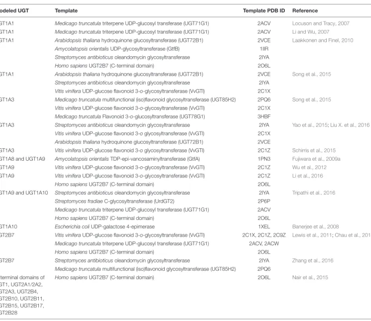
TABLE 1 | Homology modeled human UDP-glucuronosyltransferases (UGTs) and their template structures.
Modeled UGT Template Template PDB ID Reference
UGT1A1 Medicago truncatula triterpene UDP-glucosyl transferase (UGT71G1) 2ACV Locuson and Tracy, 2007 UGT1A1 Medicago truncatula triterpene UDP-glucosyl transferase (UGT71G1) 2ACV Li and Wu, 2007 UGT1A1 Arabidopsis thaliana hydroquinone glucosyltransferase (UGT72B1) 2VCE Laakkonen and Finel, 2010
Amycolatopsis orientalis UDP-glycosyltransferase (GtfB) 1IIR Streptomyces antibioticus oleandomycin glycosyltransferase 2IYA Homo sapiens UGT2B7 (C-terminal domain) 2O6L
UGT1A1 Arabidopsis thaliana hydroquinone glucosyltransferase (UGT72B1) 2VCE Song et al., 2015 Streptomyces antibioticus oleandomycin glycosyltransferase 2IYA
Vitis vinifera UDP-glucose flavonoid 3-o-glycosyltransferase (VvGTl) 2C1X
UGT1A3 Medicago truncatula multifunctional (iso)flavonoid glycosyltransferase (UGT85H2) 2PQ6 Song et al., 2015 Vitis vinifera UDP-glucose flavonoid 3-o-glycosyltransferase (VvGTl) 2C1X
Medicago truncatula Flavonoid 3-o-glucosyltransferase (UGT78G1) 3HBF
UGT1A3 Streptomyces antibioticus oleandomycin glycosyltransferase 2IYA Yao et al., 2015;Liu X. et al., 2016 Vitis vinifera UDP-glucose flavonoid 3-o glycosyltransferase (VvGTl) 2C1X
Arabidopsis thaliana hydroquinone glucosyltransferase (UGT72B1) 2VCE
UGT1A3 Vitis vinifera UDP-glucose flavonoid 3-o glycosyltransferase (VvGTl) 2C1Z Schirris et al., 2015 UGT1A8 and UGT1A9 Amycolatopsis orientalis TDP-epi-vancosaminyltransferase (GtfA) 1PN3 Fujiwara et al., 2009a UGT1A9 Vitis vinifera UDP-glucose flavonoid 3-o-glycosyltransferase (VvGTl) 2C1Z Wu et al., 2012 UGT1A9 Vitis vinifera UDP-glucose flavonoid 3-o-glycosyltransferase (VvGTl) 2C1Z Li et al., 2016
Homo sapiens UGT2B7 (C-terminal domain) 2O6L
UGT1A9 and UGT1A10 Streptomyces antibioticus oleandomycin glycosyltransferase 2IYA Tripathi et al., 2016 Streptomyces fradiae C-glycosyltransferase (UrdGT2) 2P6P
Medicago truncatula triterpene UDP-glucosyl transferase (UGT71G1) 2ACV Homo sapiens UGT2B7 (C-terminal domain) 2O6L
UGT1A10 Escherichia coli UDP-galactose 4-epimerase 1XEL Banerjee et al., 2008
UGT2B7 Vitis vinifera UDP-glucose flavonoid 3-o-glycosyltransferase (VvGTl) 2C1X, 2C1Z, 2C9Z Lewis et al., 2011;Chau et al., 2014 Medicago truncatula triterpene UDP-glucosyl transferase (UGT71G1) 2ACV, 2ACW
Homo sapiens UGT2B7 (C-terminal domain) 2O6L
UGT2B7 Streptomyces antibioticus oleandomycin glycosyltransferase 2IYA Zhang et al., 2016 Medicago truncatula multifunctional (iso)flavonoid glycosyltransferase (UGT85H2) 2PQ6
C-terminal domains of UGT1, UGT2A1/2A2, UGT2A3, UGT2B4, UGT2B10, UGT2B11, UGT2B15, UGT2B17, UGT2B28
Homo sapiens UGT2B7 (C-terminal domain) 2O6L Nair et al., 2015
Matern et al. (1982) similarly purified UGTs that catalyze chenodeoxycholic acid and testosterone glucuronidations from rat liver microsomes by a series of purification steps such as polyethylene glycol fractionation, DEAE-Sepharose CL-6B chromatography, UDP-hexanolamine-Sepharose 4B chromatography, and Bio-Gel A-1.5 m chromatography. While the molecular weight of subunit was determined to be 54 kDa in the SDS-PAGE analysis, the apparent molecular weight of the enzyme was calculated to be 316 kDa in a polyacrylamide gradient slab gel electrophoresis (Matern et al., 1982). The data indicated that rat UGTs were also present as tetrameric or even larger oligomeric forms.
Radiation Inactivation
Radiation inactivation is an analytical tool to determine molecular weights of membrane-bound enzymes in situ (Kempner and Schlegel, 1979). Enzymes are inactivated when
they are irradiated with ionizing radiation. The extent of radiation-induced inactivation of the enzymes is directly associated with the radiation dose and the molecular weight of the concerned enzymes.Peters et al. (1984)determined the molecular weights of rat UGTs by radiation-inactivation of SDS-treated lyophilized liver microsomes using a calibrated 60Co source. The radiation-inactivation analysis revealed that bilirubin mono-glucuronidation was catalyzed by a 41.5 kDa protein, while bilirubin di-glucuronidation was catalyzed by a 175 kDa protein. It was further demonstrated that proteins with molecular weight of 142 and 159 kDa catalyzed glucuronidations of testosterone and phenolphthalein, respectively (Peters et al., 1984). Similarly, the radiation inactivation analysis by Gschaidmeier and Bock (1994) revealed that molecular weights of UGTs catalyzing the glucuronidations of 1-naphthol, 6-hydroxychrysene, dihydroxybenzo[a]pyrene, and 3,6-dihydroxychrysene were 91–218 kDa. The series of radiation
fphar-07-00388 October 20, 2016 Time: 17:9 # 7
Fujiwara et al. Structure and Protein–Protein Interactions of Human UGTs
FIGURE 3 | Homology-modeled structures of human UGT1A9 and UGT1A8. Three-dimensional structures of human UGT1A9 and UGT1A8 were homology-modeled using Amycolatopsis orientalis GtfA (PDB ID: 1PN3) as template structures. The structures of UGT1A9 and UGT1A8 contain two Rossmann fold-like domains in the N-terminus and C-terminus. The structures are colored from red at the N-terminal to blue at the C-terminal. Amino acid residues that are predicted to interact with UDPGA and to be important for the catalysis are shown by red and blue licorice, respectively. The X-ray crystal structure of the C-terminal domain of UGT2B7 was retrieved from the Protein Data Bank (PDB ID: 2O6L). Adapted from Drug Metabolism and Pharmacokinetics (Fujiwara et al., 2009a).
inactivation analyses clearly demonstrated that mammalian UGTs were functional as oligomeric proteins that are composed of two to four subunits.
Cross-Linking
In intact cells, most of proteins are interacting with other protein(s) (Jeong et al., 2001). Upon the denaturing process in the SDS-PAGE analysis, such protein interactions via disulfide binding, hydrogen binding, and hydrophobic or salt-bridge interactions are disrupted, so that the proteins are separated according to their molecular weight. Cross-linkers are reagents that can introduce covalent chemical bonds between specific amino acids of proteins. Thus, proteins treated with cross-linkers can be observed as bands with higher molecular weights rather than their monomeric forms on SDS-PAGE followed by immunoblotting. Ikushiro et al. (1997) used 1,6-bis(maleimido)hexane (BMH), which is a homobifunctional cross-linker that reacts with sulfhydryl-groups of proteins. When rat liver microsomes that were incubated with BMH and were applied to the SDS-PAGE followed by the immunoblotting analysis using anti UGT1 or UGT2B1 antibodies, multiple bands with molecular masses of 50–60 kDa as well as of 120–130 kDa were observed (Ikushiro et al., 1997). They concluded that sulfhydryl group(s) of rat UGTs are located on the outside of the proteins and play a critical role in the formation of UGT dimers. Ghosh et al. (2001) used the disulfide cross-linker BMH and an amino group cross-linker dimethyl 3,30
-dithiobispropionimidate (DTBP) to demonstrate homo-oligomers of human UGT1A1, finding that both BMH and DTBT produced bands corresponding to homo-dimers of UGT1A1 on the SDS-PAGE analysis. The density of
the bands was apparently reduced when UGT1A1-expressed cells were incubated with DTBP at pH 9.0, indicating that homodimerization of human UGT1A1 can be disrupted at alkaline pH.
Affinity Purification and
Immunoprecipitations
Immunopurification and immunoprecipitation are classical methods to identify proteins interacting with a target protein. When rat liver microsomes were solubilized in buffer containing 1% Emulgen 913 and were applied to a UGT1 antibody-conjugated Sepharose 4B column, not only UGT1 isozymes but also unidentified 50-kDa protein(s) were co-eluted by eluting solution containing UGT1A-peptides (Ikushiro et al., 1997). Amino acid sequencing of the 50-kDa proteins and immunoblotting studies further revealed that the protein interacting with UGT1A was UGT2B1. Although the method used in a study by Kurkela et al. (2003)was Ni-column which is not immunopurification, they purified homo-oligomer of UGT1A9 that consisted of His- and hemagglutinin (HA)-tagged UGT1A9.
Fremont et al. (2005)conducted immunoprecipitation assays to examine protein–protein interactions between UGT1A1, UGT1A6, and UGT2B7 in human liver microsomes. When solubilized microsomes were incubated with UGT1A1-, UGT1A6-, and UGT2B7-specific antibodies, not only the antigenic proteins but also other UGT isozymes were co-immunoprecipitated. In 2007, we established cell lines that are individually or simultaneously expressing human UGT1A6 and UGT1A9 (Fujiwara et al., 2007b). A human UGT1A6-specific antibody co-immunoprecipitated UGT1A9, as well
Fujiwara et al. Structure and Protein–Protein Interactions of Human UGTs
as UGT1A6, in the UGT1A6-UGT1A9 double expression cells, although it did not immunoprecipitate UGT1A9 in the UGT1A9-expressed cells. When cyan fluorescent protein (CFP)-and HA-tagged UGT1A1s were simultaneously expressed in COS cells, anti-HA beads immunoprecipitated both CFP-and HA-tagged UGT1A1s (Operaña and Tukey, 2007). Taken together, immunoaffinity purification and immunoprecipitation assays are typical but still powerful tools to demonstrate homo-and hetero-oligomerization of UGTs. Immunoprecipitation assays by Bellemare et al. (2010) further revealed that inactive UGT1A_i2 proteins form not only homo-oligomers (i2-i2) but also hetero-oligomers with UGT1A_i1 proteins (i1-i2).
Fluorescence Resonance Energy
Transfer (FRET)
To examine UGT–UGT interactions in intact cells, Operaña and Tukey (2007) conducted a FRET analysis using cyan and yellow fluorescent proteins (CFP and YFP)-tagged recombinant human UGTs expressed in COS cells. When correction for donor and acceptor bleed through was performed and FRET signal was analyzed, it was revealed that the two fusion UGT proteins resided within ångströms from each other. Their FRET analysis demonstrated homo-oligomerization of all UGT1A isoforms as well as hetero-oligomerization of UGT1A1 with the other UGT1A isoforms. The FRET analysis conducted by a different research group indicated that not only UGT1As, but also human UGT2B7 formed homo-oligomers in SF9 cells (Yuan et al., 2015; Liu Y.Q. et al., 2016).
REGIONS AND AMINO ACID RESIDUES
RESPONSIBLE FOR OLIGOMERIZATION
OF UGTs
Importance of N-terminal Domain
To examine the region responsible for the oligomerization of UGTs, Meech and Mackenzie (1997) constructed a couple of chimeric rat UGT2B1 proteins fused with ecdysteroid glucosyltransferase (EGT), which does not oligomerize. While intact UGT2B1 formed dimers, the dimerization was not observed when the C-terminal half of UGT2B1 was fused with the N-terminal half of EGT. In 2001, two-hybrid analysis by Ghosh et al. (2001) showed that UGT1A1s that were mutated or partially deleted in their N-terminal region (L175E, C233Y, or del152-180) abolished the ability to form homo-oligomers, while UGT1A1 with partial truncation of the C-terminal (K530X) still formed homo-oligomers (Ghosh et al., 2001). These data indicated that the N-terminal domains are involved in oligomerization of mammalian UGTs.
Hydrophobic Amino Acids on the
Surface of Modeled UGT2B7
Hydrophobic amino acid residues on the surface of proteins can mediate protein–protein interactions by introducing proline
brackets and π–π interactions. To identify such hydrophobic amino acid residues of UGTs, Lewis et al. (2011) obtained a three-dimensional structure of human UGT2B7 by homology modeling (Table 1). In the homology-modeled structure of human UGT2B7, a cluster of highly hydrophobic amino acid residues on a B0
-C loop (amino acid residues 183–200) was located on the protein surface (Lewis et al., 2011). Thus, not only in vitro studies, but in silico structural analyses also supported the involvement of the N-terminal region of UGTs in oligomerization.
PROTEIN–PROTEIN INTERACTIONS OF
UGTS WITH OTHER PROTEINS
Protein Interactions with CYPs
Cytochrome P450s are phase I drug-metabolizing enzymes that are expressed in the ER membrane. To investigate the possible protein-interactions between UGTs and CYPs, Taura et al. (2000)applied solubilized rat liver microsomes to a CYP1A1-conjugated Sepharose 4B column. It was found that multiple UGT isoforms were co-eluted in a fraction where CYP1A1 was eluted. Rat UGTs were detected in immunoprecipitates when solubilized rat liver microsomes were reacted with specific antibodies against CYP3A2, CYP2B2, CYP2C11, and CYP1A2 (Ishii et al., 2007). Antibodies against human UGT2B7 and CYP3A4 immunoprecipitated not only their antigenic proteins but also CYP3A4 and UGT2B7, respectively, in solubilized human liver microsomes (Fremont et al., 2005; Takeda et al., 2005, 2009). These data indicate that mammalian UGTs interact with CYPs in liver microsomes. It remains to be determined whether such UGT-CYP interactions can still be observed in a reconstituted system.
Protein Interactions with Microsomal
Proteins
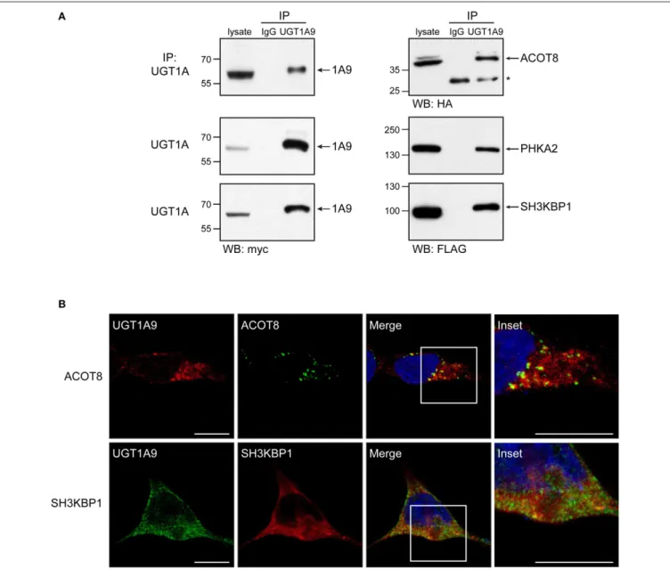
Immunoprecipitation assay with human UGT2B7 antibody was conducted using solubilized human liver microsomes. The obtained immunoprecipitate was digested with trypsin, and the resulting peptides were analyzed by LC-MS/MS to identify proteins interacting with UGT2B7 in human liver microsomes (Fujiwara and Itoh, 2014). The extensive peptide analysis showed that the peptide sequences of UGT2B7, epoxide hydrolase 1, carboxylesterase 1, alcohol dehydrogenases, and glutathione S-transferases, as well as CYPs, were included in the immunoprecipitates. It was confirmed that such peptide sequences were not detected in immunoprecipitates obtained with a control rabbit IgG antibody. Therefore, UGT2B7 might be able to form a metabolosome, which is a functional unit of metabolism (Mori et al., 2011) in liver microsomes.
Protein Interactions with Cytoplasmic
Proteins
In contrast to normal UGT1A proteins (UGT1A_i1) that are expressed in the luminal side of the ER membrane, a
fphar-07-00388 October 20, 2016 Time: 17:9 # 9
Fujiwara et al. Structure and Protein–Protein Interactions of Human UGTs
portion of UGT1A_i2 is cytoplasmic (Lévesque et al., 2007). Immunoprecipitates of solubilized human intestine and kidney homogenates with anti-UGT1A_i2 antibody were applied to a global peptide analysis (Rouleau et al., 2014). It was found that cytoplasmic catalase and peroxiredoxin 1 were co-immunoprecipitated with UGT1A_i2 proteins in both tissues, indicating that the truncated UGT1A isoform 2 is interacting with those cytoplasmic proteins. Since such protein–protein interactions were not observed when anti-UGT1A_i1 antibody was used, the interactions with cytoplasmic proteins would be specific to UGT1A_i2 proteins.
PHYSIOLOGICAL SIGNIFICANCE OF
OLIGOMERIZATION AND
PROTEIN–PROTEIN INTERACTIONS OF
UGTs
Impact of UGT–UGT Interactions on the
UGT-Mediated Glucuronidations
To investigate the effect of UGT–UGT interactions on the UGT activities,Ishii et al. (2001, 2004)cloned guinea pig UGT2B21 and UGT2B22 and examined morphine 6-O-glucuronidation in COS-7 cells expressing these UGTs. While UGT2B21 glucuronidates morphine, UGT2B22 does not have such ability to glucuronidate morphine. Morphine 6-glucuronide formation was 4.5-fold higher in COS-7 cells co-transfected with UGT2B21 and UGT2B22 compared to that in the cells transfected with UGT2B21 alone, indicating that protein–protein interactions between UGT2B21 and UGT2B22 upregulated the UGT activities (Ishii et al., 2001, 2004). This observation led us to investigate the impact of UGT–UGT interactions on the enzyme activities in humans (Fujiwara et al., 2007b; Nakajima et al., 2007). When we established stable expression systems of double human UGT1As in HEK293 cells, the S50 value of UGT1A1-mediated bilirubin glucuronidation was decreased by twofold by the co-expressions of UGT1A4 or UGT1A6 (Fujiwara et al., 2007a). A similar decrease of the S50 value was observed in the UGT1A1-mediated estradiol 3-O-glucuronidation in a co-expression system of UGT1A1 and UGT2B7 in HEK293 cells (Fujiwara et al., 2010a). These data showed that substrate-binding affinity of UGT1A1 toward bilirubin and estradiol was increased when UGT1A1 was co-expressed with UGT1A4, UGT1A6, and UGT2B7. Meanwhile, co-expression of UGT1A9 decreased the Vmaxvalue of UGT1A1-mediated estradiol 3-O-glucuronidation without affecting theS50value (Fujiwara et al.,
2007b). Kurkela et al. (2004) demonstrated that the rate of UGT1A9-catalyzed scopoletin glucuronidation was significantly decreased by co-expression of UGT1A4. Thus, UGT–UGT-interactions can modulate the catalytic rate of glucuronidation as well as the affinity of substrates toward UGTs. Importantly, the effects are dependent on interacting UGT isoforms and compounds used as substrates.
Interestingly, even though sorafenib is mainly metabolized by UGT1A9, it was demonstrated that the AUC of sorafenib was twice higher in patients with UGT1A1 variants or with
hyperbilirubinemia (Peer et al., 2012). This finding indicates that UGT1A1 might control the enzyme activity of UGT1A9in vivo by interacting with UGT1A9.
Although UGT1A_i2 proteins carry a potential UDP-glucuronic acid binding-site, they do not show substantial glucuronidation activity. As described above, the inactive UGT1A_i2 proteins form hetero-oligomers with functional UGT1A_i1 proteins. It was shown that glucuronide formation mediated by UGT1A_i1 proteins was significantly suppressed when UGT1A_i2 was co-expressed with UGT1A_i1 (Bellemare et al., 2010). The expression level and the ratio of UGT1A_i1 and _i2 are different in each tissue (Girard et al., 2007), suggesting that UGT1A_i2 can be a factor suppressing the glucuronidation in certain tissues. Interestingly, such suppression of UGT1 activity can also be caused by a truncated mutant of UGT1A1 (Gln331Stop; C to T at nucleotide 991), possibly by forming oligomers (Koiwai et al., 1996).
Impact of UGT-CYP Interactions on the
UGT Activities
While a Km value of morphine 3-glucuronide formation was 0.38 mM in UGT2B7-expressing COS-1 cells, a much higher Kmvalue (3.7 mM) was observed in UGT2B7 and CYP3A4 co-expressing cells (Takeda et al., 2005). In contrast, co-expression of CYP1A2 or CYP2C9 did not affect the kinetic parameters of UGT2B7-catalyzed morphine 3-glucuronide formations. In addition, co-expression of CYP3A4 increased a Km value of UGT1A6-mediated serotonin glucuronidation by ∼fourfold, whereas it barely affected Km values of UGT1A1-mediated 4-MU, SN-38-, or estradiol 3-O-glucuronidations as well as UGT1A7-mediated 4-MU, SN-38-, and 4-hydroxybiphenyl-glucuronidations (Ishii et al., 2014). Meanwhile, co-expression of CYP3A4 increasedVmaxvalues of UGT1A1-mediated 4-MU, SN-38-, and estradiol 3-O-glucuronidations, UGT1A6-mediated serotonin glucuronidation, and UGT1A7-mediated 4-MU, SN-38-, and 4-hydroxybiphenyl-glucuronidations, although it did not affect the UGT2B7-mediated morphine 3-glucuronidation (Takeda et al., 2005; Ishii et al., 2014). Therefore, CYP isoforms differently affect UGT-mediated glucuronidations and the effects are depending on UGT isoforms as well as their substrates.
Effect UGTs on Other Physiological
Functions
Interestingly, a more than 40-fold interindividual variability in UGT1A6-catalyzed serotonin glucuronidation was observed among individual human liver microsomes (Krishnaswamy et al., 2003, 2005). Such variability could not be explained even when the activities were normalized with the UGT1A6 content and its genetic polymorphisms, indicating that there might be some unidentified factor that is capable of modulating the UGT1A6 activity. Since CYP isoforms can modulate UGT activities in different ways, interindividual variability in the CYP expression and function might be one of the causes of these wide interindividual variabilities in UGT1A6-catalyzed serotonin glucuronidation.