UNIVERSIDADE DE LISBOA
FACULDADE DE FARMÁCIA
ROLE OF APOPTOSIS AND ITS MODULATION
IN ALZHEIMER’ DISEASE:
INSIGHTS FROM IN VITRO AND IN VIVO STUDIES
Rita Cruz Coelho de Mira Ramalho
DOUTORAMENTO EM FARMÁCIA BIOQUÍMICA
UNIVERSIDADE DE LISBOA
FACULDADE DE FARMÁCIA
ROLE OF APOPTOSIS AND ITS MODULATION IN
ALZHEIMER’S DISEASE:
INSIGHTS FROM IN VITRO AND IN VIVO STUDIES
Rita Cruz Coelho de Mira Ramalho
Research advisor:
Cecília M. P. Rodrigues, Ph.D.
DOUTORAMENTO EM FARMÁCIA BIOQUÍMICA
ROLE OF APOPTOSIS AND ITS MODULATION
IN ALZHEIMER’S DISEASE:
INSIGHTS FROM IN VITRO AND IN VIVO STUDIES
O PAPEL DA APOPTOSE E A SUA MODULAÇÃO
NA DOENÇA DE ALZHEIMER:
CONTRIBUIÇÃO DE ESTUDOS IN VITRO E IN VIVO
Dissertação apresentada à Faculdade de Farmácia da Universidade de Lisboa para obtenção do grau de Doutor em Farmácia (Bioquímica)
Rita Cruz Coelho de Mira Ramalho 2007
The studies presented in this thesis were performed at the Centro de Patogénese Molecular, Faculdade de Farmácia da Universidade de Lisboa under the supervision of Professor Cecília M. P. Rodrigues, at the Department of Medicine, University of Minnesota Medical School, Minneapolis, MN, USA, in collaboration with Professor Clifford J. Steer, and at the Department of Neurosurgery, University of Minnesota Medical School, Minneapolis, MN, USA, in collaboration with Professor Walter C. Low.
Rita Cruz Coelho de Mira Ramalho was the recipient of a Ph.D. fellowship (SFRH/BD/12641/2003) from Fundação para a Ciência e a Tecnologia (FCT), Lisbon, Portugal. This work was supported by grants POCTI/BCI/44929/2002, POCI/SAU-FCF/62479/2004, POCI/SAU-MMO/57936/2004 and PTDC/SAU-FCF/67912/2006) (to C.M.P.R.) from FCT, Portugal.
De acordo com o disposto no ponto 1 do artigo nº 40 do Regulamento de Estudos Pós-Graduados da Universidade de Lisboa, deliberação nº 961/2003, publicada em Diário da República – II Série nº 153 – 5 de Julho de 2003, a Autora desta dissertação declara que participou na concepção e execução do trabalho experimental, interpretação dos resultados obtidos e redacção dos manuscritos.
Ao Cláudio
À minha família
Contents
Preface ix
Summary xiii
Sumário xv
Abbreviations xxi
Chapter 1: General Introduction 1
Objectives 61
Chapter 2: Inhibition of the E2F-1/p53/Bax pathway by 63
tauroursodeoxycholic acid in amyloid β-peptide-induced apoptosis of PC12 cells Chapter 3: Tauroursodeoxycholic acid modulates p53-mediated 91
apoptosis in Alzheimer’s disease mutant neuroblastoma cells Chapter 4: Apoptosis in transgenic mice expressing the P301L 117
mutated form of human tau Chapter 5: Concluding Remarks 145
Acknowledgments
Curriculum Vitӕ
Preface
November 26th, 1901. Dr. Alzheimer was informed about a patient showing unusual clinical symptoms. Auguste D., 51 years old, who had never been ill prior to that time, began to suffer from delusions, having trouble remembering things, and making serious mistakes in her daily activities a few months before. Dr. Alzheimer analyzed the mental condition of Auguste D., who maintained confusing and illogical conversations. The patient showed alterations in memory, language, thought, and behavior. All symptoms worsened progressively, month by month, until 8th April, 1906, when she finally died. Her illness had lasted just over five years and Alzheimer was convinced that this was an extraordinary case. Shortly after her death, the brain was analyzed with silver impregnation staining techniques and the findings were truly surprising. Alzheimer and his co-workers, Perusini and Bonfiglio, observed lesions similar to those found in the brains of patients 70 and 80 years old suffering from dementia, but much more marked. All three firmly believed that this was an unusual case, which had never been described. Dr. Alzheimer would probably never understand the impact of his discovery. However, since then, and especially in the last decades, Alzheimer’s disease (AD) has been the focus of intensive research, to establish the abnormal molecular mechanisms that lead to the onset of the disease and to develop novel therapeutic strategies. Although considered the major cause of dementia, with prevalence increasing every year, AD is still not completely understood.
It is now established that AD can be triggered by toxic extra and intracellular aggregates formed from amyloid β and tau, respectively, but the nature of these peptides was not definitively discovered until mid 1980’s. It is becoming clear that these aggregates accumulate in selectively vulnerable regions of the brain, compromising the function and viability of neurons and glia. In the absence of the proper conditions to survive, neurons massively die, compromising the cognitive function of a brain affected by AD. A specific type of cell death, apoptosis, has brought much attention in the last few years.
The first observations of dying neurons was made in the 19th-century by a German naturalist, Carl Vogt, when studying the nervous system of toad embryos. However, it was not until 1951 when Ernst and Glucksmann discovered that cell death was an integral part of normal embryonic development. During the 1960s, much was learned about cell death at the ultrastructural level using electron microscopy. Finally, in 1972, John Kerry and co-authors described for the first time a specific process of cell death, when observing characteristic features of hepatocyte development. The process was termed apoptosis, from the Greek word αποπτοσισ, whose prefix “apo” (απο) generally means “separation”, and the suffix “ptosis” (πτοσισ) the “act of falling off”. The complete word can be translated as the falling of leaves from trees in the autumn and refers to the fragmentation of dying cells into characteristic small bodies. Decades of investigation have shown that apoptosis is an intrinsic suicide program that determines the fate of a cell. It is a common process in many types of cells and tissues. Apoptosis is not only an important event in embryonic development, but also in tissue homeostasis during adult life. In addition, its deregulation can also account for several pathological conditions, ranging from cancer to neurodegenerative disorders. In fact, recent studies suggest a critical role for apoptosis and cell death mediators in AD, even before the reduction in neuronal number.
It has become clear in recent years that prevention of cell death in disorders associated with abnormally increased levels of apoptosis may positively affect the patient outcome. Interestingly, an endogenous bile acid, ursodeoxycholic acid (UDCA) has been described as an inhibitor of apoptosis, not only in liver diseases, but also in other pathological conditions, including neurological disorders. UDCA is a major constituent of black bear bile and has been used for centuries in traditional Chinese medicine for the treatment of liver diseases. However, the mechanisms of action of the bile acid have been characterized only recently. UDCA administration can induce the protection of cholangiocytes against cytotoxicity of hydrophobic bile acids and stimulate the hepatobiliary secretion. Importantly, in 1998, Rodrigues and co-authors showed that UDCA can also have beneficial effects by inhibiting mitochondrial membrane perturbations associated with bile acid-induced apoptosis. UDCA has blossomed as a potent modulator of apoptosis, acting in a tissue-independent manner. Its effects have been tested in many pathological conditions, underscoring its potential and promising therapeutic use.
When I started the Ph.D. program in the laboratory of Professor Cecília M. P. Rodrigues, I focused on investigating the apoptotic mechanisms triggered by neurons exposed to toxic stimuli, in the specific context of AD. My first questions as a student who has just entered a new and exciting area of research, gave rise to additional new questions, which have made my last four years challenging, but rewarding. The present work provides insight into the modulation of apoptosis associated with AD, and more importantly uncovers intriguing connections and links that warrant further investigations.
The purpose of my work was to identify and characterize molecular targets for the use of taurourosodeoxycholic acid (TUDCA) as a modulator of apoptosis in AD, using in vitro and in vivo models. As a conjugated form of UDCA with
taurine, already in use for the treatment of primary biliary cirrhosis, TUDCA proved to be a potent tool in preventing apoptosis in non-hepatic diseases, such as Huntington’s and Parkinson’s disease. The use of TUDCA in AD came as a natural extension of this work. Chapter 1 provides a general, up-to-date review on the process of apoptosis. In addition, the role of bile acids as modulators of apoptosis is discussed. We also focus on describing AD and the role of apoptosis in this neurodegenerative disorder. In Chapter 2, we characterize the mechanisms of neuronal protection by TUDCA in in vitro AD. The role of cell cycle and apoptosis-related proteins in the effects of TUDCA is also presented and discussed. In Chapter 3, the role of p53, a cell cycle-related protein, in TUDCA neuroprotection is further examined, using an in vitro model of familial AD. In Chapter 4, we investigate the role of apoptosis in neurodegeneration using a transgenic mouse model of tauopathy. Apoptosis is presented as an early mechanism that contributes to increased toxicity, and eventually leads to characteristic neurological deficits of AD. Further, we confirmed the existence of a link between amyloid β and tau, via activation of apoptosis-related proteins, and its inhibition by TUDCA. Finally, Chapter 5 integrates our overall findings and discusses specific future perspectives.
The exact mechanism(s) that triggers AD is still obscure. Although more than 100 years have passed since its first description, and despite the efforts of a growing scientific community, an effective treatment is still not available. Nevertheless, in recent years, many mysteries of the disease have been unveiled, including the role of apoptosis as an important event in AD. With this thesis, we hope to contribute to a better understanding of the mechanisms of apoptosis in AD and provide evidences for the neuroprotective role of TUDCA. Ultimately, an increased knowledge of the disease and its potential modulation by bile acids may result in development of more efficient therapeutic interventions.
Summary
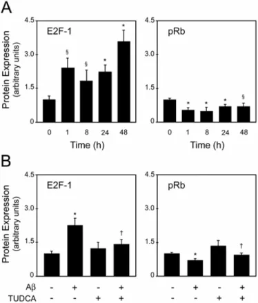
Ursodeoxycholic (UDCA) and its taurine-conjugated form, tauroursodeoxycholic acid (TUDCA), are endogenous bile acids used in the treatment of cholestatic liver disorders. Their cytoprotective effects result, in part, from their ability to modulate hepatocyte apoptosis. Interestingly, UDCA and TUDCA play a unique role in modulating the apoptotic threshold in other cell types, including neuronal cells, by interfering with classic mitochondrial pathways. In these studies, we investigated the role of apoptosis using in vitro and in vivo models of Alzheimer’s disease (AD) and determined its potential modulation by TUDCA. A hallmark pathologic feature of AD is the formation of amyloid plaques composed by aggregated amyloid β (Aβ). Our results showed that TUDCA reduced Aβ-induced apoptosis in PC12 neuronal cells, through modulation of apoptosis- and cell cycle-related proteins. In fact, TUDCA treatment resulted in inhibition of E2F-1 induction, p53 stabilization and Bax expression. Further, TUDCA protected PC12 cells against p53- and Bax-dependent apoptosis induced by E2F-1 and p53 overexpression, respectively. The role of p53 in TUDCA effects was further confirmed using an in vitro model of familial AD. In neuroblastoma cells expressing the amyloid precursor protein (APP) with the Swedish mutation (APPswe), or double-mutated human APP and PS1 (APPswe/∆E9), TUDCA modulated p53 activity, and Bcl-2 family changes. Moreover, overexpression of p53 was sufficient to induce apoptosis, which in turn
was reduced by TUDCA. Another pathologic feature of AD is the intracellular aggregation of tau into neurofibrillary tangles. Using the rTg4510 transgenic mouse model of tauopathy, expressing a mutated form of human tau, we confirmed the role of apoptosis in neurodegeneration. Increased levels of DNA fragmentation and caspase-3 activation were observed in the hippocampus and frontal cortex of young mice. These changes were associated with cleavage of tau into smaller intermediate fragments, which were often colocalized with active caspase-3. In vitro, fibrillar Aβ resulted in nuclear fragmentation, caspase activation, and caspase-3-induced cleavage of tau. Notably, incubation with TUDCA abrogated apoptosis-mediated cleavage of tau in rat cortical neurons. The results suggest that caspase-3-cleaved intermediate tau species precede cell loss in rTg54510 brains and Aβ-exposed cultured neurons. In conclusion, the work presented here underscores the role of apoptosis in neurodegeneration of AD and expands the antiapoptotic function of TUDCA. Furthermore, the results demonstrate that TUDCA regulates specific transcriptional and posttranscriptional events that impact on mitochondrial function of neurons.
Keywords: Amyloid β – Alzheimer’s disease - Apoptosis – Bcl-2 family – Bile acids – Caspases – E2F-1 – p53 – Tau
Sumário
O ácido ursodesoxicólico (UDCA) e a sua forma conjugada com a taurina, o ácido tauro-ursodesoxicólico (TUDCA), são ácidos biliares endógenos, largamente utilizados no tratamento de doenças crónicas do fígado, como a cirrose biliar primária. No entanto, só recentemente começaram a ser conhecidos e descritos os mecanismos de acção destes ácidos biliares. Actualmente, sabe-se que o efeito citoprotector do UDCA e do TUDCA se deve, maioritariamente, à capacidade destas moléculas modularem a morte celular programada ou apoptose dos hepatocitos, fenómeno que se encontra desregulado em inúmeras patologias hepáticas. De facto, através de estudos prévios, foi possível demonstrar que o UDCA e o TUDCA desempenham este papel anti-apoptótico, em parte, através da estabilização da membrana mitocondrial, prevenindo a sua despolarização e a consequente libertação de citocromo c e activação de caspases, responsáveis pela execução do processo apoptótico. Porém, desconhece-se, ainda, a maior parte dos mecanismos de sinalização iniciados por estes ácidos biliares. Uma vez que os vários produtos do metabolismo lipídico, incluíndo os ácidos biliares, possuem propriedades sinalizadoras, pensa-se que a regulação da apoptose exercida pelo UDCA e TUDCA poderá passar pela modulação a nível da transcrição génica ou mesmo a nível pós-transcricional.
Curiosamente, o papel protector do UDCA parece estender-se a outros tipos celulares e em resposta a vários agentes tóxicos. Por outro lado, após a conjugação
com a taurina e quando administrado sistemicamente, em sobredosagem, o UDCA pode ser distribuído por outros tecidos, incluíndo o cérebro, o que permite a sua aplicação em doenças não hepáticas, como é o caso de várias desordens neurológicas. De facto, os efeitos protectores do TUDCA foram já testados, in vitro e in vivo, para as doenças de Hungtington e Parkinson, assim como em modelos de acidente vascular cerebral, do tipo isquémico e hemorrágico. Muitas outras patologias associadas à desregulação da apoptose poderão, também, beneficiar desta estratégia terapêutica.
A doença de Alzheimer (AD) é uma doença neurodegenerativa progressiva, à qual estão associadas graves perdas de memória e um acentuado défice cognitivo. O cérebro de um doente de Alzheimer caracteriza-se pela presença de placas amilóides, cujo principal componente é a proteína β amilóide (Aβ), e de tranças neurofibrilhares (NFT), compostas por agregados intracelulares da proteína tau. Como resultado da formação destes agregados tóxicos, os neurónios sofrem profundas alterações, tornam-se disfuncionais e acabam por morrer em grande escala. A apoptose parece desempenhar um papel importante, como mecanismo essencial de morte celular associada à AD.
No presente estudo, investigou-se o envolvimento da apoptose na neurodegenerescência associada à AD e a possível regulação dos mecanismos apoptóticos pelo TUDCA. Foi também explorada a função de proteínas específicas da apoptose e do ciclo celular no papel anti-apoptótico do TUDCA.
Numa primeira parte do trabalho, os resultados obtidos demonstraram que, apesar do aumento da expressão da proteína anti-apoptótica Bcl-2, incubações com Aβ induzem níveis significativos de apoptose em células neuronais PC12, o que foi eficazmente inibido em pré-tratamentos com TUDCA. A inibição da apoptose induzida por Aβ parece ser feita através da via E2F-1/p53/Bax, mais especificamente pela inibição da indução do factor de transcrição E2F-1, da estabilização da proteína p53 e da expressão da Bax. De facto, o TUDCA foi
capaz de proteger as células da apoptose dependente da expressão de p53 e de Bax, após sobre-expressão de E2F-1 e p53, respectivamente.
De seguida, o papel das proteínas do ciclo celular, e mais especificamente da p53, na modulação, pelo TUDCA, da apoptose induzida por Aβ, foi confirmado num modelo das formas familiares da AD. Apesar de se manifestar numa pequena percentagem da população mundial, a AD na sua forma familiar, associada a mutações na proteína precursora da Aβ (APP) ou nas presenilinas 1 e 2, possui características muito semelhantes à forma esporádica, embora o início da doença ocorra geralmente em idades mais precoces. Utilizando células de neuroblastoma que expressam APP com a mutação Swedish (APPswe) ou duplamente mutadas na APP e na presinilina 1 (APPswe/ΔE9), observou-se um aumento dos níveis de apoptose, em consequência da produção endógena e agregação de Aβ. De facto, detectou-se fragmentação nuclear e activação das caspases -2, -6 e -8 em células APPswe e APPswe/ΔE9. Por outro lado, observou-se também um aumento da expressão de p53 e de Bax e uma diminuição da expressão de Bcl-2. Em contrapartida, a pré-incubação com o TUDCA reduziu eficazmente os níveis apoptóticos e de activação das caspases -2 e -6, restabelecendo a expressão de p53 e de proteínas da família Bcl-2. A sobre-expressão de p53 induziu, por si só, a apoptose nas células de neuroblastoma, o que, por sua vez, foi reduzido pelo TUDCA. No entanto, a inibição da via de sobrevivência fosfatidilinositol 3’-cinase reduziu a capacidade do TUDCA para proteger a apoptose induzida pela p53. Em conclusão, estes estudos demonstram que as mutações associadas às formas familiares da AD activam mecanismos apoptóticos muito semelhantes às formas esporádicas. Por outro lado, o TUDCA é capaz de reduzir a apoptose, através da inibição da p53 e da consequente modulação dos níveis de expressão das proteínas da família Bcl-2.
Por fim, numa terceira parte do trabalho, o papel da apoptose foi avaliado num modelo de tauopatia. Na AD, assim como nas tauopatias, a proteína tau deixa
de induzir a estabilização dos microtúbulos a nível do axónio, é fosforilada de uma forma anómala e agrega em NFT no corpo celular dos neurónios. Como resultado, os neurónios deixam de ser funcionais e, eventualmente, acabam por morrer. Em estudos anteriores, o modelo transgénico rTg4510, que expressa uma forma mutada de tau humana, apresentou níveis elevados de morte neuronal, em estruturas corticais e límbicas associadas à AD, atrofia cerebral e défices cognitivos. Nos nossos estudos, observou-se um aumento dos níveis de fragmentação nuclear e activação de caspase-3, especialmente em animais mais jovens, de 2,5 meses, e nas áreas do córtex frontal e do hipocampo. De facto, a apoptose aparenta ser um evento precoce nestes animais transgénicos e a activação de caspase-3 parece estar associada à clivagem de tau na sua zona C-terminal, uma vez que se observou a co-localização da caspase-3 activa e de tau clivada no cortex frontal e hipocampo de animais com 2,5 meses. A clivagem de tau pela caspase-3 foi já descrita, por alguns autores, como sendo um acontecimento essencial à sua agregação em NFT, o que vem confirmar o papel da apoptose nesta patologia. Apesar das tauopatias estarem associadas a mutações na proteína tau, não existem quaisquer mutações descritas na AD, sendo a Aβ apontada como principal responsável pelas alterações conformacionais da tau. Em estudos in vitro, por nós realizados, fibrilhas de Aβ1-42 induziram a fragmentação nuclear, a activação de caspases e clivagem de tau pela caspase-3, estabelecendo uma ligação entre as duas entidades patológicas da AD. Por outro lado, a pré-incubação com TUDCA inibiu, de forma significativa, a clivagem de tau induzida pela apoptose em neurónios corticais de rato. Deste modo, os resultados sugerem que formas intermediárias de tau, clivadas pela caspase-3, precedem a morte neuronal nos cérebros dos ratos rTg4510 e em neurónios expostos a Aβ1-42 fibrilhar.
Em suma, o presente trabalho demonstra a importância da apoptose na AD, não só como mecanismo de morte neuronal, mas também como mediadora de efeitos tóxicos. Por outro lado, estes estudos revelam o potencial papel protector
do TUDCA em modelos de AD, actuando a montante dos eventos mitocondriais, nomeadamente através da regulação transcricional da expressão de proteínas do eixo apoptótico E2F-1/p53/Bax. Ilustrou-se, ainda, a capacidade do TUDCA para inibir mecanismos tóxicos a jusante da mitocôndria, que culminam na clivagem de tau em fragmentos indutores da sua agregação.
A caracterização do ácido biliar TUDCA como modulador transcricional e pós-transcricional da apoptose na AD, consolida o papel desta molécula como uma opção terapêutica no tratamento de doenças neurodegenerativas, expandindo o seu papel protector para além das doenças hepáticas.
Palavras chave: Ácidos Biliares – Apoptose – β amilóide – Caspases – Doença de Alzheimer – E2F-1 – Família Bcl-2 – p53 – Tau
Abbreviations
Aβ amyloid β peptide
AD Alzheimer’s disease
AIF apoptosis-inducing factor
ANT adenine nucleotide translocator
Apaf-1 apoptosis protease-activating factor 1
APOE ε4 apolipoprotein ε4 allele
APP amyloid precursor protein
APPwt APP wild-type
APPswe APP with the Swedish mutation
APPswe/∆E9 APP double-mutated human APP and PS1
BH Bcl-2 homology domain
CAT chloramphenicol acetyltransferase
CDK cyclin dependent kinase
DIABLO direct IAP binding protein with low pI
DISC death-inducing signaling complex
DTT dithiothereitol
ER endoplasmic reticulum
FAD familial form of AD
FADD Fas-associated death domain
FC frontal cortex
FTDP-17 frontotemporal dementia with parkinsonism linked to chromosome 17
GR glucocorticoid receptor
GSK3β glycogen synthase kinase 3 β
HD Hungtington’s disease
IAP inhibitor of apoptosis protein
IM mitochondrial inner membrane
JNK c-Jun N-terminal kinase
MAPK mitogen-activated protein kinase
MMP mitochondrial membrane permeabilization
MPT mitochondrial permeability transition
MSN medial septal nucleus
3-NP 3-nitropropionic acid
MTT 3-(4,5-dimethylthiazol-2-yl)-2,5-diphenyl tetrazolium bromide
NF-κB nuclear factor κB
NFT neurofibrillary tangles
OM mitochondrial outer membrane
PD Parkinson’s disease
PBS phosphate-buffered saline
PI3K phosphatidylinositide 3’-OH kinase
pNA p-nitroanilide
pRb retinoblastoma protein
PS1 presenilin 1
PS2 presenilin 2
ROS reactive oxygen species
SDS sodium dodecyl sulphate
SGZ subgranular zone
Smac second mitochondria-derived activator of caspases
TNF tumor necrosis factor
TNF-α tumor necrosis factor α
TNF-R1 TNF type receptor 1
TUDCA tauroursodeoxycholic acid
TUNEL transferase mediated dUTP-digoxigenin nick-end
labeling
UDCA ursodeoxycholic acid
VDCA voltage-dependent anion channel
1
General Introduction
_________________________________________________________________________
1. Apoptosis
Apoptosis (from the Greek “falling off”) was originally described in 1972 by Kerr et al. as a common type of programmed cell death, repeatedly observed in various tissues and cell types (Kerr et al. 1972). Apoptosis is one of the most frequent phenomena occurring in multicellular organisms and is fundamental to their health. In fact, as a physiological mechanism, apoptosis has an important role in embryogenesis, synaptogenesis, immune response and tissue homeostasis. Surprisingly, per day, the human body destroys ~ 60 x 109 cells through an apoptotic process, in response to physiological, pathogenic, or cytotoxic stimuli, underscoring the relevance of this orchestrated form of cellular suicide (Reed 2002).
In contrast to necrosis, apoptosis is an active energy-dependent process, defined by a series of biochemical and morphological modifications, including condensation of chromatin, shrinking of cytoplasm and nuclear compartments, degradation of DNA into oligonucleosome-length fragments and compartmentalization of nuclear material into vesicular apoptotic bodies (Kerr et al. 1972). These are rapidly eliminated by resident phagocytic and neighboring cells, preventing the release of cellular components into the extracellular space, and consequent inflammatory response. Defects in physiological pathways of apoptosis contribute to the development of numerous medical illnesses for which adequate therapy or prevention is lacking. In fact, insufficient levels of apoptosis are implicated in cancer, autoimmune diseases and persistent infections, while excessive apoptosis can lead to neurodegenerative disorders and hepatocellular degeneration.
The highly conserved molecular basis of apoptosis was originally described in the nematode Caernorhabditis elegans (C. elegans) (Ellis and Horvitz 1986). Interestingly, 113 of the 1090 embryonic somatic cell undergo apoptosis during the
Chapter 1
_________________________________________________________________________
development of C. elegans. In a coordinated process, apoptosis is regulated by three important genes: ced-3, ced-4, that induce cell death, and ced-9, which has an antiapoptotic role (Metzstein et al. 1998).
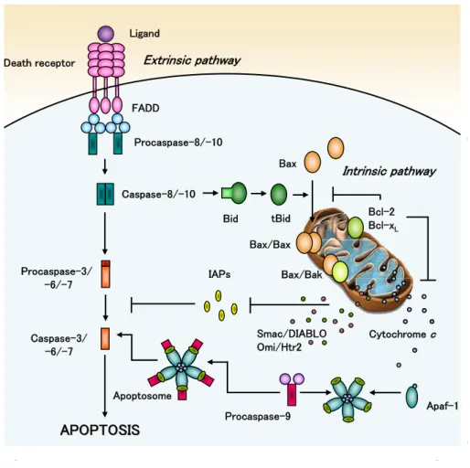
Apoptosis may occur by several molecular pathways. The best characterized and most prominent, however, are the extrinsic death receptor and intrinsic mitochondrial pathways (Fig.1).
Fig. 1. Schematic overview of extrinsic and intrinsic apoptotic pathways. In the death
receptor pathway, after interacting with their ligands, the death receptors recruit adaptor
Bax Smac/DIABLO Omi/Htr2 Ligand Apaf-1 Death receptor FADD Procaspase-8/-10 Bid tBid Procaspase-3/ -6/-7 Caspase-3/ -6/-7 APOPTOSIS APOPTOSIS Extrinsic pathway IAPs Procaspase-9 Apoptosome Bax/Bak Cytochrome c Bcl-2 Bcl-xL Intrinsic pathway Caspase-8/-10 Bax/Bax 4
General Introduction
_________________________________________________________________________ proteins such as FADD and activate caspases-8 and -10. These initiator caspases then cleave effector caspases-3, -6, and -7, which activate key downstream targets and execute the apoptotic process. In the mitochondrial pathway, death stimuli target mitochondria either directly or through transduction by proapoptotic Bax and Bak. Mitochondria release cytochrome c, Smac/DIABLO, Omi/Htr2 and other apoptogenic factors. Cytochrome c induces oligomerization of Apaf-1 that recruits and activates procaspase-9. Caspase-9 then activates effector caspases. The crosstalk between both pathways is mediated by Bid, which is truncated and activated by caspases-8. See text for more complete description.
Both pathways are characterized by an initiation phase, when a signal triggers the apoptotic process; integration/decision phase, which involves the activation of several apoptotic mechanisms; and final execution/degradation phase that culminates in cell death. Although apparently independent, the two apoptotic pathways often interact in many cell types to accomplish cell death signaling.
1.1. Mitochondrial
pathway
As the primary generators of energy and important regulators of intracellular calcium, mitochondria are essential organelles for cell survival. By coupling electron transport to the generation of proton gradients for oxidative phosphorylation, mitochondria produce ATP that is used in the metabolic activities of the cell. Thus, highly metabolic tissues such as the brain are particularly dependent on mitochondria. In the past decades, mitochondria have also emerged as critical players in cell death. In fact, all the energy that is used for maintaining life in healthy cells is completely redirected to serve a mortal purpose, under pathological conditions. After an apoptotic stimulus, such as oxidative stress, DNA damage, or protein misfolding, the levels of calcium are increased, the mitochondrial membrane is permeabilized, releasing apoptogenic factors from the
Chapter 1
_________________________________________________________________________
intermembrane space to the cytoplasm and disrupting the mitochondrial membrane potential, which culminates in cell death (Ricci et al. 2003).
1.1.1. Structural
modifications
of
mitochondria during apoptosis
Under physiological conditions, mitochondrial inner membrane (IM) is nearly impermeable to all ions including protons. This results in an electrochemical gradient, the inner mitochondrial transmembrane potential, that is essential for cellular bioenergetics (Mitchell and Moyle 1965a, b). The permeability of the mitochondrial outer membrane (OM) is also well regulated, mainly by the presence of voltage-dependent anion channels (VDAC) (De Pinto and Palmieri 1992). After an apoptotic stimulus, mitochondria undergo several modifications leading to mitochondrial membrane permeabilization (MMP), often considered as the “point of no return” (Green and Kroemer 2004). The permeability of the OM increases, allowing the release of soluble proteins that are usually retained in the intermembrane space. The IM looses its selectivity and becomes permeabilized, which results in permanent dissipation of the transmembrane potential (Marchetti et al. 1996). Far from an accidental process, the MMP is a tightly regulated phenomenon, with the involvement of dynamic pore structures and interaction of different proteins. One of the first events is the opening of the high conductance mitochondrial permeability transition (MPT) pore. In fact, excessive calcium accumulation by mitochondria during apoptosis leads to opening of the MPT pore and massive mitochondrial swelling (Bernardi 1999). Despite the uncertainty about the molecular composition, it has been suggested that the MPT pore spans the IM and the OM and is composed of proteins from membranes and matrix. The adenine nucleotide translocator (ANT), located in the IM (Brustovetsky and Klingenberg 1996), the VDAC (De Pinto and Palmieri 1992), and the cyclophilin D, from the matrix (Crompton et al. 1998), were proposed to be part of the MPT pore complex. In addition, proapoptotic proteins from the Bcl-2 family, namely
General Introduction
_________________________________________________________________________
Bax and Bak, can also engage in a close molecular cooperation with some components of the MPT complex, such as the VDAC and/or the ANT (Tsujimoto and Shimizu 2002), or form themselves pores, further enhancing mitochondrial permeabilization (Wolter et al. 1997; Kuwana et al. 2002).
1.1.2. Release of apoptogenic factors from mitochondria
As a consequence of the MMP, several apoptogenic factors are released into the cytosol, activating a family of death-inducing cysteine proteases, the caspases. Cytochrome c, a peripheral protein of the IM that functions as an electron shuttle between complexes III and IV of the respiratory chain, is a crucial factor in mediating mitochondria-dependent apoptosis (Li et al. 1997). Recent studies have identified an additional mitochondrial compartment, the intracristae space, that is formed by lamellar and tubular structures resulting from the convoluted folds of the IM cristae (Frey and Mannella 2000). Most of the cytochrome c yield (~ 85%) is contained in this compartment, which suggests that an additional step for cytochrome c release may occur. In fact, during apoptosis, cristae are remodeled, which results in the widening of junctions that delineate the intercristae space. This phenomenon facilitates the diffusion of cytochrome c to the intermembrane space and, consequently, to the cytosol through pores formed in the OM (Scorrano et al. 2002). Although not completely understood, the reorganization of cristae may require several proteins involved in mitochondrial fusion and fission processes, such as the Drp1 (Germain et al. 2005).
Once in the cytosol, cytochrome c binds and induces conformational changes to the apoptotic protease-activating factor-1 (Apaf-1), a CED-4 homolog, in the presence of ATP/dATP, recruiting procaspase-9, and forming the apoptosome. Caspase-9 then acquires the ability to trigger processing and activation of downstream caspases, finalizing the apoptotic process (Zou et al. 1999).
Chapter 1
_________________________________________________________________________
Similar to its murine homolog direct inhibitor of apoptosis proteins (IAP) binding protein with low pI (DIABLO), the second mitochondria-derived activator of caspases (Smac), is another apoptogenic factor released during MMP. Smac/DIABLO is a mitochondrial protein encoded by the nuclear genome, and is proteolytically processed within the intermembrane space to yield a mature polypeptide of 23 kDa with an IAP biding motif (Du et al. 2000). Following MMP, Smac/DIABLO is released into the cytosol, neutralizing IAPs and thus promoting caspase activation. Similar to Smac/DIABLO, the Omi/Htr2 protein is also processed in the intermembrane space into a mature form of 37 kDa (Martins 2002). Once in the cytosol, it promotes cell death either by antagonizing IAPs (caspase-dependent pathway), or via its proteolytic activity (caspase-independent pathway).
The apoptosis inducing factor (AIF) also plays an important role in the apoptotic process. In healthy cells, AIF is required for optimal detoxification of reactive oxygen species (ROS), and for the assembly or maintenance of the respiratory chain complex I (Vahsen et al. 2004). However, once released from the intermembrane space, after OM permeabilization and proteolytic maturation, AIF translocates from the cytosol to the nucleus, promoting chromatin condensation and large-scale DNA fragmentation (Susin et al. 1999). A similar role is played by endonuclease G (EndoG), a mitochondria-specific enzyme that also translocates to the nucleus during apoptosis. However, the mechanism by which EndoG cleaves DNA into nucleosomal fragments (Li et al. 2001) is not entirely known.
In addition to the above-described, many other factors are released from mitochondria during MMP. However, their precise role, if any in cell death has not yet been elucidated. Thus, further investigation is needed for the complete characterization of the mitochondrial pathway of apoptosis.
General Introduction
_________________________________________________________________________
1.2.
Death receptor pathway
Many toxic and pathological situations are associated with changes in expression and/or functioning of death receptors and their ligands, leading to caspase activation and apoptosis (Fig. 1). The tumor necrosis factor (TNF) death receptor superfamily is a group of cytokines with important functions not only in apoptosis, but also in immunity and inflammation, control of cell proliferation and differentiation (Baud and Karin 2001). Nineteen different proteins have been identified within this family, including the TNF-α and the Fas/Apo-1/CD95 receptors that are activated by binding of TNF-α and Fas ligand (FasL), respectively (Brunner et al. 1995). The members of TNF receptor family are structurally similar membrane proteins of type I. They consist of extracellular (N-terminal), transmembrane, and intracellular (C-terminal) components. Ligand-binding domains of the extracellular compenent of death receptors are characterized by the presence of 2 to 6 repeats of about 40 amino acids enriched in cysteine (Banner et al. 1993). After ligand binding, adaptor proteins such as the Fas-associated death domain (FADD) are recruited (Blagosklonny 2000), forming the death-inducing signaling complex (DISC) (Kischkel et al. 1995). Once activated, death receptors induce the cleavage and activation of procaspase8, and -10. In fact, FADD was shown to contain two death effector domains (DEDs) capable of recruiting caspase-8, and -10 (Krueger et al. 2001). Depending on cell type, caspase-8, and -10 can directly activate downstream caspases, such as caspase-3 or -7 (Peter and Krammer 2003), or transmit the death signal to mitochondria, in a cross-talk between both apoptotic pathways (Li et al. 2002) (Fig. 1). In this case, caspase-8 cleaves the inactive cytoplasmic Bid, a proapoptotic protein of the Bcl-2 family, exposing an active truncated fragment (tBid) (Scaffidi et al. 1998). Once activated, tBid translocates to mitochondria, inducing conformational changes in proapoptotic proteins, such as Bax and Bak
Chapter 1
_________________________________________________________________________
and, consequently, the MMP (Eskes et al. 2000). Moreover, tBid can also inhibit antiapoptotic proteins, such as Bcl-2, (Kim et al. 2000), or even directly permeabilize the mitochondrial OM (Goonesinghe et al. 2005), ultimately inducing caspase-3 activation and perpetuating the apoptotic process.
Finally, in the presence of ROS, TNF-R1 can also induce apoptosis via mitogen-activated protein kinase (MAPK) signaling, activating the c-Jun NH2 -terminal kinase (JNK) pathway (Shen and Pervaiz 2006). Interestingly, TNF-R1 is capable of triggering survival signals, including the activation nuclear factor κB (NF-κB) (Barnhart and Peter 2003), underscoring the relevance and complexity of death receptors.
1.3.
Other molecular intervenients in the apoptotic process
To execute apoptosis, many proteins, enzymes, and different factors are involved. In fact, to integrate death signals and perform the multiple reactions that culminate in cell death, several components, interactions, and biochemical processes are necessary, in a complex organization that defines the efficiency of apoptosis.
1.3.1. Bcl-2
family
Modulation of apoptosis is performed by the Bcl-2 family, a group of proteins that work in regulated protein-protein interactions. This family gives its name to the first identified member, over 20 years ago, at the chromosomal breakpoint of t(14;18)(q32;q21) lymphomas, B-cell lymphoma-2 (Bcl-2). Bcl-2 is an homolog of CED-9 (Tsujimoto et al. 1985). Curiously, Bcl-2 was found to inhibit cell death, rather than promote proliferation, and since then many relatives of this family have been described. In mammals, the Bcl-2 family consists of at least 20 members, including proteins that promote apoptosis, or proapoptotic proteins, and others than inhibit it, forming a complex balancing network that determines cell fate.
General Introduction
_________________________________________________________________________
Bcl-2 members can be subdivided in three groups, according to their structure and function. The antiapoptotic proteins, such as Bcl-2, Bcl-xL and Mcl-1, among others, share 3 to 4 conserved Bcl-2 homology domains (BH1-4), forming group I. Bcl-2 and its homologues potently inhibit apoptosis in response to many cytotoxic insults. The proapoptotic proteins, such as Bax and Bak, share 3 conserved domains (BH1-3), forming group II. Group III contains a subgroup of proapoptotic proteins that only have the BH3, such as Bid, Bad, Noxa and Puma, among others (Cory et al. 2003). Both types of proapoptotic proteins are required to initiate apoptosis; the BH3-only proteins usually act as damage sensors and direct antagonists of the antiapoptotic proteins, while proteins of group II act further downstream, mainly in mitochondria disruption.
Under physiological conditions, Bcl-2 binds to the cytoplasmic face of mitochondrial OM, endoplasmic reticulum (ER) and nuclear envelope, promoting the integrity of membranes, by interacting and neutralizing proapoptotic proteins (Gottlieb 2001). Bcl-2 overexpression decreases the amount of calcium mobilized from the ER to the mitochondria, inhibiting the opening of the PT pore (Baffy et al. 1993). In addition, some studies indicate that Bcl-2 and Bcl-XL can also interact with ANT and VDAC, inhibiting the formation of the MPT pore (Marzo et al. 1998; Shimizu et al. 2000).
Proapoptotic Bax, under normal circumstances, resides as a monomer in the cytoplasm (Hsu et al. 1997), while Bak is attached to the mitochondrial OM as an integral membrane protein (Griffiths et al. 1999). Following induction, an increased expression of proapoptotic proteins occurs, changing the balance between anti- and proapoptotic factors. Consequently, Bax undergoes conformational changes, translocates to the mitochondria, oligomerizes and inserts into the OM (Wolter et al. 1997). Once attached to mitochondria, it is thought that Bax, alone or associated with Bak, can provoke or contribute to permeabilization of the OM, allowing the release of cytochrome c (Kuwana et al. 2002). The
Chapter 1
_________________________________________________________________________
mechanism by which proapoptotic proteins permeabilize the OM, however, is still controversial and not entirely understood. Some studies suggest that Bax and Bak can themselves form hydrophobic pores in the membrane (Wolter et al. 1997; Kuwana et al. 2002), or simply destabilize membrane lipid bilayers (Basanez et al. 2002). Alternatively, Bax might interact with proteins of the MPT pore, such as VDAC and ANT, inducing the MMP (Tsujimoto and Shimizu 2002). Further, Bax and Bak can enhance the loading of the ER calciumstore, boosting the calcium load to mitochondria (Scorrano et al. 2003).
Finally, BH3-only proteins act as sentinels that when activated trigger apoptosis in response to developmental cues or intracellular damage. These proapoptotic proteins exert their action by two different mechanisms. In fact, they can interact with antiapoptotic proteins, dissociating them from other BH-3-only or from BH1-3 proteins, and promoting MMP (e.g. Bad), or they can directly activate the BH1-3 proteins to initiate MMP, either by stimulating the translocation of Bax to mitochondrial OM or by local effects on Bak (e.g. tBid) (Letai et al. 2002).
1.3.2. Caspases
Caspases are a family of cysteine proteases that play an important role in the execution of apoptosis, cleaving a restricted set of target substrates after an aspartate residue in their primary sequence (Thornberry and Lazebnik 1998). In 1993, the gene ced-3 was discovered in C. elegans, showing great similarities with caspase-3; a connection between caspases and apoptosis was established for the first time (Yuan et al. 1993). Since then, many other caspases have been described in mammalian and non-mammalian species.
Caspases share similarities in amino acid sequence, structure, and substrate specificity. In healthy cells, they are present in the cytosol as inactive precursors, called zymogens. Caspases contain three domains, including a N-terminal prodomain, a large subunit with the active cysteine, and a C-terminal small unit
General Introduction
_________________________________________________________________________
(Thornberry and Lazebnik 1998). After an apoptotic signal, the zymogen is exposed to two cleavage events. The first proteolytic cleavage divides the chain into large and small caspase subunits, and a second cleavage removes the N-terminal prodomain (Wolf and Green 1999). The cleavage of the zymogen is not always an obligatory requirement for caspase activation. However, all activated caspases can be detected as cleaved fragments in apoptotic cells (Degterev et al. 2003).
According to their function, caspases are grouped as upstream proteases termed initiator caspases (caspases-2, -8, -9 and -10), and their downstream targets known as effector or executioner caspases (caspase-3, -6 and -7) (Thornberry and Lazebnik 1998). The initiator caspases function as signal integrators for apoptotic or proinflammatory stimuli. They contain larger prodomains and specific sequence motifs, such as the caspase recruitment domain (CARD), for caspase-2 and -9, or a pair of DEDs, for caspase-8 and -10 (Hofmann et al. 1997; Ashkenazi and Dixit 1998). These domains mediate the recruitment of zymogen to death signaling complexes, leading to its auto-catalytic activation. Moreover, the homodimerization of zymogens appears to be a crucial step for the activation of initiator caspases in contrast to executioner caspases.
Caspase-9 is a key component of the mitochondrial pathway and its activation occurs after cytochrome c release and formation of the apopotosome. The zymogen is recruited to the complex through CARD-CARD interactions, and rapidly processed into active caspase-9, which in turn is responsible for the activation of downstream caspases, such as caspase-3 (Thornberry and Lazebnik 1998). On the other hand, caspase-8 has a key role in the death receptor pathway and is activated in the presence of external death signals. In response to the activation of receptors of the TNF family, the zymogen is recruited to the DISC via binding to FADD, resulting in caspase-8 activation and subsequent activation of downstream caspases (Boldin et al. 1996; Varfolomeev et al. 1998).
Chapter 1
_________________________________________________________________________
Caspase-10 is structurally very similar to caspase-8, but its function in apoptosis is not entirely known (Degterev et al. 2003). In fact, some studies suggest that caspase-10 may have a function that overlaps with caspase-8 in Fas ligand-mediated apoptosis. Caspase-10 might be recruited to the Fas DISC, cleave Bid, and activate the mitochondrial pathway.
Finally, caspase-2 was one of the first caspases discovered, but its physiological function and activation remain obscure. Recent results suggest that caspase-2 can be activated by dimerization (Butt et al. 1998) or by recruitment of a large complex similar to the apoptosome, named the PIDDosome (Tinel and Tschopp 2004). It is often localized in the cytosol, the nucleus (Colussi et al. 1998), and the Golgi (Mancini et al. 2000), although its protein targets in these compartments remain largely unclear. Nevertheless, it was demonstrated that in some cells caspase-2 is responsible for the mitochondrial OM permeabilization and the release of apoptogenic factors in response to DNA damage (Zhivotovsky and Orrenius 2005). It can also associate with the Fas DISC, but apparently it is not required for Fas-induced cell death (Lavrik et al. 2006). Interestingly, caspase-2 can also function independently of its protease activity, such as by activation of MAPK and NF-κB signaling pathways (Lamkanfi et al. 2005).
Once activated, initiators caspases cleave effector caspases, which lack the long prodomain and the ability to self-activate. Effector caspases are responsible for cleaving most of the cellular substrates, finalizing the apoptotic process. Caspase-3 is the main downstream effector caspase, which can be activated via the death receptor pathway, following activation by caspase-8, and through the mitochondrial pathway, by caspase-9 (Porter and Janicke 1999). It is responsible for cleavage of many substrates, including nuclear lamins and cytoskeletal proteins, such as fodrin and gelsolin that are associated with morphological changes in apoptotic cells (Kothakota et al. 1997). In addition, caspase-3 cleaves the inhibitor of caspase-activated DNAse (ICAD), promoting the activation of the
General Introduction
_________________________________________________________________________
endonuclease CAD, which induces the characteristic nucleosomal DNA fragmentation (Sakahira et al. 1998). Caspase-3 is also responsible for the cleavage of poly(ADP-ribose) polymerase (PARP), inhibiting its capacity to repair DNA (Rosen and Casciola-Rosen 1997).
Caspase-7 is highly homologous to caspase-3, with similar substrate specificity and redundant functions in the majority of general apoptotic events. It can be activated by caspase-8 (Hirata et al. 1998) and caspase-9 (Li et al. 1997), and has also a specific role in the ER-stress response pathway (Rao et al. 2001). Caspase-6, although structurally similar to caspase-3 and -7 has different substrate specificities. Its function and activation are still not entirely understood. However, some caspase-6 substrates have been already described and include lamin A (Takahashi et al. 1996).
Even with apparently similar functions, effector caspase-3, -6 and -7 have different relevance to the apoptotic process. In fact, depletion of caspase-3 in a cell-free apoptotic system inhibited most of the downstream events, including DNA fragmentation and chromatin condensation, while elimination of caspase-6 and -7 did not produce the same effects (Slee et al. 2001). Thus, caspase-3 appears to be the primary effector caspase, with more specialized functions. Nevertheless, if caspase-3 is missing or not functioning, the other effector caspases can compensate the catalytic mechanisms, creating alternative and novel networks (Zheng et al. 2000).
Other caspases play important roles in the inflammation process, such as caspase-1, -5 and -11. In fact, caspase-1 is involved in proinflammatory cytokine maturation (Ghayur et al. 1997), while caspase-5 is associated with the formation of the inflammasome. This protein complex is responsible for the activation of inflammatory caspases (Martinon et al. 2002). Caspase-11 was proposed to be the murine functional orthologue of human caspase-5 (Lin et al. 2000). Finally,
Chapter 1
_________________________________________________________________________
caspase-12 also appears to have a distinct role in ER-stress mediated pathway, which is correlated with disruption of calcium homeostasis (Lamkanfi et al. 2004).
Overall, caspases are the key executioners of apoptosis, with different functions, integrating and terminating the mechanisms that lead to cell dysfunction and death. Their expression and activation are spatially and temporally regulated, depending on cell type and development stage, underscoring versatility and wide spread function. Giving their importance and power to destroy cells, caspases are tightly regulated in normal cell function. The IAPs are the primary inhibitors of caspase activation, whose homologues have been subsequently described in all eukaryotes, from yeast to humans (Crook et al. 1993). The p53 protein (Clem et al. 1991) and CmrA (a cytokine response modifier gene) (Ray et al. 1992) can also regulate the activation of caspases. In the last years, several peptide and non-peptide inhibitors have been developed, providing novel therapeutic tools in prevention of apoptosis associated with pathogenic situations, such as neurodegenerative and infectious diseases, and ischemia-reperfusion disorders. However, most studies did not result in less cell death, since the use of caspase inhibitors often sensitizes cells to necrosis and/or autophagy (Vandenabeele et al. 2006). Moreover, cells can also undergo apoptosis in caspase-independent pathways, involving several other proteases such as cathepsins, calpains and granzymes. This compromises the expected regulation and inhibition by therapeutic drugs, challenging science to discover and develop better solutions.
1.3.3. Cell cycle-related proteins
The balance between cell death and proliferation may be the most important phenomenon in tissues homeostasis. Typically, eukaryotic cells replicate with a complexity of events involving a large number of proteins. However, after stress stimuli or DNA-damaging events, cells undergo several modifications to induce
General Introduction
_________________________________________________________________________
either cell cycle arrest and DNA repair, or apoptosis when injury compromises survival (Fig. 2).
Fig. 2. Schematic representation of modulation of apoptosis by cell cycle-related proteins.
Under normal conditions, the transcription factor E2F-1 and p53 are downregulated by pRb and Mdm-2, respectively. Following an apoptotic stimulus, E2F-1 is released and either activates or represses its target genes, including those encoding for p73 and p14ARF, and NF-κB. Consequently, p14ARF inhibits Mdm-2 and indirectly stabilizes p53. p53 regulates
the expression of proapoptotic Bax, Noxa, Puma, among others. In addition, it can transcriptionally repress Bcl-2 and induce Apaf-1 expression, further enhancing the apoptotic response. p53 also regulates the mitochondrial death pathway, in a
transcription-Cytochrom e c Bcl-2 Bcl-xL Bax/Bax Bax/Bak p73 pRb E 2F-1 p14ARF Mdm-2 p53 p-pRb NF-κB P p53 Bax Puma Noxa Bcl-2 Apaf-1 Nucleus 17
Chapter 1
_________________________________________________________________________ independent manner, by inhibiting Bcl-2 and Bcl-xL, and activating Bax and Bak. See text
for more complete description.
A central player in protecting the integrity of the genome is the tumor suppressor p53, a transcription factor that regulates the expression of a large number of target genes. The protein p53 is present at low levels under physiological conditions but becomes rapidly stabilized and activated in response to a variety of stimuli. The p53 network is activated through at least three independent pathways. These include DNA damage via the protein kinase ataxia telangiectasia mutated (ATM) and Chk2; aberrant hyperphosphorylation and cell cycle re-entry triggered by oncogenes Ras or Myc and by p14ARF, and by cytotoxic stimuli such as chemotherapeutic agents, in a ATM, Chk2 or p14ARF-independent pathway (Vogelstein et al. 2000). Once activated, p53 can either cause cell cycle arrest by transactivation of p21, or induce apoptosis by both transcription-dependent and -intranscription-dependent mechanisms (Steele et al. 1998).
The precise mechanisms by which p53 becomes stabilized are not entirely clear, but may involve post-translational modifications of p53 and its repressor Mdm-2. Under unperturbed conditions, p53 is tightly regulated by Mdm-2, an E3 ubiquitin ligase that binds to and poly-ubiquitinates p53, targeting it for degradation (Iwakuma and Lozano 2003). Moreover, Mdm-2 is itself a transcriptional target of p53, in a negative feedback loop that maintains low physiological levels of p53 (Zauberman et al. 1993). In addition, several proteins have recently been shown to cooperate with Mdm-2 in p53 regulation, such as the homolog MdmX protein (Parant et al. 2001). In toxic conditions that lead to activation and increased levels of p53, the Mdm-2/p53 interaction is affected by conformal changes and phosphorylation of p53 in specific residues (Lakin and Jackson 1999). Once stabilized, p53 accumulates in the nucleus, regulating
General Introduction
_________________________________________________________________________
expression of numerous proapoptotic genes, such as Bax (Miyashita and Reed 1995), Noxa (Oda et al. 2000), and Puma (Nakano and Vousden 2001). In addition, it can also transcriptionally repress Bcl-2 (Miyashita et al. 1994) and induce Apaf-1 expression (Robles et al. 2001), further enhancing the apoptotic response. Moreover, p53 is capable of transactivating genes involved in the death receptor apoptotic pathway, such as FasL and Fas (Vogelstein et al. 2000). Recent evidence also indicates that p53 regulates the mitochondrial death pathway, in a transcriptional-independent, non-nuclear mechanism. In fact, the results suggest that p53 binds and inhibits Bcl-2 and Bcl-xL, and activates proapoptotic and multi-domain Bax and Bak, inducing permeabilization of the mitochondrial OM (Schuler and Green 2005). Thus, it is clear that p53 is more than a transcription factor, working in a varied and complex manner to promote efficient elimination of malfunctioning cells.
E2F-1 is also a transcription factor, member of the E2F family that comprises six elements with the ability to regulate many target genes involved in the control of cell proliferation. Regulation of E2F-1 is mediated primarily by interaction with unphosphorylated retinoblastoma protein (pRb) that masks and inhibits the transactivation domain of E2F-1. Following phosphorylation of pRb by cyclin-cyclin dependent (CDK) complexes, E2F-1 is released and free to mediate activation of target genes (Dyson 1998). Interestingly, E2F-1 can also regulate apoptosis by at least three different mechanisms. In fact, E2F-1 stabilizes p53 by induction of p14ARF, a possible direct target of E2F-1, which binds to Mdm-2 and prevents p53 degradation (Kamijo et al. 1998). On the other hand, EMdm-2F-1 transcriptionally upregulates p73, a homolog of p53 that shares the ability to induce apoptosis (Irwin et al. 2000). E2F-1 can also inhibit antiapoptotic factors, such as NF-κB, thus promoting cell death (Phillips et al. 1999). It is thought that different apoptotic pathways induced by E2F-1 occur simultaneously in cooperative actions that further enhance the death signal.
Chapter 1
_________________________________________________________________________
Thus, after a toxic stimulus and under appropriate conditions, cells have mechanisms to either arrest cell cycle and repair DNA, or trigger a complex and efficient mechanism that culminates in death.
2. Role
of
bile
acids in apoptosis
Bile acids are produced in the liver and secreted into the intestine, where they play crucial biological roles such as the solubilization of lipids in the intestinal lumen, among many others. However, certain hydrophobic bile acids are cytotoxic molecules that can increase cell proliferation in the intestinal tract (Bayerdorffer et al. 1993) and/or induce cell death by necrosis and apoptosis (Patel and Gores 1995). In contrast, more hydrophilic species can be cytoprotective (Heuman et al. 1991).
2.1.
Bile acid biosynthesis and physiology
Bile acids are the major components of bile, synthesized in the liver from neutral sterols by a complex series of chemical reactions (Russell and Setchell 1992). They are a class of acidic steroids with a cyclopentanoperhydrophenanthrene nucleus (ABCD-ring) containing 19 carbons, and most commonly a C5 side chain with a terminal carboxylic acid (Rodrigues et al. 2004). In humans and most animal species, bile acids are produced primarily from the cholesterol metabolic pathway. The complete synthesis of bile acids requires approximately seventeen enzymes. The expression of these enzymes is tightly regulated by nuclear hormone receptors and other transcription factors, which ensure a steady supply of bile acids to a highly demanding metabolic environment. Importantly, the initial and rate-limiting step for the major bile acid biosynthetic pathway is the 7α-hydroxylation of cholesterol, catalyzed by the cytochrome P450 enzyme,
General Introduction
_________________________________________________________________________
cholesterol 7α-hydroxylase (CYP7A1). Different bile acid species have diverse degrees of hydrophobicity, as determined by their biochemical and physicochemical properties. The amphipathic structure allows these water-soluble compounds to interact with proteins and insert into lipid bilayers. These effects will have severe influences on cell function and structure, particularly when intracellular concentrations of bile acids exceed certain limits, as it is the case in cholestasis.
Primary bile acids are synthesized in the liver, conjugated with the amino acids glycine or taurine, and then secreted via the bile ducts and gallbladder into the lumen of small intestine (Russell and Setchell 1992). Bile acids act as detergents to emulsify dietary lipids and fat-soluble vitamins, but they can also solubilize bilirubin and other catabolites. Furthermore, the expression of genes that synthesize cholesterol, fatty acids, and bile acids are regulated by intermediates and/or end-products of the bile acid pathway itself (Repa and Mangelsdorf 1999). While emulsified nutrients are taken up by enterocytes in the proximal segments of the gut, bile acids re-enter the liver via the portal vein, and are transported back into the gallbladder for use in the next feeding cycle (Russell and Setchell 1992).
The biliary bile acid pool also includes secondary bile acids, such as deoxycholic and lithocholic acids. These bile acids are not formed in the liver, but rather result from the metabolism of primary bile acids by intestinal bacteria. Biotransformations of primary bile acids include also the formation of ursodeoxycholic acid (UDCA), by oxidation of chenodeoxycholic acid to 7-oxolithocholic acid, followed by reduction yielding the 7β-isomer.
Chapter 1
_________________________________________________________________________
2.2.
Bile acids and apoptosis
2.2.1. Bile acid-induced apoptosis
Accumulation of toxic bile acids is a common feature of several chronic human liver diseases, resulting from interruption in bile flow. This pathological condition, known as cholestasis, can promote liver cell death, leading to cirrhosis (Hofmann 2002). It was thought that hydrophobic bile acids, such as glycochenodeoxycholic and taurochenodeoxycholic acids could induce cytotoxicity by acting as detergents on cell membranes. However, other evidence suggests that basic cellular mechanisms of hepatocyte injury might be primarily involved (Schmucker et al. 1990), ultimately causing cell death by either necrosis or apoptosis. Importantly, the presence of classic Councilman bodies and cell failure suggests that apoptosis may play a key role in cholestasis. Bile acid-induced apoptosis has been shown in vivo as well as in primary rat hepatocytes and human hepatoma HuH-7 cells (Rodrigues and Steer 2000). However, the predominant type of liver injury may depend upon several factors, such as the cell type, level of exposure, and metabolic status of the cell.
The mechanisms by which bile acids induce apoptosis in hepatocytes are still not entirely known. Several studies have shown that caspase activation, mitochondrial dysfunction, and cellular distribution of Bcl-2-related proteins determine the fate of hepatocytes in models of cholestasis (Maher 2004). In addition, toxic bile acids can also induce ligand-dependent and -independent death receptor pathways, via Fas- and tumor necrosis factor-related apoptosis inducing ligand (TRAIL) receptors (Faubion et al. 1999; Higuchi et al. 2003). Subsequently, FADD is recruited and activates caspase-8 and Bid, which results in downstream activation of effector caspases and cathepsin B (Roberts et al. 1999; Sokol et al. 2001). The activation of death receptors invariably signals the mitochondrial pathway of apoptosis in hepatocytes. In fact, deoxycholic acid
General Introduction
_________________________________________________________________________
(DCA) was shown to induce the MPT pore formation in isolated mitochondria (Rodrigues et al. 1998a), as well as mitochondrial depolarization, increased ROS production, translocation of Bax to mitochondria and cytochrome c release (Rodrigues et al. 1999; Rodrigues et al. 2003a). Further, MPT was prevented by antioxidants and cyclosporine A, an inhibitor of the megapore channel (Botla et al. 1995; Rodrigues et al. 1998b; Sokol et al. 2001).
Curiously, the liver has the ability to limit apoptosis during cholestasis by triggering specific mechanisms. Although it has been shown that Bcl-2, Bcl-xL, and Bax are expressed in the liver, only cholangiocytes and not hepatocytes normally express antiapoptotic Bcl-2. However, induction of cholestasis by bile duct ligation leads to Bcl-2 expresion in hepatocytes, which may represent an adaptative phenomenon to protect hepatocytes (Kurosawa et al. 1997). In addition, the activation of NF-κB and subsequent regulation of antiapoptotic genes (Schoemaker et al. 2003), as well as the cytoplasmic sequestration of p53 (Oh et al. 2002) are complementary mechanisms triggered by the liver to inhibit or modulate apoptosis induced by toxic bile acids.
2.2.2. Inhibition of apoptosis by ursodeoxycholic acid
In contrast to toxic hydrophobic bile acids, UDCA improves liver function in patients with hepatobiliary disorders (Lazaridis et al. 2001). It is normally present in human bile in a low concentration, representing only 3% of total bile acids. In black bears, however, UDCA is the major biliary bile acid. Bear bile has been used for centuries in traditional Chinese medicine as a remedy for liver disorders (Hagey et al. 1993). In the Western world, UDCA has been used for a few decades as a therapeutic agent for chronic cholestatic liver diseases. At the present, it is the only drug approved by the United States Food and Drug Administration for the treatment of primary biliary cirrhosis (Lazaridis et al. 2001; Paumgartner and Beuers 2004).
Chapter 1
_________________________________________________________________________
Both unconjugated UDCA and its amidated conjugates, tauroursodeoxycholic acid (TUDCA) and glycoursodeoxycholic acid (GUDCA) are effective modulators of toxicity induced by more hydrophobic bile acids (Rodrigues and Steer 2000). The mechanisms of action of UDCA in cholestasis may involve the protection of injured cholangiocytes, stimulation of impaired biliary secretion, detoxification of hydrophobic bile acids, and/or inhibition of hepatocyte apoptosis (Paumgartner and Beuers 2004). It is not clear which of these mechanisms play a primary role for the beneficial therapeutic effects of UDCA. Most likely, UDCA acts in a coordinated process involving several effects, depending on the type and stage of the disease.
The role of UDCA in preventing apoptosis in cholestasis has been intensely studied . In fact, toxic bile acids fed to rats induced apoptosis in the liver, while UDCA inhibited this effect in vivo, in part by preventing translocation of proapoptotic Bax from the cytosol to the mitochondria (Rodrigues et al. 1998b) (Fig. 3). These studies were subsequently extended to show that UDCA plays a unique role in modulating apoptosis in different cell types, in response to a variety of agents, acting through different apoptotic pathways (Rodrigues et al. 1998a). The antiapoptotic effect of UDCA appears to involve the mitochondrial membrane. In fact, UDCA and its conjugates prevent the release of cytochrome c, caspase activation, and PARP cleavage associated with mitochondrial depolarization and Bax channel formation induced by apoptotic stimuli (Rodrigues et al. 1999). Moreover, UDCA partially prevented apoptosis via the death receptor pathway in primary mouse hepatocytes co-cultured with fibroblasts expressing the Fas ligand, possibly by its direct effects at the mitochondrial membrane (Azzaroli et al. 2002)
Additional mechanisms of action for UDCA may also be engaged, where the bile acid interferes with alternate molecular targets (Fig. 3). In fact, DNA microarray analysis showed that UDCA can significantly modulate the expression of 96 different genes, most of them involved in apoptosis, but also in cell cycle
General Introduction
_________________________________________________________________________
regulation and proliferation (Castro et al. 2005). Apaf-1 was found to be downregulated in rat hepatocytes in response to UDCA incubations. Importantly, UDCA can also interfere with molecular targets upstream of the mitochondria. UDCA inhibited TGF-β1-induced E2F-1 transcriptional activation, p53 stabilization and p53-associated Bax expression, independently of its effect on the mitochondria and/or caspases (Solá et al. 2003b). UDCA also inhibited the downregulation of Bcl-2 by TGF-β1, which is consistent with decreased p53 stabilization and/or of NF-κB degradation. Furthermore, recent evidence showed that both UDCA and TUDCA reduce transcriptional activation and expression of cyclin D1 in primary rat hepatocytes incubated with deoxycholic acid (Castro et al. 2005; Castro et al. 2007). The modulation of cyclin D1 expression appears to contribute to the antiapoptotic effects of the bile acid, in part through a p53-dependent mechanism. In fact, UDCA modulates the E2F-1/Mdm-2/p53 apoptotic pathway in hepatocytes via a nuclear steroid receptor (NSR)-dependent mechanism (Solá et al. 2004). UDCA upregulated both glucocorticoid (GR) and mineralocorticoid (MR) receptor expression in hepatocytes during TGF-β1-induced apoptosis. Moreover, it was shown that UDCA promotes GR/hsp90 dissociation, inducing subsequent NSR translocation (Solá et al. 2005). The deletion of the C-terminal region of GR inhibited the capacity of UDCA to induce GR/hsp90 dissociation, GR translocation and modulation of apoptosis, indicating that the ligand binding domain of GR is required for the antiapoptotic function of UDCA. These results strongly suggest that UDCA translocates to the nucleus, in a complex with GR, where it may modulate apoptosis-related genes. In contrast, recent evidence suggests that the modulation of induced neuronal apoptosis by TUDCA requires an interaction with MR (Solá et al. 2006).
TUDCA was also shown to regulate the ER stress-mediated pathway in HuH-7 cells, reducing the calcium efflux and the activation of caspases-12 (Xie et al. 2002) (Fig. 3). Moreover, treatment of obese and diabetic mice with TUDCA