Faculdade de Farmácia
Novel Antiretroviral Therapeutic Strategies for HIV
Rita Ferreira da Cunha
Mestrado Integrado em Ciências Farmacêuticas
Faculdade de Farmácia
Novel Antiretroviral Therapeutic Strategies for HIV
Rita Ferreira da Cunha
Monografia de Mestrado Integrado em Ciências Farmacêuticas apresentada à Universidade de Lisboa através da Faculdade de Farmácia
Orientador:
Prof. Doutora Andreia Ascenso, Professora AssistenteCo-Orientador:
Prof. Doutora Quirina dos Santos Costa, Professora AssistenteGeneral Index Table Index ... 5 Figure Index ... 6 Abbreviation List ... 7 Abstract ... 9 Resumo ... 10 Methods ... 11 1. Introduction ... 12
1.1. Genomic Structure of HIV ... 12
1.2. Viral Particle Structure ... 13
1.3. HIV tropism and co-receptors ... 14
1.4. Virus entry, infection and replication in target cells ... 15
1.5. HIV transmission and immunological response ... 17
1.6. Viral latency ... 18
2. Therapeutic Targets vs Commercial Antiretrovirals ... 19
2.1. Nucleotide/nucleoside Reverse Transcriptase Inhibitors (NRTI) ... 21
2.2. Non-nucleoside Reverse Transcriptase Inhibitors (NNRTIs) ... 21
2.3. Integrase Inhibitors (IIs) ... 22
2.4. Protease Inhibitors (PIs) ... 22
2.5. Fusion inhibitors: Enfuvirtide ... 23
2.5.1 T20 derived lipopeptides ... 25
2.6 Pharmacokinetic enhancers: Cobicistat ... 26
2.7 CCR5-antagonist ... 27
3. New Drugs and Vaccines – Overview of Phase III Clinical Trials ... 29
3.1 Post-attachment inhibitors: Ibalizumab ... 29
3.2 Long acting (LA) injectable Cabotegravir/Rilpivirine formulation ... 30
3.3 Fostemsavir ... 32
3.4 Leronlimab (PRO 140) ... 33
3.5. UB-421 ... 34
3.6 Vaccines ... 34
4. Novel Therapeutic Strategies ... 36
4.1 New Transdermal Drug Delivery Systems ... 36
4.1.1 Transdermal delivery system for Tenofovir Alafenamide ... 36
4.1.2 Transdermal delivery of Enfuvirtide (T20) via ultrasounds ... 37
4.2 Nanosystems for Drug Delivery ... 38
6. References ... 44
ANNEX... 54 Table Index
Table 1:Classification of commercial antiretroviral drugs for HIV and therapeutic advantages and disadvantages.………. 20 Table 2:Examples of combination antiretroviral therapy for HIV ………...28 Table 3: Examples of main outcomes from in vitro studies of ARV loaded nanocarriers for HIV management.………...42 Table 4: Examples of drugs in preclinical, phases I and II clinical trials………...54
Figure Index
Figure 1: Genomic structure of the HIV virus………...13 Figure 2: HIV-1 Viral particle structure………..14 Figure 3: HIV replication cycle ……….15
Abbreviation List
AIDS: Acquired Immunodeficiency Syndrome ABC: Abacavir
ADCC: Anti-body dependent cellular cytotoxicity
ART: antiretroviral therapy ARV: antiretroviral ATZ: Atazanavir AZT: Zidovudine CA: Capsid protein
CAP: Cellulose Acetate-Phthalate CNS: Central Nervous System CR: cabotegravir
c-ART: combination antiretroviral therapy cDNA: complementary DNA
CD4 bs: CD4 binding site CHR: C-terminal heptad repeat DLV: Delaviridine
DTG: Dolutegravir EFV: Efavirenz
EMA: European Medicines Agency Env: Envelope Glycoprotein ETR: Etavirine
FDA: Food and Drug Administration FDC: Fixed dose combination FI: Fusion Inhibitors
FP: Fusion peptide
FPPR: Fusion peptide proximal region Gag: group specific antigen
GALT: gut-associated lymphatic tissue GFR: Glomerular filtration rate HeLa: human cervical cells
HIV: human immunodeficiency virus IN: integrase
II: Integrase Inhibitors
INSTI: Integrase Strand Transfer Inhibitors LA: Long acting
LC.MS/MS: liquid chromatography/electrospray ionization mass spectrometry
3TC: Lamivudine LTR: long-term repeat MA: Matrix protein
mAb: Monoclonal antibodies
MPER: Membrane proximal external region MVC: Maraviroc
nAb: Neutralizing antibodies NC: Nucleocapsid
ND: nanodiamond
NHR: N-terminal heptad repeat
NRTI: Nucleotide/nucleoside Reverse Transcriptase Inhibitors
NSI: Non-syncytium inducing
OBT: Optimized background therapy PGLA: poly (lactide co-glicolide) NFV: Nelfinavir
NNRTI: Non-nucleoside reverse transcriptase Inhibitors
NRC: non-randomized cohort NVP: Nevirapine
PBD: Pocket-binding domain
PBMC: Peripheral blood mononuclear cells PI: Protease Inhibitors
PIB: Polyisobutylene Pol: Polymerase
PETIM: poly (propyl ether imine) PR: Protease
pM: picomolar
PSA: Pressure sensitive adhesives RAL: Raltegravir
RC: Randomized cohort RPV: Rilpivirine
RT: reverse transcriptase SI: syncytium inducing 6 HB: Six-helix bundle
SIV: Simian immunodeficiency viruses SQV: Saquinavir
TAF: Tenofovir Alafenamide
TAR: Trans-activation response element TCR: T-cell receptor
TDDS: Transdermal Drug Delivery System TMC114: Darunavir
TMD: Transmembrane domain TNF: Tenofovir disoproxil fumarate TRM: triptophan rich motif
Abstract
When the first cases of HIV infection first surfaced in the 1980s, AIDS was a deadly disease without any therapeutic alternatives. Currently, there is still no cure for the most cases mainly due to the multiple tissues that act as a reservoir for this virus besides the high viral mutagenesis leading to antiretroviral drug resistance. Throughout the years, multiple drugs with specific mechanisms of action on distinct targets have been approved. Nowadays, a standard antiretroviral therapy must be composed of several antiretrovirals targeting multiple phases of the virus’ replication cycle. Although the combination antiretroviral therapy (c-ART) is effective in reducing viral load to undetectable levels, it also presents few disadvantages regarding side effects, frequency of administration and the possibility of drug resistance. In this context, several new drugs (including novel targets) and vaccines are being now tested in pre-clinical and clinical trials for HIV management. Regarding the drug delivery, an attempt to change the route of administration of some conventional antiretrovirals has proven to be successful and surpassed some issues related with patient compliance. Accordingly, nanotechnology has brought a new approach to overcoming these inconveniences as well as certain formulation obstacles (e.g. drug solubility and biodistribution). The encapsulation of antiretroviral drugs into nanosystems has shown improved drug release and pharmacokinetic profiles. In this review, the most recent phase III clinical studies and other research therapies as advanced transdermal antiretroviral nanodelivery systems will be discussed on HIV context.
Keywords: HIV; Clinical Trials; New Antiretrovirals; Vaccines; Advanced Transdermal
Resumo
Aquando do aparecimento dos primeiros casos de infeção por HIV na década de 1980, tratava-se de uma doença letal para a qual não existiam alternativas terapêuticas. Atualmente, ainda não existe uma cura, sobretudo devido aos tecidos que servem como reservatórios do vírus. Ao longo dos anos, muitos fármacos foram aprovados, com diferentes alvos para diversificar a estratégia terapêutica. Atualmente, um regime terapêutico convencional deverá ser composto por vários fármacos, que atuem sobre diversas fases do ciclo de replicação do vírus. A terapêutica antirretrovírica combinada, apesar de eficaz em reduzir a carga viral para níveis indetetáveis, tem algumas desvantagens, nomeadamente, efeitos adversos, a frequência de administração e a possibilidade de resistência aos fármacos, devido à elevada taxa de mutação do HIV. Neste sentido, vários novos fármacos e vacinas estão a ser testados em ensaios clínicos e pré-clínicos para o controlo da infeção. Além de novos fármacos pertencentes às classes terapêuticas já conhecidas, existem novas classes de fármacos a ser estudadas em ensaios clínicos, com novos alvos e novos mecanismos de ação. Além disto, existem novas vacinas a ser testadas como uma possível solução para erradicar reservatórios virais e possivelmente eliminar a infeção por completo. No que toca à veiculação de fármacos, está a ser feita uma tentativa de modificar a via de administração de alguns antirretrovirais convencionais no sentido de ultrapassar alguns problemas dos doentes em relação a adesão à terapêutica. Por fim, a nanotecnologia trouxe uma nova abordagem para ultrapassar obstáculos de formulação, nomeadamente a solubilidade do fármaco. A encapsulação de antirretrovirais em nanosistemas tem-se revelado promissora no que toca a melhorar a libertação dos fármacos e o seu perfil farmacocinético. Nesta monografia serão abordados os ensaios clínicos mais recentes, bem como algumas terapias ainda sob investigação, como nano sistemas de veiculação transdérmica.
Palavras-chave: HIV; Ensaios Clínicos; Antirretrovirais; Vacinas; Nanosistemas de
Methods
The research for this literature overview was based on electronic sources, mostly from the PubMed database, the EMA database, and the ClinicalTrials.gov database. Most of the research was performed between early March and October 2019, and sporadically, outside this period. Based on the analysis of literature, the most relevant articles were selected. Regarding article selection, there were no restrictions with language; nevertheless, all sources cited in this review were written in English. The publication dates of these sources ranged from 2001 to 2019. During research and selection of the information, there was always a concern with obtaining the most reliable information published in journals, reports and web pages. At the end, 97 references were included in this review.
1. Introduction
The human immunodeficiency virus (HIV) is still a very prominent issue in worldwide health. AIDS can now be considered a chronic infection since patients are living longer due to several options of antiretroviral therapy (1). The number of new cases (incidence) has decreased from 3.3 million in 2002 to 1.6 million in 2012, nevertheless, as of 2018, an estimated 37.9 million people are living with HIV and 1.7 million are newly infected (1,2).
The transmission of this virus was originated from several different zoonotic infections combined with simian immunodeficiency viruses (SIV) in African primates, such as chimpanzees from Central Africa and sooty mongabey monkeys from West Africa in the case of HIV-1 and HIV-2, respectively. Reports indicate that the infection of the human population with this virus occurred between 1920 and 1940 probably with bushmeat hunters (1,3)
Phylogenetically, HIV is part of the genus Lentivirus, the family Retroviridae and the sub-family Orthoretrovirinae. In terms of genetic and viral characteristics, it can be divided into two types, HIV-1 and HIV-2 (3).
There are four different groups of HIV-1: M; N; O and P. Group M can be further subdivided into subtypes A to D, F to H, J and K (3). Subtype C is more prevalent in Africa and India, whereas subtype B predominates in Americas, Australia and Western Europe. HIV-2, in turn, is mostly circumscribed to West Africa (1). The genetic diversity of HIV-1 can be explained by the fault-prone functioning of the reverse transcriptase, which leads to a very high mutation rate and an increased incidence of circulating recombinant subtypes. This diversity represents a hard obstacle to the efficacy of the current therapies (1).
1.1. Genomic Structure of HIV
The HIV genome is formed by two identical single chain RNA molecules, confined to the core of the virus particle. This virus particle produces an enzyme, known as reverse transcriptase, responsible for the transcription of the viral RNA into the pro-viral (double-chained) DNA, which can be integrated into the human genome, due to the function of another viral protein, the integrase (IN). The original viral RNA is then degenerated (3).
The viral genome is delimited at both ends by long term repeat sequences (LTR) (Fig.1). One end at the 5’ region codes for the promotor of viral genes transcription. It is followed by the group-specific antigen (gag) gene (direction 5’ to 3’), which codes for the outer core matrix protein (MA/p17), the capsid protein (CA/p24), the nucleocapsid and a nucleic-acid stabilizing protein. The gag gene is followed by the polymerase (pol) gene, which is responsible for the production of the reverse transcriptase (RT), the RNase H, and viral integrase which degrades the viral RNA and incorporates the viral DNA into the human genome, respectively. Next envelope (env) gene codes for two membrane proteins gp140 (transmembrane protein, TM) and gp120 (surface glycoprotein SU) (3).
Fig.1: Genomic structure of the HIV virus. LTR: long-term repeat sequences, which are not translated into proteins.
The one end at the 5’ region works as a promotor of the transcription of all the other genes. gag: group-specific antigen; pol: polymerase; env: envelope. In HIV-2, the vpx takes the place of the vpu gene. The HIV-1 genome is composed of 9.600 nucleotides whereas the HIV-2 genome has roughly 9.800 nucleotides. R.C.2019.
Besides these main proteins, there are others with important functions regarding the initiation of the viral replication, such as tat, the transactivator protein which activates the transcription of viral genes, and rev that regulates the splicing process of the viral RNA (3). On the other hand, Vpu, Vpr and Vif play an important role on viral replication and infectiousness. The fact that HIV-2 encodes for Vpx instead of Vpu, explains its diminished pathogenicity compared to HIV-1 (3).
1.2. Viral Particle Structure
The viral HIV particle is round and measures about 100 nm in diameter. Its outer lipid membrane, the envelope, is formed by a lipid bilayer and trimers of the envelope proteins, more specifically, trimers of gp120, are connected to the membrane through
trimers of gp41 (Fig.3). Theenvelope covers the external capsid membrane, which in turn is composed by the matrix protein (MA). Inside this membrane, there is a cone shaped capsid formed by the capsid protein p24. Within the capsid lays the genetic material of the virus composed by two single-stranded RNA molecules, surrounded by viral enzymes, such as RT, RNase H and IN, which are directly linked to the nucleic acid. Finally, some oligopeptides can be also found within the capsid upon the release of virions from the infected cell, through a phenomenon called proteolytic processing (3).
Fig.2: HIV-1 Viral particle structure. The HIV virion is composed of many structural proteins such as gp120 and
gp41 in the envelope, which protects the outer lipoprotein membrane, composed of matrix protein (MA or p17). Inside outer core membrane is the nucleocapsid, to which the capsid protein (CA) is anchored. Inside the nucleocapsid is the viral RNA, reverse transcriptase (RT), integrase, protease, and a nucleic acid stabilizing protein Adapted from (4).
1.3. HIV tropism and co-receptors
HIV is a virus with high genetic diversity since the viral reverse transcriptase (RT) can easily originate errors in replication. When it comes to the viral tropism, HIV strains can be classified either as macrophage-tropic (M-tropic) or as T-cell line tropic viruses. M-tropic viruses are non-syncytium inducing (NSI), which means that these viruses do
not lead to the formation of multinucleated cells known as syncytia, whereas T-tropic viruses are syncytium inducing (SI) (5).
Regarding the type of co-receptors used for the cell binding process, the HIV virions can be classified as R5 viruses if they have tropism for the CCR5 co-receptor, or as X4 viruses, if they interact with the CXCR4 co-receptor. It might be possible that a viral particle uses both co-receptors to infect the target cell, and in this case, it would be classified as a R5X4 virus. Generally, in early stages of infection, R5 viruses are more abundant, while X4 or R5X4 viral strains predominate in the late stages of infection (6,7).
The most common change in tropism is the alteration from R5 to X4, usually followed by CD4+ cell counts dropping, even though the reverse can also occur. It is known that X4 viruses can be found in lower quantity at earlier stage of infection, and then, became more frequent at later stages, which means that its evolution might happen due to immunodepression. T-tropic X4 viruses are linked to lower circulating CD4+Tcells in patients who are both experienced and naïve to antiretroviral therapy (ART) (7). M-tropic viruses typically infect peripheral blood mononuclear cells (PBMC) throughout all stages of the disease, despite being associated with a slower progression (7).
1.4. Virus entry, infection and replication in target cells
The first step for a virion to successfully infect a target cell is its binding and subsequent entry (Fig.2). This process is mediated by the envelope protein (Env), a complex macromolecule formed by a trimer of gp120 and gp41 heterodimers, which are significantly glycosylated. Its gp120 subunit is composed by five variable regions (loops V1 to V5) and five preserved regions (C1 to C5) (6).
Fig.3: HIV replication cycle. The virus replicates in a multi-step process that starts with the connection of the virion
to the CD4 bs, followed by the connection to the co-receptor, fusion of the envelope and cellular membrane, liberation of the viral mRNA, reverse transcription, integration, transcription, viral protein synthesis and finally cleavage of polypeptides, viral assembly and release from the host cell. According to the different phases of viral replication, there are several different antiretroviral drugs. Adapted from: (1).
Initially, Env binds to the host protein CD4 of T helper cells, macrophages, astrocytes and dendritic cells by interacting with the CD4 binding site (CD4 bs) through its gp120 subunit. In turn, this interaction causes conformational changes in the variable regions, V1, V2 and V3. The conformational change in V3 and the formation of β sheet induce a conformational change that exposes a new site for the gp120 subunit to bind to the co-receptor (3,6).
The co-receptor binding causes a change in the gp41 subunit by disclosing a gp41 fusion protein (one for each gp41 in the trimer), which will connect to the target cell membrane due to its extremely hydrophobic nature, thereby enabling the fusion of both viral envelope and host cell membrane (6). The fusion occurs due to the connection of the amino-terminal helical regions (HR-N) to the host cell membrane, with the carboxy-terminal helical regions (HR-C), thus forming a six-helix bundle (6HB). The 6HB brings both membrane and envelope together, creating a fusion pore, which allows the delivery of viral genetic material into the host cell (6).
Following the fusion of both virion and host cell, the viral capsid is then absorbed by an endosome. Inside the phagocyte, H+ ions are released, causing a decrease in pH value, and inducing the delivery of the capsid into the cytoplasm. The viral RT, after being activated, will convert the viral single stranded RNA into complementary DNA (cDNA), forming a pre-integration complex, which does not possess introns, and therefore, can be integrated into the host cell genome, while the RNA strand is destroyed by RNase H (3).
The next step is integration. The cDNA is transported through the cytoplasm and into the host cell nucleus via the nucleopores, with the intervention of the Vpr protein. The process is fundamentally mediated by the integrase protein (IN), which starts by removing nucleotides from the 3’ ends of the proviral DNA, and then proceeds to catalyze a nucleophilic attack to the phosphodiester bonds of the DNA chains, thus forming a covalent bond between viral and host DNA. This is the essential step in viral replication that allows the viral proteins to be produced along with the ones necessary
for cellular function, as if they were part of the cell’s own DNA. After the integration process is finished, persistent infection is fully established for the infected cell and the ones resulting from its division (8,9).
Once integration is completed, several viral proteins can now be produced through transcription using the host cell’s RNA polymerase II. Transcription factors bind to the LTR regions and, after a chain of chemical reactions, the regulatory proteins such as Tat, Rev and Nef are produced. Tat will activate transcription by connecting its trans-activation response element (TAR) element to the LTR region (8,9).
The Gag-Pol protein resulting from the translation of mRNA is transported and becomes imbedded into the host-cell’s plasma membrane. This budding process is mediated through the gag protein’s L-domains (10).
Meanwhile, all the other structural viral proteins are being produced and ensembled together in the plasma membrane, forming a multiprotein structure. This leads to the activation of the most important protein in this entire process, the viral protease (PR), which will allow the release of all structural proteins, such as MA, CA and NC, as well as their reorganization into mature virions that can now go on to infect other cells (9). In order to the budding process be successful, there must be a great decrease in CD4 molecules at the surface of the host cell preventing the synergy between them and the gp120. Nef, Vpu and Env are the proteins responsible for this task (8).
1.5. HIV transmission and immunological response
The primary infection with HIV generally occurs following the exposure of the mucosa to the virions, usually after sexual contact with an infected partner. It can also happen by vertical transmission, where an infected mother transmits the virus to her child during child-birth, or more rarely, by endovenous transmission during a transfusion of infected blood or illicit drug use (1,11,12).
The period between the infection of the first host-cell and the moment when the virus becomes detectable in the blood is called the eclipse phase, and usually lasts from 7 to 21 days. After the infection of the first cell, the virus continues to replicate in the mucosa, submucosa and adjacent lymphatic tissue. The replication concentrates in the GALT (gut-associated lymphatic tissue) very early (11,12).
What follows is an exponential rise of the viral loading, with the CD4+ cell count decreasing rapidly. This results in a symptomatic phase characterized by flu-like very non-specific clinical signs that usually last between 7 to 10 days. After a few weeks, the host’s immune system is able to generate a response (4,8).
First starts the cellular immune response, with the activation of CD8+ cytotoxic lymphocytes. Their T-cell receptor (TCR) will bind to viral proteins, which are in turn connected to MHC I, the antigen-presenting molecule, in order to eliminate the infected cells (8). Generally, after 3 to 5 weeks, a humoral response starts to produce specific neutralizing antibodies that will destroy the virions via phagocytosis. The convergence of both types of immune response leads to a decrease in viremia and a new rise in CD4+ cell counts. The period in which there is an infection but without antibodies is called the “serological window period” (4).
Even though this is an asymptomatic phase, and the viral loading is somewhat controlled, there is still a loss of immune cells since the virus continues to replicate in the lymphatic tissue (its reservoir), destroying its structure. Eventually, without antiretroviral therapy, the viral loading becomes higher as the CD4+ cell count diminishes, leading to the beginning of the AIDS stage. In this stage, the patients are more susceptible to opportunistic infections such as Mycobacterium tuberculosis, Cytomegalovirus, Herpes Zooster and Kaposi’s Sarcoma, among many others (4)
1.6. Viral latency
There are several aspects that make it difficult to eradicate the virus completely once a person is infected. One of them is related to the absence of proof-reading activity in the viral RT, causing for a great number of mutations and genetic diversity in the HIV genome. The other one is about the ability of the virus to infect resting memory or naïve cells, leading to a latent viral state (4,8). The huge problem with viral latency is that it can occur even after patients have undergone antiretroviral therapy reducing viremia to an undetectable level (8).
2. Therapeutic Targets vs Commercial Antiretrovirals
Based on the replication cycle of the HIV, several therapeutic targets and antiretroviral drugs have been developed over the years. In general, antiretrovirals can be classified into eight major types, according to their mechanism of action (13,14):
2.1. Nucleotide/Nucleoside Reverse Transcriptase Inhibitors 2.2. Non- Nucleotide Reverse Transcriptase Inhibitors 2.3. Integrase Inhibitors
2.4. Protease Inhibitors 2.5. Fusion Inhibitors
2.6. Pharmacokinetic Enhancers 2.7. CCR5 Antagonist
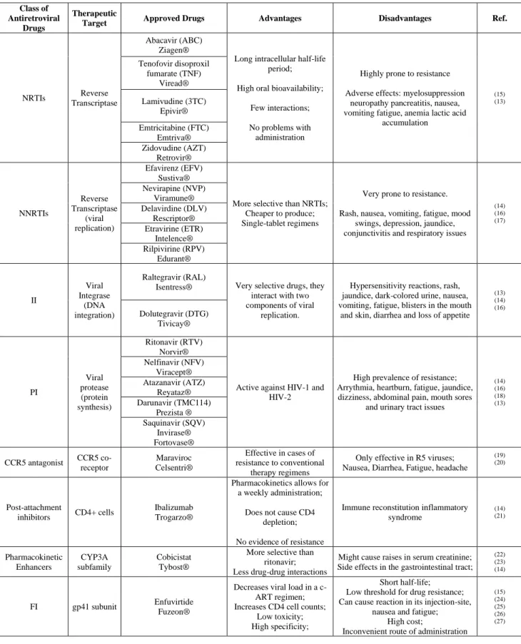
Table 1- Classification of commercial antiretroviral drugs for HIV and therapeutic advantages and disadvantages.
Class of Antiretroviral
Drugs
Therapeutic
Target Approved Drugs Advantages Disadvantages Ref.
NRTIs Reverse
Transcriptase
Abacavir (ABC) Ziagen®
Long intracellular half-life period;
High oral bioavailability;
Few interactions;
No problems with administration
Highly prone to resistance
Adverse effects: myelosuppression neuropathy pancreatitis, nausea, vomiting fatigue, anemia lactic acid
accumulation (15) (13) Tenofovir disoproxil fumarate (TNF) Viread® Lamivudine (3TC) Epivir® Emtricitabine (FTC) Emtriva® Zidovudine (AZT) Retrovir® NNRTIs Reverse Transcriptase (viral replication) Efavirenz (EFV) Sustiva®
More selective than NRTIs; Cheaper to produce; Single-tablet regimens
Very prone to resistance.
Rash, nausea, vomiting, fatigue, mood swings, depression, jaundice, conjunctivitis and respiratory issues
(14) (16) (17) Nevirapine (NVP) Viramune® Delavirdine (DLV) Rescriptor® Etravirine (ETR) Intelence® Rilpivirine (RPV) Edurant® II Viral Integrase (DNA integration) Raltegravir (RAL)
Isentress® Very selective drugs, they interact with two components of viral
replication.
Hypersensitivity reactions, rash, jaundice, dark-colored urine, nausea, vomiting, fatigue, blisters in the mouth
and skin, diarrhea and loss of appetite (13) (14) (16) Dolutegravir (DTG) Tivicay® PI Viral protease (protein synthesis) Ritonavir (RTV) Norvir®
Active against HIV-1 and HIV-2
High prevalence of resistance; Arrythmia, heartburn, fatigue, jaundice, dizziness, abdominal pain, mouth sores
and urinary tract issues
(14) (16) (18) (13) Nelfinavir (NFV) Viracept® Atazanavir (ATZ) Reyataz® Darunavir (TMC114) Prezista ® Saquinavir (SQV) Invirase® Fortovase® CCR5 antagonist CCR5 co-receptor Maraviroc Celsentri® Effective in cases of resistance to conventional therapy regimens
Only effective in R5 viruses; Nausea, Diarrhea, Fatigue, headache
(19) (20) Post-attachment inhibitors CD4+ cells Ibalizumab Trogarzo®
Pharmacokinetics allows for a weekly administration;
Does not cause CD4 depletion;
No evidence of resistance
Immune reconstitution inflammatory syndrome (14) (21) Pharmacokinetic Enhancers CYP3A subfamily Cobicistat Tybost®
More selective than ritonavir; Less drug-drug interactions
Might cause raises in serum creatinine; Side effects in the gastrointestinal tract;
(22) (23) (14)
FI gp41 subunit Enfuvirtide Fuzeon®
Decreases viral load in a c-ART regimen; Increases CD4 cell counts;
Low toxicity; High specificity;
Short half-life; Low threshold for drug resistance; Can cause reaction in its injection-site,
nausea and fatigue; High cost;
Inconvenient route of administration
(15) (24) (25) (26) (27)
2.1. Nucleotide/nucleoside Reverse Transcriptase Inhibitors (NRTI)
The first ones to be developed were the Nucleotide/Nucleoside Reverse Transcriptase Inhibitors (NRTI). Their structure is very similar to the one of the viral nucleosides, except for the absence of a hydroxyl group in the 3’ position of their deoxyribose sugar, thereby preventing a phosphodiester bond between the NRTIs and the next 5’nucleosides. As a result, the nucleoside chain is interrupted and the proviral DNA is not formed. These drugs are formulated and delivered as prodrugs, which need to be phosphorylated in order to become active (13,16). NRTIs are classified as competitive inhibitors of the RT, as they bind to the enzyme’s active site being integrated into the viral DNA chain (18).
Regarding pharmacokinetics, this class of drugs is known to have a long intracellular half-life period, good oral bioavailability, no restrictions of administration and improbable interactions with other drugs (15).
On the other hand, few side effects have been documented regarding the NRTIs. Some of them include myelosuppression, pancreatitis and neuropathy. Also, the NRTIs are very prone to inducing resistance mechanisms (13,15).
The first NRTI to be approved was Zidovudine (AZT) in 1987. The last one to be approved was Emtricitabine, in 2003(28).In 1996, studies had already proven the efficacy of using a combination of antiretroviral drugs as opposed to monotherapy. Nowadays, any initial therapy regimen must have at least three different drugs with distinct therapeutic targets (14).
2.2. Non-nucleoside Reverse Transcriptase Inhibitors (NNRTIs)
When it comes to reverse transcriptase inhibitors, there are also the Non-nucleoside Reverse Transcriptase Inhibitors (NNRTIs), which bind directly to the HIV RT in a non- competitive inhibition process (14). NNRTIs bind to a specific pocket of the viral RT, away from the RT’s active site, thereby inducing a conformational change that inhibits the enzyme’s activity (18). NNRTIs do not inhibit the RT of any other retroviruses, nor the one of HIV-2 (16). NNRTIs are quite cheap to produce and permit single tablet regimens (17).
As for adverse effects, NNRTs can cause, among others, rashes, nausea, vomiting, fatigue, mood swings depression, jaundice, conjunctivitis and respiratory issues (14).
Just like the NRTIs, NNRTIs are very prone to inducing resistance. A simple change in an aminoacid in the NNRTI binding pocket of the RT can lead to great resistance, with very little changes in viral replication (16).
The first NNRTI to be approved was Nevirapine (NVP), in 1996, the same year when combined antiretroviral therapy (c-ART) was first introduced (17). The most recent drug to be approved in this class was Doravirine approved in November 2018 for use across the European Union (29).
2.3. Integrase Inhibitors (IIs)
In the last decade, the viral integrase has been successfully used as a therapeutic target. The integration of viral DNA into the host’s genome is composed of two steps: the first one occurs when integrase cleaves the nucleotides at the 3’ end of the host DNA strands creating “loose ends”. This is called the 3’ processing reaction. The second step of integration is the strand transfer reaction, where integrase, allows the transfer of viral DNA to the nucleus, where it will be incorporated into the host genome (13).
The integrase inhibitors designed until now are really strand transfer reaction inhibitors (INSTIs). These inhibitors are quite selective drugs since, through a very unique biochemical mechanism, they interact with the co-factors (metallic cations) at the enzyme’s active site as well as with the complex formed by viral DNA and integrase. The pharmacophore binds to the metallic cations whereas a lipophilic group interacts with the viral DNA-integrase complex (16).
Regarding side effects, INSTIs can cause hypersensitivity reactions, rash, jaundice, dark-colored urine, nausea, vomiting, fatigue, blisters in the mouth and skin, diarrhea and loss of appetite (14).
Raltegravir (RAL) was the first integrase inhibitor in 2007. The most recent approval in this class of antiretroviral drugs was Elvitegravir in 2014 (17). However, in 2016, the drug’s marketing authorization for the European Union was withdrawn for commercial reasons (30).
2.4. Protease Inhibitors (PIs)
As mentioned in the first chapter, the viral protease is a crucial component of the viral replication cycle, because it is responsible for the processing (cleavage) of several
precursors, such as gag-pool or gag into mature proteins and thus, the formation of mature virions (13,16).
Protease inhibitors bind to this enzyme in a competitive manner in the same place as its natural substrate. The inhibition of this enzyme’s activity leads to immature virions and less viral spreading (16,18).
The main issue with these antiretroviral drugs is that the protease gene, despite being quite small, is very prone to mutation, which can very easily lead to resistance. Nevertheless, resistance to PIs is largely a result of the build-up of both primary and compensatory mutations acquired in order to restore viral capacity (16). These mutations can occur in the protease gene or at the protease’s substrate cleavage sites (16).
Regarding adverse reactions, PIs are known to cause arrhythmia, heartburn, fatigue, jaundice, dizziness, abdominal pain, mouth sores and urinary tract issues such as kidney stones and dark-colored urine (14).
Saquinavir (SQV) was the first PI to be approved in 1995 by the FDA and 1996 by the EMA (29,31). The most recent approval in this drug category was Darunavir, approved by the FDA in 2006 and by the EMA in 2007 (28,29).
2.5. Fusion inhibitors: Enfuvirtide
Enfuvirtide is the first drug to be approved as a fusion inhibitor. The fusion between viral envelope and CD4 cell membrane is, as discussed previously, mediated by the Envelope Glycoprotein (Env), which is composed by the gp120 and gp41subunits. On one hand, gp120 enables receptor and co-receptor binding, whereas gp41 allows for the concrete fusion of both viral envelope and cell membrane (24).
Gp41 is the enfuvirtide’s main therapeutic target. The intracellular area is formed by a cytoplasmic tail (Cyt). Its extracellular region is composed of several subdomains such as the fusion peptide (FP), the fusion peptide proximal region (FPPR), the N-terminal heptad repeat (NHR), the C-terminal heptad repeat (CHR), the membrane proximal external region (MPER) and the transmembrane domain (TMD) (24,25).
The connection of gp120 to the target cell co-receptors, either CCR5 or CXCR4, brings about changes in the gp41 configuration. The FP will now be unprotected and will then enter the target cell membrane, originating a prehairpin structure, which will then shift
its conformation to become a “hairpin- like structure”(24,25). Afterwards, the three CHR domains are rearranged into the three NHR domains’ trimeric “coiled-coil” structure in an anti-parallel sense, forming the 6 helix-bundle (6HB) or “core” (24,25).Structurally, enfuvirtide is a synthetic peptide partially similar to the CHR region. This gp41 component can be sub-divided into an NHR-binding domain which is analogous to Fuzeon®, and a pocket-binding domain (PBD), with hydrophobic pockets that bind to the NHR exterior (24).
Fuzeon® acts by stopping the connection between NHR (also referred to as HR-1(26)) and CHR (also referred to as HR-2 (26)) thereby preventing the formation of the 6-HB and ultimately the membrane fusion with the viral envelope. Nevertheless, Fuzeon® is believed to interfere with other regions of gp41, given that other similar peptides still seem to decrease the viral load of some Fuzeon® resistant strains (24).
Enfuvirtide was approved in 2003 as a treatment option for patients infected with HIV-1 (given that it is not effective in treating HIV-2), who are in an advanced stage of disease progression and are resistant to other antiretroviral drug classes (26). It is taken by subcutaneous injection and the standard regimen for an individual older than 16 is 90 mg twice a day (27).
The TORO clinical trials demonstrated a few benefits in the administration of enfuvirtide in cases where patients have shown resistance to therapy with NRTIs; NNRTIs and PIs, with significant decreases in viral load and increases in CD4 cell counts (27).
TORO compared viremia and CD4 cell counts in patients taking both enfuvirtide and an optimized conventional ART regimen versus the conventional ART regimen on its own. After 48 weeks of treatment, 30% of the enfuvirtide patients had their viremia reduced to <400 copies/ml versus 12% in the conventional therapy arm. At a 95 week follow-up, 17.5% of patients taking enfuvirtide had their viral load at under 50 copies/ml (27). Despite being a promising therapeutic option, Fuzeon® has few disadvantages. On one hand, it has an inconvenient route of administration, which might bring about issues with adherence to therapy (24,27). On the other hand, it has quite a low threshold for resistance, which can limit its use in a clinical setting. For the most part, resistance to this drug occurs due to mutations in the HR-1 region of gp41, even though some cases are explained by mutations in HR-2 (15,27).
The most common side effects associated with enfuvirtide use are reactions near the injection site, nausea, diarrhea, fatigue and eosinophilia (27).
2.5.1 T20 derived lipopeptides
Despite having a different mechanism of action from other ARV drugs, T20 has limited clinical use nowadays for several reasons. Firstly, T20 has a very short half-life, which leads to an inconvenient posology (90mg twice a day via subcutaneous injection). Also, it has a very low antiretroviral activity and is only active against HIV-1. Finally, T20 has a very low threshold for resistance (25,32).
In order to surpass these issues, several T20 based lipopeptides have been synthesized over the years. A few examples include LP40, LP46, LP52 and LP80. LP46 was synthesized based on another lipopeptide, T1249, which was tested as a second-generation fusion inhibitor. Although it presented higher antiviral activity than T20, its phase II clinical trial was interrupted due to reports of serious side effects (33). In order to produce LP46, T1249’s tryptophan rich motif at the C-terminal (TRM) was replaced with palmitic acid, a C16 fatty acid (25,32). The result was a T20-derived lipopeptide with a much lower IC50 against not only T20 resistant strains, but also some HIV-2 strains and two SIV strains (32). Also, LP46 was found to be more stable than T20 and with higher affinity to the target site (32).
Taking this into account, other T20 derived lipopeptides have surfaced, such as LP52. This molecule was designed based on LP46 and LP40 exhibiting a higher threshold for drug resistance and capable of inhibiting T20 resistant strains. Additionally, it has a longer half-life and higher antiviral activity with IC50 values as low as picomolar (pM)
(33).
In a more recent study (34), several new lipopeptides were developed, using fatty acids with different lengths. LP80, with an 18 carbon long fatty acid chain, was found to be a quite potent fusion inhibitor against T20 resistant strains, according to a single- cycle infection assay. It showed cytotoxicity as LP52 and T20 when tested with PBMCs and different cell lines, such as TZM-bl cells, HEK293T cells and MT-4 cells. It also showed favorable results in pharmacokinetics studies in healthy rhesus macaques (34). When administered at 3mg/kg to rhesus macaques chronically infected with simian-human immunodeficiency virus (SHIV), once a day for two weeks, viral load was
reduced to below the detection limit after 4 days of treatment, in 3 out of 5 monkeys in the study (34).
2.6 Pharmacokinetic enhancers: Cobicistat
Ever since combination antiretroviral therapy (c-ART) was firstly implemented in 1996, it is known that PIs and NNRTIs are metabolized by isoenzymes belonging to the CYP450 family, and therefore, interfere with the metabolization process of other very commonly taken drugs (23).
Also in 1996, it was proven that ritonavir could constrain the metabolization process, and therefore, allow for higher concentrations of other antiretrovirals and larger intervals in their administration. Thus, ritonavir started to be progressively co-administered with other first generation PIs such as lopinavir or saquinavir, and afterwards with second generation PIs, such as atazanavir and darunavir, always at sub- therapeutic doses of 100mg (23).
Over the years, investigators came to the conclusion that ritonavir is capable of interfering with several enzyme subfamilies, such as CYP2C19, CYP2C9 CYP2D6 and P-glycoprotein, being responsible for a huge number of interactions with other drugs as well as very unpleasant adverse effects regarding the gastrointestinal tract (23). A few years ago, in 2012, cobicistat was approved as a pharmacokinetic booster in co-administration with other antiretrovirals. Contrarily to ritonavir, it has no specific antiretroviral activity. However, it is substantially more selective than ritonavir, considering that it inhibits only the CYP3A isoenzyme subfamily, which makes patients less prone to drug interactions (22,23).
By inhibiting the CYP3A isoenzymes, cobicistat allows an increasing in the plasma concentrations of other antiretrovirals, such as PIs and NNRTIs, thereby enabling higher intervals in administration with a lesser pill burden and improved adherence to therapy (23).
Some clinical trials showed an increase in serum creatinine levels and a decrease in glomerular filtration rate (GFR) in patients taking cobicistat. Nevertheless, in a phase I study, these abnormalities were explained by cobicistat use causing a decline in the activity of certain transporters, such as SLC22A2, which are essential for the elimination of creatinine. This means that cobicistat causes higher serum creatinine levels, thereby altering the GFR estimation calculated using the Cockcroft-Gault
formula. Thus, cobicistat does not really alter glomerular filtration (22,23). It is now known that this drug restrains some cation renal transporters. However, no dosage adjustments are required for patients with a defective liver or kidney function (22). Regarding CYP3A4 inducers such as carbamazepine and rifabutin, cobicistat is not as effective as ritonavir when administered simultaneously with these drugs, since it mostly metabolized by CYP3A4 and CYP2D6 (22).
2.7 CCR5-antagonist
The CCR5-antagonist Maraviroc (MVC), was approved by both the Food and Drug Administration (FDA) in the US and the European Medicines Agency (EMA) in Europe in 2007 (29). CCR5- antagonists were the first ever class of antiretroviral drugs to target host cells and not virions (19). Their advent rose from the need to introduce other drugs with different therapeutic targets, in order to solve the drug resistance with other molecules (19).
As already discussed, CCR5, along with CXCR4, is one of the co-receptors present in TH4 lymphocytes that in turn allows the HIV virions to attach to it, and thereby, enter the immune cells. CCR5 is also the most common co-receptor, being present in the majority of target cells, which makes it a very promising therapeutic target (19).
MVC will bind to the co-receptor’s hydrophobic pocket and induce a conformational change avoiding its recognition by viral gp120, thereby limiting virion entry into peripheral blood mononuclear cells (PBMC) (13,35).
MVC is mostly metabolized in the liver by CYP450 enzymes, such as CYP3A4 and CYP3A5, which is why it should be administrated cautiously with other CYP450 inducing or inhibiting drugs, such as PIs (35).
As previously mentioned, in the early stages of HIV progression, CCR5 is the dominant co-receptor. However, as the disease progresses, CXCR4 becomes more and more frequent, and therefore, MVC will not be as effective as in the early stages (19), Furthermore, it is known that some mechanisms of resistance involve mutations which cause a higher affinity of gp120 towards CCR5 already connected to MVC (35). As for adverse effects, among the most common ones are nausea, diarrhea, fatigue and headaches (20).
At least, some examples of combination antiretroviral therapy (c-ART) can be seen in
Table 2.
Table 2- Examples of combination antiretroviral therapy for HIV. Approved
Drugs Active Substances References
Genvoya® 150mg elvitegravir/ 150 mg cobicistat/ 200mg emtricitabine/ 10
mg tenofovir (36)
Atripla® 600mg efavirenz/ 200mg emtricitabine/ 245mg tenofovir-DF (37)
Rezolsta® 800mg darunavir/ 150mg cobicistat (38)
Triumeq® 50mg dolutegravir/ 600mg abacavir/ 300mg lamivudine (39)
Evotaz® 300mg atazanavir / 150mg cobicistat (40)
Descovy® 200mg emtricitabine / 10mg tenofovir alafenamide
3. New Drugs and Vaccines – Overview of Phase III Clinical Trials
Nowadays the wide range of therapeutic options and c-ART regimens available have allowed for HIV to become a chronic infection, rather than a deadly condition, as it was considered initially (42). Although c-ART is the best therapeutic strategy at the moment, is not the ideal solution, given that patients experience several side effects and are forced to life-long medication with a considerable risk of emergence of a resistant viral strain and rebound in viral load due to the virus’ reservoir (43,44).
When discussing a cure for the HIV virus, it is important to establish the difference between a “sterilizing cure” and a “functional cure”. A “sterilizing cure” consists of the elimination of both active infected cells and the virus’ latent reservoir, whereas a “functional cure” implies a prolonged suppression of viremia to an undetectable level and sustaining a normal CD4+ cell count (42).
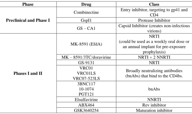
At the moment there are several new drugs and vaccines being tested in pre-clinical and clinical trials for HIV treatment or prophylaxis. In this chapter, most recent clinical studies on phase III will be further discussed.
3.1 Post-attachment inhibitors: Ibalizumab
Ibalizumab was approved in March 2018 by the FDA and granted a “breakthrough therapy status” in 2015 (45). Its approval by the EMA is still pending at this time, although the agency has issued a positive opinion, recommending the granting of a marketing authorization for this drug on June 25th 2019 (46).
At the moment, Ibalizumab is approved for treatment in combination with other drugs and in non-naïve patients, who have already been treated with other classes of antiretrovirals unsuccessfully, due to drug resistance. It is the first drug to target the CD4 receptor, and therefore, the first to be considered a post-attachment inhibitor (45,47).
In molecular terms, as an IG4 monoclonal antibody (IG4 mAb), ibalizumab connects to domain 2 of the CD4 receptor and leads to conformational changes, thereby preventing HIV virions from entering the host cells (48).
In order to a viral particle enters a host cell, HIV must firstly bind to the T-cell through the CD4 T cell receptor. This is followed by the previously described interactions and conformational changes in the gp120 protein and its connection with the co-receptors
(48). Ibalizumab, in its turn, induces conformational changes in the CD4 receptor which avert the gp120 – co-receptor interactions, and consequently preventing viral entry. This action mechanism also explains why ibalizumab is effective against both R5 and X4 viruses (48).
There are few particular aspects that make ibalizumab an innovative therapeutic option. It does not cause CD4 depletion, due to important factors. Firstly, the specific epitope of the CD4 receptor to which it binds is distant from the allocation site of the MHC II molecule (21,48). Furthermore, ibalizumab has an IgG4 structure, which means that it has very low ADCC (anti-body dependent cellular cytotoxicity), hence a very low rapport towards cytotoxic immune responses. Therefore, CD4+ cells will not be destroyed through cytotoxic pathways (48).
At the moment, the FDA recommends the subcutaneous administration of 2000mg loading dose, and subsequently, 800mg maintenance dose every 2 weeks (45).The drug’s half-life is estimated to range between 3 and 3.5 days (21,48). Ibalizumab is generally well tolerated, with adverse effects such as rash, headaches, nausea and depression being reported in a very low percentage of patients (48).
In summary, ibalizumab presents as a very promising and safe therapeutic option for patients suffering from resistance to the recommended ARV regimens, and might also be more convenient when it comes to administration intervals, allowing for higher adherence to therapy and patient autonomy (21).
3.2 Long acting (LA) injectable Cabotegravir/Rilpivirine formulation
Cabotegravir (CR) is an INSTI, structurally similar to dolutegravir (49,50). It is currently being developed as a tablet, to be administered once daily as well as a long acting injectable formulation, in combination with rilpivirine (50,51). CR’s half-life is particularly long, ranging between 20 to 40 days (50). CR is strongly bound to albumin and eliminated through the liver, however no dose adjustments are required for patients with an impaired hepatic function. (50). Rilpivirine is a NNRTI. It has also a long half-life being a long-acting formulation and ranging from 30 to 90 days. It can cause headaches, nausea, dizziness and fatigue and it might also interfere with liver and pancreatic enzymes (50).
Recently, the results from Phase III clinical trials on this formulation have been published (51). There are presently two trials regarding the impact of this formulation: FLAIR and ATLAS (50).
FLAIR is an open-label Phase III trial. All the accepted patients were ART-naïve. Aside from K103N, none of them presented any other NNRTI resistance associated mutations (RAMs) (50).
This trial was divided into two phases. The induction phase lasted for 20 weeks in which all patients took the oral fixed dose combination (FDC) of abacavir/lamivudine/dolutegravir. At week 20 began the maintenance phase, and all patients with less than 50 copies/ml were randomly distributed as to either remain with the initial therapy or start a different oral therapy regimen with 30mg cabotegravir + 25 mg rilpivirine once a day for 4 weeks. Following the 4 week period, the patients were given a loading dose of 600mg cabotegravir + 900mg rilpivirine intramuscularly and afterwards a maintenance dose of LA cabotegravir 400mg + 600mg LA rilpivirine once a month (50,52,53).
After 48 weeks, the assessment showed that 7 patients out of 283 doing oral therapy (2.5%) had over 50 copies of viral RNA/ml as opposed to 6 patients out of 283 taking the LA formulation (2.1%). However, there were patients in both arms of the trial that abandoned the study due to ineffectiveness and few other patients had confirmed virologic failure, which means that these drugs failed in keeping viral load under 200 copies/ml. These results were enough to prove non-inferiority of LA injectable therapy in relation to oral therapy (47,50,52,54). Patients were generally satisfied with this new therapy regimen and injection site reactions (ISRs) were mostly mild and constrained (50).
For the ATLAS study, also open-labeled and randomized, all the participants followed an oral therapy regimen with 2 NRTI + 1 INSTI, NNRTI or PI for a minimum of six months and all of them had less than 50 copies/ml of HIV RNA. The patients were distributed randomly to either continue with their usual regimen or enter the LA therapy arm. Similar to the FLAIR study, LA therapy patients received 30mg LA cabotegravir + 25mg LA rilpivirine for 4 weeks. Afterwards, they were given through intramuscular injection 600mg of LA cabotegravir + 900mg LA rilpivirine as a loading dose and a
maintenance dose of 400mg LA cabotegrvir + 600mg rilpivirine, taken once a month, through the same route of administration (50,55,56).
The results showed that after 48 weeks only 5 out of 308 patients doing LA therapy had over 50 copies of viral RNA/ml (1.6%), compared to 1.0% (3/308) in the control group. This means that LA cabotegravir + rilpivirine was considered non-inferior to triple oral therapy. Contrary to the FLAIR study, there was a higher incidence of adverse reactions (47,50).
With both studies, it has been proven that LA injectable therapy has significant advantages when compared to oral therapy, like the low frequency of the injections. Most patients found this option preferable, however, there are still some concerns about this treatment option, such as viral resistance, how to proceed when they miss an injection or the possibility that this type of regimen might make patients less compliant to a medication schedule (50). Additionally, it is known that rifampicin activates CR’s metabolism, reducing its concentration in the plasma. This might cause a problem in HIV positive patients co-infected with Tuberculosis (TB), since rifampicin is commonly used to treat it (50).
Despite these promising results, there are still some questions to be answered, such as LA therapy’s effect on women, and its use during pregnancy. At the moment, there are ongoing studies, such as MOCHA aimed to provide information regarding the pharmacokinetics and safety of using LA CR on adolescents from 12 to 18 years old and ALTAS 2M, which is looking into a different regimen of 600mg LA CR + 900mg LA rilpivirine every 8 weeks (50).
3.3 Fostemsavir
Fostemsavir was developed as a prodrug of temsavir, an attachment inhibitor. Its mechanism of action is innovative, as it connects to the viral glycoprotein gp120. Gp120 is then unable to bind to the CD4 receptor, and therefore viral entry is prevented (49,57,58). At the moment, the BRIGHTE study is an ongoing phase III clinical trial assessing the efficacy of this new drug. All participants are infected with multi-resistant HIV-1, not responding to therapy, and with a viral load of over 400 copies/ml (58,59). Patients were assigned into two cohorts: one randomized (RC) and another non-randomized (NRC). In the RC, patients were given either placebo or 600mg fostemsavir twice a day along with their failing ART drugs for 8 days. From the 9th day forward all
patients received 600mg fostemsavir twice daily along with optimized background therapy (OBT), just like the patients in the NRC (58,59).
The results showed that there was a low prevalence of adverse effects, being the most frequent headaches, nausea and diarrhea (58,59). However, there were few patients who experienced more severe adverse effects such as acute renal failure and hepatocellular injury, among others (58). Fostemsavir has no registered major interactions with other classes of ARV drugs (58). Nevertheless, it is still needed more information about this new drug’s potential side effects and interactions. After 24 weeks, 146 out of 272 patients in the RC (54%) had less than 40 copies of viral RNA/ml. In the NRC, the percentage of patients who had their viral load less than 40 copies/ml was 36% (36/99) (58,59).
3.4 Leronlimab (PRO 140)
Leronlimab is a humanized IgG4 monoclonal antibody. Its target is the CCR5 chemokine receptor, since it binds to the extracellular domains of this molecule, such as loop 2 and the N-terminus (60,61). By this way, PRO 140 prevents virions from entering and infecting other host cells (60,61). Besides PRO 140, maraviroc’s therapeutic target is also the CCR5 co-receptor. However, there are few differences between these two drugs. Maraviroc binds to the transmembrane hydrophobic pocket of the co-receptor (60), and this difference in binding sites suggests that these drugs can be used synergistically together (62).
There are currently ongoing phase IIb/III trials aiming to evaluate the safety and effectiveness of leronlimab. On one hand, there is CD 02, which evaluates leronlimab’s efficacy in patients with an unsuccessful ART regimen. They were given either 350mg of PRO 140 subcutaneously or placebo for 1 week along with their regular medication. One week later, participants were given PRO 140 with an optimized regimen. Unfortunately, the results were not very promising, since the failure rate was 76% due to the emergence of X4 viral strains, for which PRO 140 was ineffective (60). On the other hand, there is CD 03, which is currently recruiting participants. This trial intends to test the transition of stable patients being treated with conventional therapy into a weekly regimen of subcutaneous leronlimab in monotherapy, by assessing the patients’ viral load for 48 weeks (63).
Nevertheless, this drug’s high tolerability and high threshold to resistance might make it suitable for pre and post exposure prophylaxis (60).
3.5. UB-421
UB- 421 is an Fc-aglicosylated humanized IG1 antibody that targets the CD4 receptor. It prevents attachment and viral entry by attaching to domain 1 of the receptor and blocking it in a competitive manner (64).
At the moment, UB-421’s safety and efficacy are being assessed in several clinical trials. One of them is a phase II/III trial that started in September 2019. In this study, UB-421 is set to be assessed along with a failing ART regimen in an initial stage, which will last one week, and later, it will be evaluated with an optimized background regimen for 24 weeks. The primary outcome measure will be the viral loading log10 difference from the baseline (65).
Additionally, there is a phase III study set to start in January 2020, in which the main goal is to evaluate the drug’s efficacy, safety and tolerability as a monotherapy agent. Participants will be distributed into two cohorts. Cohort 1 will consist of a control group taking standard c-ART, while in cohort 2 patients will take only UB-421 at 25mg/kg intravenously for 26 weeks bi-weekly. All patients will then enter a follow-up phase receiving standard c-ART. The primary outcome measure will be the number of patients without virologic failure (66).
3.6 Vaccines
The eminent need for an effective vaccine against HIV has been discussed over several years. However, despite various attempts, this goal has not been achieved yet. To this date, there have been few effective clinical trials for vaccines against HIV. The most promising one was a phase III RV144 trial, but it showed a low level of protection against the virus (67,68). One the main reasons for this viral resistance is based on the elevated variability of this virus which hinders finding a target that is common to so many different strains (67,69).
Considering this extreme viral variability, a vaccine design based on live inactivated virus or attenuated virus may not be a viable strategy. Alternatively, the vaccines that have been currently tested, use attenuated viral proteins (such as gp120) in order to induce an immune response with the production of broadly neutralizing antibodies (70,71).
The target for neutralizing antibodies (nAb) is Env, and consequently, this is a suitable immunogen when designing a vaccine taken into account its role in viral targeting and entry. Another important aspect to consider is the delivery system for the vaccine. For example, nanoparticles have been shown as suitable carriers for vaccines (70,71). The vesicular stomatitis virus has already been tested as a viral vector for expression of heterologous genes from other viruses, such as Ebola and malaria (72). It is now being tested as a vector for the expression of the env glycoprotein of HIV in order to induce the production of neutralizing antibodies (73).
Accordingly, there is an ongoing clinical trial aiming to test a therapeutic vaccine for HIV in which a multiantigen DNA plasmid combined with an IL-12 plasmid (acting as adjuvants), and a vesicular stomatitis virus vector expressing the HIV-1 gag gene are tested. The goal of this vaccine is to increase humoral immune response in infected individuals, and possibly reactivating memory CD4 cells to eradicate latent reservoirs (74).
When designing an HIV vaccine, synthetic proteins that are similar to the env trimer are also quite interesting, since they have been used to attempt the induction of an immune response against HIV. Gp140 is similar to env with exception of the gp120 subunit and the external part of the gp41 subunit (75).
A phase III clinical trial will be set to start later this year to assess the efficacy of a new vaccine containing mosaic antigens, such as env, gag-pol and gp140 (composed of gp120 plus the ectodomain of gp41) obtained from an adenovirus serotype 26 vector (76). This is an innovative challenge to formulate a “global vaccine” and overcome the major obstacle that is genetic diversity of HIV (77).
4. Novel Therapeutic Strategies
4.1 New Transdermal Drug Delivery Systems
Since most ARV are administered orally, it is important to identify the main adversities of oral ART such as bioavailability, frequency of administration and the hepatic first pass metabolism. Transdermal drug delivery systems (TDDS) are currently being investigated in order to diminish these issues (78–80).
Thus, TDDS present as an alternative to the conventional oral ARV regimens, since this administration system allows for a controlled and continuous release of the drug, avoiding the pharmacokinetic variations of the oral administration. This means that it may be possible to use drugs with a short half-life, and find a simpler regimen for patients, and therefore, a higher compliance will certainly be achieved (78,79,81). In order to obtain a successful TDDS, it is important to understand the morphology of the skin. Briefly, the outer layer of the epidermis, the stratum corneum, is composed of dead queratinized cells, whose main function is to act as a barrier for any external agents (78,79).
Transdermal drugs usually penetrate the skin mostly by diffusion and permeate through the stratum corneum, across the lipidic layers of the dermis, and finally, into the microvesssels of the skin. The ability of a certain drug to diffuse through the skin, or its skin permeability, is influenced by its physicochemical properties (78,79). The main ones include the drug concentration, pH, molecular weight (which should be low, ideally around 500 Da), log P (preferably ranged between 1 and 3) and the melting point (78–80).
There are certain components that are essential in order to ensemble a TDDS. Besides the drug itself and its reservoir, a TDDS should have: a) permeation enhancers to increase the permeability through the stratum corneum (e.g. pyrrolidones, surfactants, phospholipids and solvents); b) pressure sensitive adhesives (PSA) to keep contact between the skin and the device; c) a release liner, to cover the patch, which could be considered part of primary packaging, and d) a backing layer which should not allow the diffusion of any excipients (78,79).
4.1.1 Transdermal delivery system for Tenofovir Alafenamide
Tenofovir Alafenamide (TAF) is a NRTI commonly used for HIV treatment as part of oral therapy regimens. A controlled matrix delivery system for this drug has been
studied recently due to its low oral bioavailability (80). In this system, the active substance is dispersed in a polymer matrix and a suitable solvent which later evaporates, forming a drug reservoir. The reservoir is then shaped and interconnected with several layers. The overall system is composed of an adhesive layer that controls the release rate of the drug and is in contact with the skin, followed by the drug reservoir and a second adhesive layer, connected to an exterior impermeable laminate (78–80).
Accordingly, the goal of this study (80) was to formulate a patch able to release TAF at 8mg/day consistently for one week. Two types of patches were produced as silicone based or polyisobutylene (PIB) suspension patches and acrylate solution patches. For each patch, several formulations were prepared varying the components, TAF concentration and permeation enhancers. Each group of patches was evaluated for specific criteria, such as: a) crystallization of TAF in each matrix; b) effect of TAF particle homogenization; c) coat weight and TAF amount in each patch after exposure to stress conditions and d) in vitro skin permeation studies using Franz diffusion cells. Overall, the suspension silicone patch formulated with 15% TAF (w/w), silicone, oleic acid, oleyl alcohol and mineral oil as permeation enhancers revealed to be the most suitable one, since it presented the target flux permeation rate of 7 µg/cm2/h in a 50 cm2 patch area for an entire week, reaching a daily dose of 8.4mg TAF. Additionally, this formulation was stable overtime, non-irritating to the skin and convenient when peeled off (80). Despite these positive results and the promising chance of a new route of administration for TAF, further studies are still needed regarding both pharmacokinetics and safety of this new device (80).
4.1.2 Transdermal delivery of Enfuvirtide (T20) via ultrasounds
As previously discussed, enfuvirtide (T20) is an entry inhibitor, usually administered by subcutaneous injection, 90mg twice a day, which makes it rather inconvenient in terms of patient compliance. Thus, its transdermal delivery could be an interesting option. However, there are some obstacles such as enfuvirtide’s high molecular weight (4492 Da), which could compromise the diffusion through the stratum corneum (79,82). Nevertheless, ultrasound technique could be a way to surpass this problem, since it modifies the barrier function of the stratum corneum, enabling the diffusion of large molecules through the skin (83,84). Ultrasound waves are known to contribute to a fluid state of lipid skin layers, which in turn, promotes the transcellular pathway. Moreover,
ultrasound waves also create pores that allow large molecules to cross the epidermis (84,85).
A recent study has assessed the effects of transdermal delivery of enfuvirtide using a low-frequency and low power ultrasound transducer patch in porcine models. The models were divided into 3 separate groups for 30 days as follows: one control group receiving injectable enfuvirtide twice a day; another group undergoing ultrasound treatment with saline solution and a third one being treated with transdermal enfuvirtide via ultrasounds (82).
During the study, T20 was in direct contact with the skin. The final device was obtained using wound dressing patches and a silicone ring that served as a reservoir, over which the ultrasound transducer was placed (82).
All groups were evaluated in order to understand the differences between them regarding skin health and bioavailability. The skin health criteria were based on histologic cuts and trans-epidermal water loss. Bioavailability of T20 was evaluated through liquid chromatography/electrospray ionization mass spectrometry (LC-MS/MS) (82).
Regarding skin health, there were no significant differences between the saline group and the transdermal T20 group which indicates that ultrasound by itself did not affect the skin. The histologic cuts showed mild signs of inflammation in the active patch group. As for pharmacokinetics, the animals from the active patch group had a longer tmax and a lower Cmax when compared to the injectable T20 group. The plasma concentrations were generally lower with the transdermal treatment as expected (82).
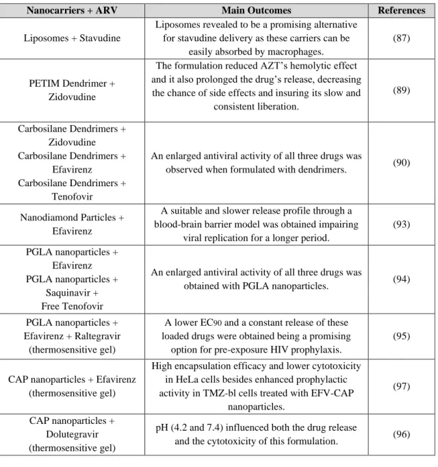
4.2 Nanosystems for Drug Delivery
Nanotechnology has contributed for several applications in drug delivery through different routes of administration being quite useful in overcoming formulation obstacles, such as solubility, bioavailability and stability of the active substance (86). Additionally, nanosystems could be a strategy for targeted therapy since they allow the encapsulation of drugs or specific genes that could be transported not only to infected cells but also to reservoir tissues such as central nervous system (CNS) and lymph nodes, to potentially eradicate the virus (86).