BJRS
RADIATION SCIENCES
07-02A (2019) 01-13ISSN: 2319-0612
Accept Submission: 2018-08-13
Characteristics of Pb
2+doped CsI matrix under gamma
and neutron excitations
Maria da Conceição Costa Pereira, Tufic Madi Filho, José Roberto Berretta, Lucas
Faustino Tomaz and Marlene Cristina Pinto
Instituto de Pesquisas Energéticas e Nucleares (IPEN / CNEN - SP) Av. Professor Lineu Prestes 2242
05508-000 São Paulo, SP – Brazil
ABSTRACT
In recent years, there has been an increasing interest in finding new fast scintillating material or improve the characteristics of known scintillators for the demand of high energy physics, industrial and nuclear medical ap-plications. Divalent lead ions Pb2+ built in some crystal structures are efficient emission centers and their
appli-cations in scintillators were and are still the reason of an intensive study of emission properties of different com-pounds containing these ions. In this context, the crystals of Pb2+ doped CsI matrix were grown by the vertical
Bridgman technique and subjected to annealing in vacuum of 10-6 mbar and constant temperature of 350°C, for
24 hours, and then they were employed. To evaluate the response of the CsI:Pb scintillator crystal to gamma radiation, radioactive sources of 137Cs (662 keV), 60Co (1173 keV and 1333 keV), 22Na (511 keV and 1275 keV)
and 133Ba (355 keV) were used. The operating voltage of the photomultiplier was 2700 V for the detection of
gamma rays and the accumulation time in the counting process was 600 s. The scintillator response to neutron radiation from a radioactive source of AmBe with energy range of 1 to 12 MeV was available. The activity of the AmBe source was 1 Ci Am. The emission rate was 2.2 x 10⁶ neutrons / second. The operating voltage of the
pho-tomultiplier tube was 1300 V. The accumulation time in the counting process was 600 s. With the results obtai-ned, it may be observed that the crystals are sensitive to these radiations.
1. INTRODUCTION
The scintillation method is widely used for detection of various types of ionizing radiation. A scin-tillator consists of material able to convert energy of absorber ionizing radiation into a flash light. The flash light , usually visible or ultraviolet, emitted by a scintillator, is called scintillation. Any scintillator should be transparent to the wavelength of its own emission. The use of scintillation to detect radiation is a century old. The discovery of scintillator materials may be divided into three periods. The first period included the earliest scintillators: CaWO4, first used in the year following
Rontgen’s discovery for X-rays; uranyl salts used by Becquerel to discover radioactivity and by Rutherford to study alpha particle scattering. The second period began with Hofstadter’s deve-lopment of the thallium activated NaI. The third period, the past two decades, has witnessed a veri-table renaissance in research and development of scintillator materials, prompted in large part by the need for scintillators for precision calorimetry in high energy physics and for high light output scintillators for medical imaging, geophysical exploration, tomography, astrophysics and numerous other scientific and industrial applications.[1,2]
There is still considerable interest in finding inorganic materials with perfect scintillation properties or improving the traditional scintillators, including alkali halides. In order to enhance the characte-ristics of a known scintillator or to obtain a new crystal, it is necessary to solve many problems, such as crystalline structure, chemical binding or impurity concentration, which is more advantage-ous for high light output of a crystal.[3]
Scintillators based on cesium iodide are the leading ones among the materials available for radiation detectors. Cesium iodide based scintillators have high luminescence yield accompanied by high energy resolution and radiation stability. Cesium iodide has elements of ionic radius, namely: the cesium ion (Cs+) radius is 1.652 Å, electronegativity 0.7 eV and the iodine ion (I-) radius is 2.256 Å; electronegativity 2.5 eV the equilibrium value lattice separation is 4.512 Å. This facilita-tes the incorporation of a large number of cationic impurities. Various impurities have been inclu-ded into alkali halides and their effects on electrical, magnetic and optical properties have been in-vestigated. The CsI matrix is poorly hygroscopic, its constituents have a high atomic number, are easy to handle and have relatively low cost.[4] Babin et al. [5] mention the interest in CsI: Pb
crys-tals as possible materials for scintillators, since their luminescence is practically not studied. Some data reported by different authors are quite contradictory. This indicates a complexity of this system and a strong dependence on the optical characteristics of the crystals that may be related to the method used for growth. According to Keszthelyi et al. [6], the scintillation characteristics of the crystals are also influenced by the growth method used.
Divalent lead ions Pb2+ in some crystal structures are efficient emission centers and their application in scintillators was and is still the reason of an intensive study of emission properties of different compounds containing these ions. The study of pure and doped cesium iodide has been the theme of search in our laboratory. In continuation, a systematic investigation has been carried out in Pb2+ doped cesium iodide crystals under gamma and neutron excitations and the results are discussed in this paper.
2. MATERIALS AND METHODS
Lead doped CsI crystals were grown using the vertical Bridgman technique. [7,8,9,10,11] The grown crystals were subjected to annealing. In this procedure, vacuum of 10-6 mbar and constant temperature of 350° C, for 24 hours, were employed. The crystals were cut into cylindrical form and their faces were polished with ethylene glycol p.A. (C2H6O2). The side surfaces were left
unpol-ished to enhance internal reflection.
The luminescence decay time of each crystal was determined under excitation of 662 keV gamma rays from a 137Cs source. The scintillation photons were detected using a bi-alkaline photomultiplier tube (model RCA 8575, 21 pins). The conformation of the signal from the photomultiplier tube was obtained using the RC (resistor-capacitor) circuit having a time constant of approximately 5 ns and fed directly to the oscilloscope (Philips PM3295A), resulting in the measurement of the decay of the scintillation pulses. In Fig. 1 the electronic system used for luminescence decay time measurements is schematically shown.
Figure 1: Diagram of the electronic system used for luminescence decay time measurements.
In the gamma ray response study, the crystals were cut with dimensions of 20 mm diameter and 20 mm high, polished and coupled directly to the bi-alkaline photomultiplier tube (model RCA 8575, 21-pin) using silicone grease (Dow Corning) of 0.5 McStokes viscosity as optical interface. This ensures a uniform refractive index across the contact surface between crystal and photomulti-plier tube. The sides of the crystal which were not in contact with the photosensor were covered with several layers of polytetrafluoroethylene (PTFE) tape to assure good reflection of light. The radioactive sources were positioned in the center of the upper face of the crystal. Conventinal OR-TEC eletronic modules constitute the electronics for the treatment of the signals from the photomul-tiplier tube. Gamma radiation sources with energies in the range of 335 keV to 1333 keV were used. The operating voltage of the photomultiplier was 2700 V and the accumulation time in the counting process was 600 s. In Fig. 2, the diagram of the measuring system is showed.
Figure 2: Schematic representation of the electronics associated with the CsI:Pb scintillator crystal coupled to the photomultiplier tube.
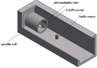
The AmBe source with energy range of 1 to 12 MeV was used to study the neutron radiation res-ponse. The activity of the AmBe source was 1Ci Am. The emission rate was 2.6 x 10⁶ neu-trons/second. The operating voltage of the photomultiplier tube was 1700 V; the accumulation time in the counting process was 1800 s. The scintillator used was cut with dimensions of 20 mm diame-ter and 10 mm high.
The detection efficiency of the scintillator crystal was measured with the AmBe source positioned at a distance of 100 mm from the photomultiplier tube. A foil of cadmium (Cd) was placed around the detector and the photomultiplier tube in order to avoid the scattered neutron contribution, as showed in Fig.3. Paraffin shielding to protect the worker, was used.
Figure 3: Exploded schematic representation of the array used for measurements.
3. RESULTS AND DISCUSSION
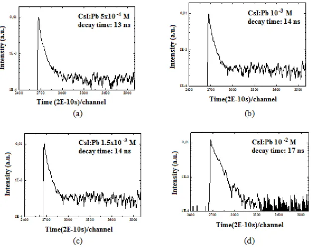
In Fig. 5, the luminescence decay curves of the CsI:Pb crystals are presented. For comparison pur-poses, the luminescence decay constant was determined for the pure CsI crystal, as shown in Fig. 4.
Figure 5: Luminescence decay time spectra of CsI: Pb crystals at various concentrations: (a) 5x10⁻⁴ M, (b) 10-3 M, (c) 1,5x10⁻³ M and (d) 10-2 M.
Fig. 5 and Fig.4 show the luminescence decay curves for the CsI:Pb and pure CsI crystals. As it
may be observed in these curves, there were no significant differences for the various concentrations of lead in the crystals evaluated. The incorporation of the Pb into the CsI matrix, did
not, practically, change the luminescence decay time compared to that obtained for pure CsI, whose values are between 13 ns and 17 ns. These results are very promising, i. g. to obtain crystals having shorter decay time comparable to pure CsI scintillator crystal. The crystal of pure CsI produced in this work, for comparison purposes showed a luminescence decay constant of 12 ns in accordance with the literature [12,13,14].
The luminescence decay time parameter is one aspect of crystal quality, especially for applications requiring high radiation rates or high energy radiations. The luminescence decay time results found in this work are encouraging since, with the introduction of dopant ions such as Tl+ or Na+ in the CsI matrix, it results in increased luminescence decay time for 1 s and 630 ns respectively. The CsI:Tl crystal is one of the most efficient scintillators in gamma ray detection per unit volume, but the long decay time, in the order of 1s, is one of its disadvantages, increasing the probability of oc-currence of overlapping signals, when more intense radioactive sources are used. The decay time in the order of 1s, practically makes it unfeasible rapid temporal studies, such as those of the decay structure performed in nuclear physics. The CsI: Na crystal presents good light production, but the luminescence decay time is still high for specific applications and its main disadvantage is the hy-groscopy [1,2].
Fig. 6 to 9 show the gamma radiation spectroscopy results of 137Cs (662 keV), 60Co (1173 keV and 1333 keV), 22Na (511 keV and 1275 keV) and 133Ba (355 keV) obtained with the CsI:Pb crystals. It
may be observed that the crystals are sensitive to these radiations; however, the photopeak of these radionuclides have not been identified.
Figure 6: Spectra obtained for the radiation of
137Cs, 60Co, 22Na and 133Ba sources with CsI:Pb
5x10⁻⁴ M crystal.
Figure 7: Spectra obtained for the radiation of
137Cs, 60Co, 22Na and 133Ba sources with CsI:Pb
Figure 8: Spectra obtained for the radiation of
137Cs, 60Co, 22Na and 133Ba sources with CsI:Pb
1.5x10⁻³ M crystal.
Figure 9: Spectra obtained for the radiation of
137Cs, 60Co, 22Na and 133Ba sources with CsI:Pb
10-2 M crystal.
From the curves obtained for the CsI:Pb crystals, using gamma radiation from the sources of 137Cs,
60Co, 22Na and 133Ba (Figs. 6-9) it was not possible to extract the resolution of these detectors. The
low resolution capacity of the crystals of CsI:Pb may be attributed to the self- absorption of the lu-minescence in the crystals themselves, due to the presence of Pb in their structures. However, for applications where the energy resolution is not a relevant item, the CsI:Pb crystals may be used when a radiation detector with good mechanical resistance, relatively low cost of production and different sizes and geometries is desired. In electromagnetic calorimeters, the need for large-scale high-energy particle detectors has led to international collaboration to develop, optimize and pro-duce scintillators for this purpose. The Compact Muon Solenoid (CMS) experiment in the Large Hadron Collider (LHC) accelerator at CERN uses approximately 80,000 PbWO4 (lead tungstate)
crystals, totalling approximately 100 tonnes [15]. Another project, ALICE, also in the accelerator (LHC) will use 18,000 PbWO4 crystals, about 13 tons [16]. The crystal scintillator PbWO4 presents
light output of 250 photons / MeV and decay time of 5 to 15 ns [16,17].
Fig. 10 shows of measurements performed using the CsI:Pb crystals with concentrations 10-2 M, 10-3 M and 5x10⁻⁴ M and the neutron source.
Figure 10: Results of the measurements using CsI:Pb crystal and neutron source.
CsI:Pb crystals, when excited with radioactive source of AmBe with energy range of 1 to 12 MeV, showed sensitivity to fast neutron, and the highest counting efficiency was obtained in the concen-tration of 10-2 M.
4. CONCLUSION
The CsI: Pb crystals presented short decay time values, demonstrating the viability of their use for measurements of high dose and high energy rates. These results have shown to be very promising to obtain crystals having short decay time, when compared to the pure CsI scintillator crystal. The crystals of CsI:Pb showed sensitivity to gamma radiation detecting the energies of the radioac-tive sources used, although it was not possible to identify the corresponding photopeak.
The addition of the lead ions (Pb2+) to the CsI matrix resulted in crystals with promising results, when excited with neutron radiation. The crystal showed to be sensitive to fast and thermal neutron. In the dopant concentration range (Pb) studied, the best neutron detection efficiency was obtained for the molar fraction 10-2.
The results showed the feasibility of using the crystals developed in this work for application in high energy physics. In our work environment, they may be applied as area of a neutron detector and used in the personal dosimeter for the workers of the IEA-R1 and IPEN/MB-01 nuclear research reactors.
REFERENCES
1. KNOLL,G. F. Radiation Detection and Measurement, 4th ed. New York, NY, USA:John Wiley & Sons, 2010.
2. TSOULFANIDIS, N. Measurement and Detection of Radiation, New York, N.Y.: Harper
& Row, 1983.
3. ZAZUBOVICH, S. Physics of halide scintillators. Radiation Measurements, v. 33, p. 699-704, 2001.
4. SELVASEKARAPANDIAN, S.; BRAHMANANDHAN, G.M.; MALATHI, J.; JOSEPH, V. Thermoluminescence and photoluminescence studies on gamma irradiated CsI:Pb2+ crystals.
Radiation Effects & Defects in Solids, v.161, n°9, p.559-570, 2006.
5. BABIN, V.; KRASNIKOV, A.; NIKL, A.; NITSCH, K.; STOLOVITS, A.; Zazubovich, S. Luminescence and relaxed excited state origin in CsI:Pb crystals. Journal of Luminescence, v.101, p.219-226, 2003.
6. KESZTHELYI, L.S.; FOLDVÁRI, I.; VOSKA, R.; FODOR, Z.; SERES, Z. Decay time mea-surements on pure CsI scintillators prepared by different methods. Nucl Instrum Methods Phys
Res, v.A303, p. 374-380, 1991.
7. BRIDGMAN, P. W. Proc. Amer. Acad. Arts Sci., v. 60, p. 303-383, 1925.
8. PEREIRA, M.C.C.; FILHO, T.M. Scintillation Characteristics of CsI Crystal Doped Br under Gamma and Alpha Particles Excitation. Materials Sciences and Applications, v. 05, p. 368-377, 2014.
9. PEREIRA, M.C.C.; FILHO, T.M.; HAMADA, M.M. The effect of Pb 2+ dopant in the crystal of CsI and its application as scintillation detector: a study of alpha particles. Radiation Effects and
Defects in Solids, v. 167, p. 921-928, 2012.
10. MARGULIES,M.; WITOMSKI,P.; DUFFAR,T. Optimization of the Bridgman crystal growth process. Journal of Crystal Growth, v.266, p. 175-181, 2004.
11. RAVI, B.; RAJARAJAN,G. Study on growth and optical, scintillation properties of thallium doped cesium iodide scintillator crystal, Oriental Journal of Chemistry, v. 30, p. 581-586, 2014.
12. HAO,G.R.; HUI,Z.S.; CHUAN,Z.Z.; KAN, Z.; FAN, Y.; YING,L.H.; FENG, C.X. Luminescence and decay time properties of pure CsI crystals. Journal of Inorganic Materials, v.32 (2), p.169-174, 2017.
13. AMSLER, C.; GROGLER, D.; FOFFRAIN, W.; LINDELOF, D.; MARCHESOTTI, M.;
NIEDERBERGER, P.; PRUYS, H.; REGENFUS, C.; RIEDLER, P.; ROTONDI, A. Temperature dependence of pure CsI: scintillation light yeld and decay time. Nucl Instrum Methods Phys Res., v.A480, p.494-500, 2002.
14. MIKHAILIK, V. B.; KAPUSTYANYK, V; TSYBULSKYI, V.; RUDYK, V.; KRAUS, H.
Luminescence and scintillation properties of CsI: A potential cryogenic scintillator. Phys Status
15. R. Novoyny, “Inorganic scintillators-a basic material for instrumentation in physics”, Nucl.Instrum. Methods Phys. Res. v.A537, pp.1-5 (2005).
16. CMELCHER, C. L. Perspectives on the future development of new scintillators. Nucl
Instrum Methods Phys Res, v.537, p. 6-14, 2005.
17. KOBAYASHI, M.; USUKI, Y.; ISHII, M.; ITOH, M.; KIKL, M. Further study on different dopings into PbWO4 single crystals to increase the scintillation light yield. Nucl Instrum Methods