João Paulo Caseiro Baixinho
Degree in Cell and Molecular Biology
Development of curcumin lipid
formulations for food applications:
transport, permeability and safety
evaluation on a mucus-secreting intestinal
epithelial cell model
Dissertation to obtain master degree in
Biotechnology
Supervisor: Ana Matias, PhD, IBET
Co-Supervisor: Vanessa Gonçalves, PhD, IBET
“Copyright”
Development of curcumin lipid formulations for food applications: transport, permeability and safety evaluation on a mucus-secreting intestinal epithelial cell model.
João Paulo Caseiro Baixinho, FCT/UNL e UNL
VII
AcknowledgmentsGostaria de agradecer a todos as pessoas que, direta ou indiretamente, contribuíram para a realização deste trabalho.
Em primeiro lugar, à Doutora Ana Matias, por me ter dado a oportunidade de desenvolver este trabalho no laboratório de Nutracêuticos e Libertação Controlada. Agradeço toda a orientação prestada, a sua preocupação e apoio constante.
À Vanessa Gonçalves, agradeço toda a orientação, conhecimento e apoio. Obrigado acima de tudo pela tua disponibilidade.
Agradeço a todos os membros e colegas do laboratório de Nutracêuticos e Libertação Controlada e de uma forma especial:
À Joana Guerreiro, por toda a ajuda prestada nos ensaios celulares. Muito obrigada pela tua disponibilidade e paciência.
Ao Agostinho Alexandre, por todos os momentos passados juntos. Pelas críticas e conhecimento transmitido. Obrigada pela tua preocupação e companhia.
À Liliana Rodrigues por estar sempre disponível para ouvir as minhas dúvidas e problemas. Obrigado pelo teu apoio e assertividade!
À Carolina, Naiara, Ana Nunes e Ana Roda. Obrigada pela vossa amizade e preocupação. Agradeço de uma forma especial aos meus amigos e colegas, Nuno, Pedro e Miguel por todo o apoio e companhia nos momentos mais difíceis. Um especial obrigado ao Miguel pela entreajuda e companheirismo nos mais diversos momentos. Sem dúvida que contigo isto ficou mais fácil!
Um especial obrigado ao Tiago Filipe “Baladeiro” e ao João “Potter” Ribeiro por transformarem
cada dia de Sporting num dia de descontração, diversão e amizade. Obrigado pelas distrações que precisava.
IX
PrefaceThis work was performed at the Nutraceuticals and Bioactives process technology laboratory, Instituto de Biologia Experimental e Tecnológica (IBET), under the scope of the project NanoMaxiSafe (PTDC/AGRTEC/5215/2014), financially supported by the Fundação para a Ciência e Tecnologia (FCT), Portugal. Part of the work presented in this thesis has been included in poster communications during the year.
Poster communication:
J. N. Guerreiro, J. C. Baixinho et al. Improving bioactives intestinal absorption using lipidic
nanodelivery systems in food applications: evaluation of toxicity and bioavailability. 1st UNGAP
Meeting, 2018, Leuven, Belgium.
J. N. Guerreiro, J. C. Baixinho et al.Cytotoxicity and bioavailability evaluation using an in vitro
triple co-culture intestinal cell culture: improving bioactives absorption using novel lipidic nanodelivery systems in food applications. BioBarriers, 27st-28st August 2018, Saarbrucken,
XI
AbstractCurcumin is the main phenolic pigment extracted from turmeric, the powdered rhizome of
Curcuma longa. It has been shown to exhibit antioxidant, antimicrobial, anti-inflammatory and
anticarcinogenic activities however as health promoting agent, curcumin, is limited by its poor solubility in aqueous solution and its low bioavailability and therefore cannot be widely used in food and pharmaceutical processing industry.
The challenge addressed in this work was to produce curcumin formulations to enhance its characteristics and evaluate its permeability, transport and cytotoxicity on a stablished in vitro cell
co-culture model that mimics the intestinal epithelium.
Solid lipid nanoparticles (SLN) and microparticles (SLM) have been visualised as a promising platform on development of formulations for food applications. Since traditional production methods
possess a series of limitations, the processing by “green technologies” like supercritical carbon dioxide
(scCO2) has been widely investigated. Through Particles from Gas Saturated Solutions (PGSS®)
process, beeswax microparticles loaded with curcumin (9:1 (w/w)) were produced and characterized in terms of physicochemical properties: size, morphology, curcumin content and particles dispersion index. Operation process parameters were optimized and defined via response surface methodology and the best response was achieved at 160 bar, 73°C and 10% curcumin load. Under these conditions, encapsulation efficiency was 89.75 ± 2.23 % with a curcumin load of 8.98 % (w/w). Curcumin formulations underwent a digestive process and were tested for their cytotoxicity in Caco-2 monolayer.
A triple co-culture has been established and characterized for use as an in vitro intestinal model.
To closely mimic the intestinal epithelium the production of mucus by the HT29-MTX-E12 cell line cultured together with the Caco-2 enterocytes and M-cells like phenotype was observed. The model was used to evaluate the transport and permeability of free and encapsulated curcumin and its permeability was stablished as a value of 1.0 x 10⁻⁷ cm/s.
Keywords:
XIII
ResumoA curcumina, o principal pigmento fenólico extraído da curcuma apresenta diversas propriedades antioxidantes, antimicrobianas, anti-inflamatórias e anticarcinogénicas, no entanto a sua utilização é limitada pela sua fraca solubilidade e biodisponibilidade no epitélio intestinal humano, o que consequentemente provoca a sua limitada utilização na indústria alimentar e farmacêutica.
O desafio abordado neste trabalho foi produzir formulações de curcumina para despoletar as suas características e avaliar a sua permeabilidade, transporte e citotoxicidade num modelo de co-cultura celular estabelecido para mimetizar o epitélio intestinal.
Partículas lipídicas solidas (SLP) têm sido visualizadas como uma plataforma promissora no desenvolvimento de formulações para aplicações alimentares. Como os métodos tradicionais de
produção possuem uma série de limitações, o processamento por “tecnologias verdes” como o dióxido
de carbono supercrítico (scCO2) tem sido amplamente investigado. Através do processo Particles from
Gas Saturated Solutions (PGSS®), micropartículas de cera de abelha carregadas com curcumina (9:1
(m/m)) foram produzidas e caracterizadas em termos das suas qualidades físico-químicas: morfologia, tamanho, quantidade de curcumina e índice de polidispersão. O processo de produção foi otimizado e definido através da metodologia de superfície de resposta sendo que a melhor resposta foi obtida a 160 bar, 73 °C e 10 % de curcumina. Nestas condições, a eficiência de encapsulação foi de 89,75 ± 2,23 % com uma encapsulação de 8,98 %. As micropartículas foram submetidas a um processo digestivo e testadas quanto à sua citotoxicidade em monocamada de células Caco-2.
Para mimetizar o epitélio intestinal uma co-cultura tripla foi estabelecida e caracterizada observando-se a produção de muco pela linha celular HT29-MTX-E12 cultivada em conjunto com os enterócitos Caco-2 e o fenótipo das células M. O modelo foi utilizado para avaliar o transporte e a permeabilidade da curcumina livre e encapsulada sendo o transporte estabelecido nesta co-cultura como 1,0 x 10⁻⁷ cm/s.
Palavras-chave:
XV
List of ContentsAcknowledgments ... VII
Preface ... IX
Abstract ... XI
Keywords: ... XI
Palavras-chave: ... XIII
List of Contents ...XV
List of Figures ...XIX
List of Abbreviations ... XXV
1. Introduction ... 1
1.1. Food industry – Functional foods and Nutraceuticals ... 1
1.1.1. Bioactive Compounds ... 1
1.1.1.1. Curcumin as a nutraceutical ... 1
1.2. Bioactives formulation ... 3
1.2.1. Nanoemulsion-based delivery systems ... 4
1.2.2. Solid lipid particles ... 4
1.3. Supercritical fluid technology ... 4
1.3.1. Supercritical-based precipitation methods ... 5
1.3.1.1. Particles from Gas Saturated Solutions ... 6
1.4. Concepts and models for bioactives permeability studies ... 6
1.4.1. Bioactive compounds oral administration ... 6
1.4.2. Intestinal composition and permeability ... 7
1.4.2.1. Caco-2 cell model ... 8
1.4.2.2. M Cells ... 8
1.4.2.3. Caco-2/HT29-MTX/Raji B Model ... 9
1.5. Aim and rational of the thesis ... 11
2. Materials ... 13
3. Methods ... 15
XVI
3.1.1. Curcumin Nanoformulations ... 15
3.1.1.1. Nanoemulsions preparation ... 15
3.1.1.2. Solid lipid nanoparticles preparation ... 15
3.1.2. Curcumin microparticles ... 15
3.1.2.1. Melting point measurements ... 15
3.1.2.2. Particles from gas saturated solutions ... 16
3.2. Particles’ characterization ... 18
3.2.1. DSC measurements ... 18
3.2.2. Encapsulation efficiency ... 18
3.2.3. Particle size and morphology ... 18
3.2.4. Curcumin formulations sterilization ... 19
3.3. In Vitro Release of Curcumin ... 19
3.4. In vitro Digestion ... 19
3.4.1. Oral Phase ... 19
3.4.2. Gastric Phase ... 20
3.4.3. Small Intestine Phase ... 20
3.5. In vitro cell-based assays ... 20
3.5.1. Cell Culture and sub culturing ... 20
3.5.1.1. Adherent cells culture ... 20
3.5.1.2. Non-adherent cells culture ... 21
3.5.2. In vitro cell models ... 21
3.5.2.1. Cytotoxicity assay - MTS ... 21
3.6. Triple co-culture validation and morphology ... 22
3.6.1. Alcian Blue assay ... 22
3.7. Curcumin transport assay ... 22
3.7.1. Cell monolayer integrity ... 22
3.7.2. Co-culture permeation assay ... 23
3.7.3. Curcumin measurements ... 23
4. Results and discussion ... 25
XVII
4.1.1. Particles from gas saturated solutions ... 25
4.1.1.1. Melting point measurements ... 25
4.1.1.2. Modelling of curcumin precipitation through PGSS® ... 26
4.1.2. Solid lipid nanoparticles and nanoemulsions ... 28
4.1.3. Physical chemical characterization ... 28
4.1.3.1. Size and morphology ... 28
4.1.3.2. Zeta potential ... 30
4.1.3.3. Thermal behaviour – DSC measurements ... 30
4.1.3.4. Dissolution study... 31
4.1.3.5. In vitro digestion: impact on SLM and emulsions ... 31
4.1.3.6. Cytotoxicity assay ... 33
4.2. Validation and characterization of the in vitro model of human intestinal epithelium 37 4.2.1. Mucus identification ... 37
4.2.2. Morphological features of M-like cells ... 38
4.3. Curcumin permeability studies ... 39
5. Conclusions ... 41
6. References ... 43
7. Appendix ... 49
Appendix A – Supercritical fluids properties. ... 49
Appendix B – High-performance liquid chromatography ... 50
XIX
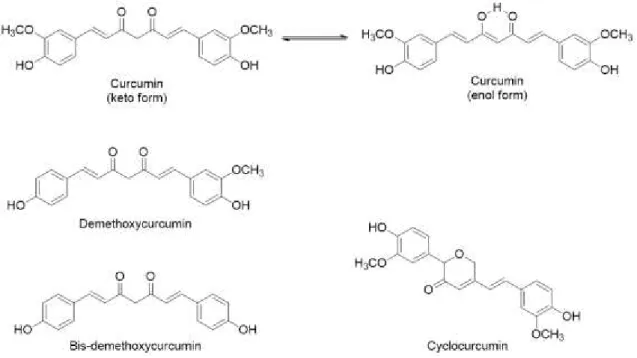
List of FiguresFigure 1.1| Chemical structures of curcuminoids. Adapted from Shiyou Li et al. 19 ... 2
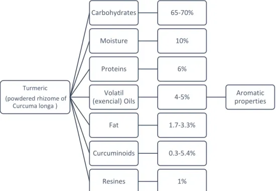
Figure 1.2| Turmeric constitution. Approximate ranges are shown based on supporting references 8, 10
and 11. The focus it’s curcuminoids composition that include curcumin, demethoxycurcumin, bis -demethoxycurcumin, and cyclocurcumin. ... 3
Figure 1.3| Structures of solid lipid particles according to the distribution of drug molecules (pink dots) in the lipid matrix (brown filling). The grouped pink dots symbolize drug molecules not soluble in the matrix but dispersed in other hand the dispersed ones represent drug dispersed on molecular form. (A-D) capsules; (E-F) spheres. Adapted from A. Sao Pedro. 123 ... 4
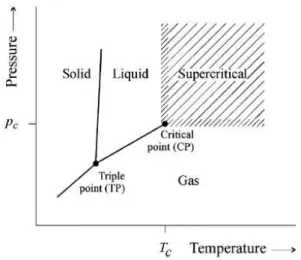
Figure 1.4| Phase diagram of a compound (PT). 37... 5
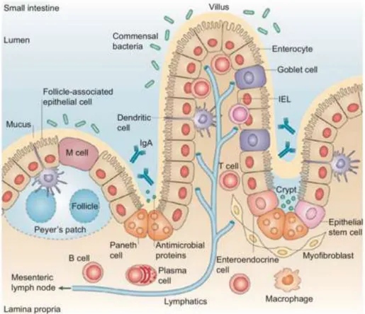
Figure 1.5| Representation of the organization of the small intestine epithelium. Adapted from Maria T. Abreu. 124 ... 7
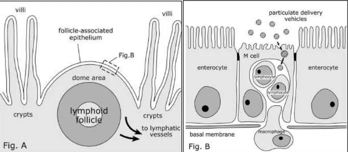
Figure 1.6| Schematic sections of Peyer's lymphoid follicle and follicle-associated epithelium (FAE), showing the transport of particulate delivery vehicles by M cells. The lymphoid follicle is situated under the projecting area in the intestinal lumen between the villi. Covered by FAE, this epithelium is characterized by the presence of specialized antigen-sampling M cells (Fig. B). Adapted from Clark et al, 2001. 72 ... 9
Figure 1.7| Schematic representation of a Transwell® co-culture model. Caco-2/HT29-MTX cells are
seeded on the polycarbonate inserts at the apical side while Raji B are added later to the basolateral compartment. Image adapted from Hubatsch et al, 2007.125 ... 9
Figure 1.8| Illustration of A) Caco-2 monolayer and B) Caco-2/HT29-MTX/Raji B co-culture model setup. Adapted from C.Pereira et al.52 ... 10
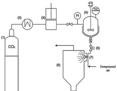
Figure 3.1| PGSS® experimental setup: (1) CO2 cylinder, (2) cryostate, (3) pneumatic piston pump, (4)
stirred vessel (electrically thermostated), (5) automated depressurization valve, (6) recovery vessel, (7) nozzle. Adapted from V.S.S. Gonçalves et al. 89 ... 16
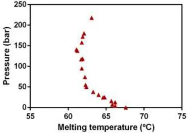
Figure 4.1| Melting points of Beeswax in the presence of CO2. ... 25
Figure 4.2| Curcumin solid lipid particles produced by PGSS® whit different curcumin load. A) 1%
Curcumin; B) 5,5% Curcumin; C) 10% Curcumin. ... 26
Figure 4.3| Fitted response surfaces to the A) EE, as a function of curcumin load and pressure and B) PS, as a function of temperature and pressure. Response surfaces plotted for two variables with the other fixed at middle settings. ... 27
XX
Figure 4.5| SEM micrographs of blank and loaded curcumin particles produced by PGSS® (nozzle
diameter d=250μm); A) blank SLM at 1000x and B) 1500x magnification; C) curcumin loaded SLM at 1000x and D) 1500x magnification; Effect of operating conditions. ... 29
Figure 4.6| DSC thermographs of pure compounds and Solid lipid microparticles. Products of supercritical encapsulation of curcumin, loaded and not loaded with curcumin. ... 31
Figure 4.7| Dissolution profiles of curcumin (C) and curcumin encapsulated microparticles (P). A) in SIF, SGF and B) SGF plus SIF at 37 ± 0.2 °C during 72h at constant agitation (50 rpm)... 31
Figure 4.8| Cytotoxicity assay using MTS reagent. Incubation of curcumin formulations in Caco-2 cell line during 24h at 37 ºC and 5% CO2 humidified atmosphere. Solution of 100 % (v/v) of culture medium in cell culture was used as a positive, none cytotoxic, control. A) Free curcumin; B) Curcumin solid lipid microparticles blank and loaded produced by PGSS® technique. ... 33
Figure 4.9| Cytotoxicity assay using MTS reagent. incubation of all digested microparticles, loaded and empty, in Caco-2 cell line during 24h at 37 ºC and 5% CO2 humidified atmosphere. Solution of 100 % (v/v) of culture medium in cell culture was used as a positive, none cytotoxic, control. ... 34
Figure 4.10| Cytotoxicity assay using MTS reagent: incubation in Caco-2 cell line during 24h at 37 ºC and 5% CO2 humidified atmosphere. Solution of 100 % (v/v) of culture medium in cell culture was
used as a positive, none cytotoxic, control. A) MCT based emulsions; B)LCT based emulsions. 34
Figure 4.11| Cytotoxicity assay using MTS reagent: incubation in Caco-2 cell line during 24h at 37 ºC and 5% CO2 humidified atmosphere. Solution of 100 % (v/v) of culture medium in cell culture was used as a positive, none cytotoxic, control. A) MCT and B) LCT oil excipient. ... 35
Figure 4.12| Cytotoxicity assay using MTS reagent: incubation of all nanoemulsions compounds in Caco-2 cell line during 24h at 37 ºC and 5% CO2 humidified atmosphere. Solution of 100 % (v/v) of culture medium in cell culture was used as a positive, cytotoxic, control. A) SLM non-digested and B) non-digested ones. ... 35
Figure 4.13| Staining of mucus present in Caco-2 and HT29-MTX monolayer and co-culture. Staining at 7, 14 and 21 days on a Transwell® plate. ... 37
Figure 4.14|SEM analysis of the triple culture. (H) Mucus-secreting HT29-MTX cells were observed in the triple culture and were properly identified through the layer of mucus they produced. Compared with the caco-2 cells (C) with its characteristics tight junctions and microvilli. ... 38
Figure 4.15| SEM analysis of the triple culture. M cells (M) were identified due to their lack of microvilli in contrast to Caco-2 cells. ... 39
Figure 7.1| Curcumin samples of two distinct suppliers: Alfa Aesar (black) and Sigma (blue). ... 50
XXI
XXIII
List of TablesTable 3.1| Three-factor, three-level face-centered cube design used for RSM. ... 17
Table 3.2| Real values of the variables for the coded ones. ... 18
Table 3.3| HPLC method used to curcumin quantification. Flow = 0.3mL / min. ... 24
Table 4.1| Summary of The CCFC design for the three independent variables and experimental results. ... 27
Table 4.2| Model equations for the response profiles fitted to the values of EE and PS as a function P, T and %C, and respective R2 and Radj2. ... 28
Table 4.3| Microparticles characterisation. ... 29
Table 4.4| Characterization of the emulsion-based curcumin nanoparticles. ... 30
Table 4.5| Characterization of solid lipid nanoparticles before and after IVD. ... 32
Table 7.1| Critical Properties of Fluids of Interest in Supercritical Processes. 35 ... 49
XXV
List of Abbreviations
Abbreviation Full form
%C Curcumin load
Caco-2 Caco-2 human colorectal adenocarcinoma cell line
CCFC Central Composite Face-Centered cube design
DLS Dynamic light scattering
DMEM Dulbecco’s Modified Eagle’s Medium
DSC Differential Scanning Calorimetry
EE Encapsulation efficiency
FBS Fetal Bovine Serum
FFA Free fat acids
GI Gastrointestinal
HBSS Hanks Balanced Salt Solution
HPLC High-performance liquid chromatography
IVD In vitro digestion
LCT Low chain triglycerides
LD Laser diffraction
M cells Microfold cells
MCT Medium chain triglycerides
MEM-NEAA Non exencial aminoacids
M-like cells Microfold-like cells
MTS CellTiter 96® AQueous One Solution Cell Proliferation Assay reagent
MTX Methotrexate
NE-LCT MCT Based nanoemulsions
NE-MCT MCT Based nanoemulsions
O/W Oil in Water
P Pressure
XXVI
PBS Phosphate Buffer SolutionPDI Particles dispersion index
P-gp P-glycoprotein
PGSS® Particles from Gas Saturated Solutions
PPs Peyer’s Patches
PS Particle size
PST Penicillin-Streptomycin
R2 Correlation coefficient
Radj2 Adjusted correlation coefficient
RSM Response Surface Methodology
ScCO2 Super critical carbon dioxide
SD Standard deviation
SEM Scanning electron microscopy
SFE Supercritical fluid extraction
SGF Simulated gastric fluid
SIF Simulated intestinal fluid
SLM Solid lipid microparticles
SLP Solid lipid particles
SSF Simulated salivary fluid
T Temperature
TEER Transepithelial electrical resistance
TP Triple point
1
1. Introduction1.1. Food industry – Functional foods and Nutraceuticals
Nowadays, the world population are understandably more interested in the potential benefits of nutritional support for disease control or prevention and tends to eat more nutritional foods.1 These diets
are known to have a lower incidence of cardiovascular diseases and certain types of cancer. With the increase of this concern and through epidemiological and clinical studies an evident relationship between diet and health is observed. 1,2 In the past few years, many food bioactive constituents,
functional ingredients (vitamins, antimicrobials, antioxidants, colorants, etc.), have been commercialized in form food supplements that incorporate food extracts to obtain a product with a beneficial physiological function attributed.3 With these modifications these resulting products cannot be classified
as ‘food’ and in in 1989 the term ‘‘nutraceutical’’ was formed by the Foundation for Innovation in Medicine
(New York, US). A new hybrid term that represents both nutrients and pharmaceuticals to designate the
new foods created. The term ‘‘functional food’’ are known by the Institute of Medicine of the National Academy of Sciences (1994) as any food or food ingredient that, when consumed regularly, may provide a health benefit beyond their nutritional properties.4
The concepts of nutraceuticals, functional foods, or dietary supplements are confusing and most often they can be used interchangeably. Dietary supplements which, through a bioactive agent, are presented as a non-food matrix and used with the function of improving health through dosages that exceed those that could be obtained from of normal food.2,3,5,6
The aim of nutraceuticals is significantly different from functional food, with emphasis on prevention and treatment of diseases. Various nutraceuticals are used in nutritional therapy, which is mainly based on scientific research and with more clear information about chemical structures, biological functions and clinical information.7 Therefore, these characteristics attracted the attention of people to
seek a better diet and the production of new functional foods meeting the needs of growing Human population.
1.1.1. Bioactive Compounds
Across cultures there are many different dietary patterns, some of which promote health and others that increase risk of chronic disease. Despite cultural differences there are some shared characteristics of healthy dietary patterns. These compounds are extra nutritional constituents that typically occur in small quantities in foods. 3
1.1.1.1. Curcumin as a nutraceutical
Curcumin [(E,E)-1,7-bis(4-hydroxy-3-methoxy-phenyl)-1,6-heptadiene-3,5-ione] (Figure 1.1), a hydrophobic polyphenol, is the main phenolic pigment extracted from turmeric, the powdered rhizome of Curcuma longa (Figure 1.2), widely cultivated in several tropical areas in Asia and despite the use in
2
The medicinal importance of powdered coloured extracts of dried roots, often called turmeric, is well documented. It has been used for many diets in particularly as an anti-inflammatory agent, however curcumin, identified as the active principle, has been shown to exhibit antioxidant, antimicrobial, anti-inflammatory, anticarcinogenic activities, etc. 8,13–15
Curcumin and turmeric products have been characterized as safe by health authorities such as the Food and Drug Administration in United States of America, the Natural Health Products Directorate of Canada, and the Expert Joint Commit- tee of the Food and Agriculture Organization/World Health Organization on food additives.11 While in EU it bears a status of food ingredient (E100) and is present
in many food additives. 16,17
The turmeric contains three principal curcuminoids: 75-80% curcumin, 15-20% demethoxycurcumin, 3-5% bis-demethoxycurcumin. In recent studies cyclocurcumin appears as the fourth most abundant curcuminoid in turmeric (Figure 1.3). 18–20 Curcumin, first isolated in 1815, is found
as a crystalline powder insoluble in water under acidic or neutral conditions but soluble in ethanol, alkali, ketone, acetic acid and chloroform. Is unstable undergoing rapid hydrolytic degradation in neutral or alkaline conditions to feruloyl methane and ferulic acid. Since it is insoluble in aqueous medium and has poor stability towards light, alkalinity, enzymes and heat, it cannot really be widely used in food and pharmaceutical processing industry. 10,19,21
As health promoting agent, curcumin, is limited by its poor solubility in aqueous solution and its low bioavailability. 13 Due to these limitations, studies have been developed with the aim to reduce the
rapid degradation and increase curcumin’s bioavailability. These studies are based on encapsulation
techniques and nanotechnological approaches, however the increased bioavailability of curcumin will depend mainly on the physicochemical properties of the carrier.
3
1.2. Bioactives formulationParticle formation and encapsulation technologies are widely employed in the pharmaceutical, cosmetic and food industries. However, the development of suitable delivery systems is fundamental to overcome barriers to bioactive usefulness.
Many formulations consist in comprising a core material (the active component) surrounded by a coating material (typically a bio-polymer) (Figure 1.3). The resulting materials and systems can be designed to exhibit novel and significantly improved chemical and biological properties. 22,23
In the field of particulate delivery systems, the ability to control size, morphology and release of encapsulated compounds is fundamental to good targeting but is often held back by severe processing conditions or inadequate methods. These methods presuppose two important requirements: the encapsulation system must preserve stability of the bioactive compounds during processing and storage and to prevent undesirable interactions with food matrix, which should be depends on the type of molecule and carrier.22,24 There is a multitude of possible ways to encapsulate bioactives. 22,23,25
Triglycerides based-nanoemulsions and supercritical fluids encapsulation techniques are the one type of formulations explored in this work.
Turmeric (powdered rhizome of
Curcuma longa )
Carbohydrates 65-70%
Moisture 10%
Proteins 6%
Volatil
(exencial) Oils 4-5%
Aromatic properties
Fat 1.7-3.3%
Curcuminoids 0.3-5.4%
Resines 1%
Figure 1.2| Turmeric constitution. Approximate ranges are shown based on supporting references 8, 10 and 11. The
4
1.2.1. Nanoemulsion-based delivery systemsEmulsions of O/W are regarded as useful tools with a great potential in the food sector to incorporate food ingredients. 26
In general, nanoemulsions can be produced using a variety of methods within two different approaches: high energy or low energy approaches. High energy methods result from the application of high disruptive forces through mechanical devices, with the aim of causing the rupture of oil droplets dispersing them in the aqueous phase. The second approach, low energy, depends on the spontaneous formation of tiny oil droplets within mixed oil-water-emulsifier systems when changing the solution or ambient conditions, such as composition or temperature. 27–29
The formulation of nanoemulsions consist of a mixture of immiscible liquids. Briefly, a lipid phase, that generally acts as a carrier of lipophilic active compounds, is dispersed in an aqueous phase in the form of nanometric scaled droplets (<100nm). 27 The oil phase can be constituted by different
non-polar compounds, normally triglycerides are used. Besides the lipid and aqueous phases, the formulation of nanoemulsions requires the use of stabilizers such as emulsifiers to prevent the breakdown of the nanoemulsion structure. 28,30
1.2.2. Solid lipid particles
Solid lipid particles have emerged in the last two decades as an alternative to liposomes, emulsions or polymer particles as potential release vehicles (Figure 1.3). 31 Usually comprised of
biodegradable and non-toxic lipids, solid at room and body temperature, promote the release of the incorporated compound through a prolonged profile after administration at a constant concentration of the molecule of interest in the blood stream. 32,33 In addition, these vehicles have the advantage of
increasing particle uptake by epithelial cells and can promote sustained drug release.
1.3. Supercritical fluid technology
Solvents are used in large amounts in the chemical, pharmaceutical, food, and natural product industries. The call for new products with singular features and design of new environmentally friendly
Figure 1.3| Structures of solid lipid particles according to the distribution of drug molecules (pink dots) in the lipid matrix (brown filling). The grouped pink dots symbolize drug molecules not soluble in the matrix but
5
and sustainable technologies is emerging in society and shifting the technological processes towards high pressure. 32
A pure component is considered to be in a supercritical state if its temperature and its pressure are higher than the critical values. The critical point represents the end of the vaporization curve in the PT phase diagram (Figure 1.4). These types of fluids add a new important property to conventional (liquid) solvents: their density; liquid-like densities and, therefore, solvating characteristics equivalents that can be easily tuned to the process needs. But, at the same time, they present gas-like properties such as very low surface tensions, low viscosities, and moderately high diffusion coefficients. In general, the physical properties in the critical region enhance mass and heat transfer processes. 34–37
The most widely used SCF is scCO2, which is nontoxic, non-flammable, cheap, widely available
and easy recyclable. Once its critical properties can be achieved at moderate pressures and
near-ambient temperatures (Table 1), is one of the most fluid used as “green solvent” for bioactives
encapsulation. Furthermore, the use of ScCO2 eliminates or reduces the use of toxic organic solvents,
in the process, and allows to obtain solvent-free products due to the high solubility of most organic solvents in the fluid. 36,38,39
1.3.1. Supercritical-based precipitation methods
The use of scCO2 provides several advantages in comparison with conventional techniques or
even comparing with other supercritical fluids. It can be used as a solvent, antisolvent, as extracting agent or even to improve the spraying process in different techniques. The solvent properties of scCO2,
i.e. the solubility of polymers in scCO2 and the solubility of CO2 in the polymers, are two key fundamental
subjects in this field.
The solvent power of supercritical scCO2 can be used to dissolves non-polar or slightly polar
compounds and decreases with the increase of compounds molecular weight. Has high affinity with oxygenated organic compounds and exhibit low solubilities for free fatty acids, glycerides and water (<0.5 wt%), at temperatures below 100 °C. One of the most interesting characteristic is the capability to
6
separate compound that are less volatile, have a higher molecular weight and/or are more polar, as pressure increases. However, proteins, polysaccharides, sugars and mineral salts are insoluble. 34,36,37
1.3.1.1. Particles from Gas Saturated Solutions
The PGSS® technique was patented by Weidner et al 40–42 and allows to form particles from
substances that are non-soluble in supercritical fluid. Briefly, compounds mixture absorb a large amount of CO2 that cause a decrease of melting point (glass transition temperature for amorphous polymers) or
a swelling effect on the substance.40,43,44 The major advantages of this process, in comparison with other
supercritical-based precipitation methods, is the low intake of carbon dioxide and the possibility to process thermolabile substances.
The polymeric or lipidic solution formed, which can contain between 5-50 wt% of the compressed gas, is expanded through a nozzle causing the release of CO2 with large cooling effect,
due to the energy consumption, leading to a quasi-instantaneous solidification of the droplets. This rapid cooling of the gas during the expansion process is due to the Joule-Thomson effect. In few words, since an expansion of a gas from high to low pressure through a throttle valve is an isenthalpic process, it leads to a significant temperature drop. 45–47
The morphology, size and apparent density of the produced particles may depend on several parameters such as the structure and viscosity of the compounds to be precipitated, the operating conditions, and even the equipment used to perform the PGSS® process. 36,44,48,49
1.4. Concepts and models for bioactives permeability studies
Prediction of human intestinal absorption, bioavailability and compounds cytotoxicity is a major goal in the optimization and production of drugs and formulated products intended for non-invasive delivery. Oral bioavailability is a highly desirable property for molecules of particulate interest nonetheless poor permeability and/or absorption make a molecule unsuitable for further development, and there is interest in finding ways to avoid the situation of having a potent, yet impermeable molecule.
50,51 The determination of oral bioavailability is as important as identifying the potential therapeutic uses
of nutraceuticals. Consequently, various techniques have been currently employed to predict bioactive compounds absorption in the different phases of discovery and development. 52
1.4.1. Bioactive compounds oral administration
Over recent years, a major challenge in compounds delivery has been the design of appropriate vehicles for the oral administration of bioactives. Bioavailability represents both the quantity of the active component absorbed through the blood circulation and the rate of this phenomenon. However, is extremely conditioned for enzymatic degradation and poor penetration of the intestinal membrane. 53
7
drug reaching the general circulation depends on many different factors and is mostly determined by its physicochemical properties. To predict the compounds intestinal behaviour strategies for modulating tight-junction permeability increasing paracellular transport of molecules has been implemented. 54,55
1.4.2. Intestinal composition and permeability
In oral administration absorption of compounds by passive diffusion in the intestinal epithelium (Figure 1.5) is the major transport mechanism presented. Nonetheless to be absorbed, the molecules must diffuse across a series of separate barriers. Some of them includes the mucosal side, the mucus gel layer, the intestinal epithelial cells, the lamina propria and the endothelium of the capillaries. However, only the epithelial cells barrier is the most significant to compounds absorption. 56–59 Now,
several in vitro experimental models with high-throughput capacity and adequate predictability of absorption potential in human epithelial are available for evaluating intestinal permeability and transport.
60,61
Because the oral route is the most commonly administration route used, the intestinal barrier has received much attention. In order to simulate the intestinal barrier, Caco-2 cell line have gained
massive approval as a reliable and high throughput in vitro model for evaluating a large number of
candidates for their intestinal absorption potential. 55,62,63
Figure 1.5| Representation of the organization of the small intestine epithelium. Adapted
8
1.4.2.1. Caco-2 cell modelCaco-2 cells, a human colon adenocarcinoma, has been used extensively for the high throughput screening of compounds permeability. Allows effective in vitro prediction of compound permeability and absorption while considering all the physical, chemical, and biological events during intestinal transportation. 64
They undergo spontaneous enterocytic differentiation in culture and when they reach confluency, on a semipermeable porous filter, express many primary-like qualities of enterocytes, both functional and morphological, and can fully polarize into differentiated monolayers displaying brush border regions (microvilli) and cell-cell (tight junctions).65–68 Still, this system presents some limitations
when compared to the human intestinal epithelium, specifically the overexpression of P-gp, an efflux pump, and the formation of strong tight junctions that could lead to a decrease in the paracellular permeability. Nevertheless, these monocultures only characterize absorptive cells, which do not mimic completely the human intestinal epithelium as it is a combination of different cell types: absorptive cells or enterocytes, goblet cells (mucus producer cells), endocrine cells and M cells. Being the enterocytes the most abundant cells, followed by goblet cells. 55,69–71
Studies regarding membrane transport are still present as inconsistent due to the high requirement on the quality of the monolayer. Factors such as cell condition, passage number, length of cultivation and circumstance under which the study is performed diverge among laboratories and affects the viability of the results. 65
1.4.2.2. M Cells
In the intestine, PPs are the major sites of antigen and microorganism sampling. M cells (Figure 1.6) are responsible for the transepithelial transport of foreign material and microorganisms from the external environment to the lymphoid follicles. 72,73 These cells typically contain a small number of
microvilli in the apical zone and a basolateral cytoplasmic invagination in which the lymphocytes and macrophages are located. Passage of antigens and microorganisms through M cells is an essential step for the development of mucosal immune responses and the pathology of many infectious diseases. Particle delivery vehicles are largely prevented from passing between epithelial cells by tight junctions. However, since M cells have a relatively high transcytotic capacity compared to enterocytes, can represent an efficient route for the transport of nutraceuticals carried by particulate carriers through the intestinal epithelial barrier. Kernéis et al.74 constructed the first intestinal in vitro co-culture model based
on Caco-2 cells and mouse Peyer’s patch lymphocytes. The interaction of the epithelial cell line Caco -2 with lymphocytes triggered the formation of cells-like M cells that resemble intestinal M cells functions and morphology. 75 Later, to overcome the use of primary murine lymphocytes, Gullberg et al. 75
developed a similar model based on a co-culture of Caco-2 cells and human Raji B lymphocytes, a cell
line that is originated from a human Burkitt’s linfoma, since Raji cells exhibit B cell markers, which are
major inductive of M cells phenotype. 76 Results based on the work of des Rieux et al. 54,77 exhibited that
9
Like described for Antunes et al. 60 to obtain the phenotype M-cell like, one common approach
is to use a 21 days in vitro model that consists in a co-culture of Caco-2 cells seeded on Transwell®
membranes inserts (normal oriented) (Figure 1.7) and human Raji B lymphocytes added, to the basolateral chamber of the insert, after 14 days. Whit this method it was possible to detect that some cells developed M-cell like morphology.
1.4.2.3. Caco-2/HT29-MTX/Raji B Model
The mucus layer covering GI tract, represents a significant barrier to particles absorption. Mucus is a viscoelastic adhesive gel that coats all exposed epithelial surfaces not covered by skin.78 Mucus
protects the underlying epithelium by both lubricating the surface and trapping and removing foreign particulates. Has been shown to be highly adhesive to pathogens and particulate systems, offering many opportunities for the development of adhesive interactions with small polymeric particles. 79,80
Figure 1.6| Schematic sections of Peyer's lymphoid follicle and follicle-associated epithelium (FAE), showing the transport of particulate delivery vehicles by M cells. The lymphoid follicle is situated under the projecting area in the intestinal lumen between the villi. Covered by FAE, this epithelium is characterized by the presence of specialized antigen-sampling M cells (Fig. B). Adapted from Clark et al, 2001. 72
Figure 1.7| Schematic representation of a Transwell® co-culture model.
10
Since the early 1980s, several methods have been developed to differentiate the adenocarcinoma cell line HT29 into mature intestinal cells under appropriate culturing conditions. 81
Through selective pressure with MTX a distinct subpopulation of mucus-secreting HT29 cells that maintain their ability to differentiate under normal culture conditions were isolated by Lesuffleur et al. 82
At late confluence, HT29-MTX monolayers show a dense mucus layer on their apical surface and a morphology identical to human intestinal goblet cells. However, as opposed to Caco-2 cells, does not express P-gp and do not form TJs as tight which promotes the increase in paracellular transport pathways. These mucus layers is labile and could be removed by extensive and rough washings. 81,83,84
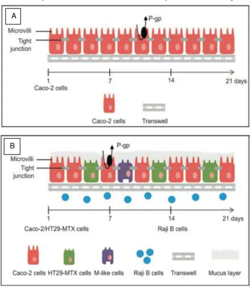
Considering the above-mentioned culture models all their advantages and drawbacks, a cell-based model based on the combination of Caco-2, mucus-producing HT29 cells and Raji B lymphocytes was stablished resulting in a more promising experimental results reproducing the intestinal epithelium in a more complete way like illustrated in figure 8. 55,61
Figure 1.8| Illustration of A) Caco-2 monolayer and B) Caco-2/HT29-MTX/Raji B co-culture model setup. Adapted from C.Pereira et al.52
11
1.5. Aim and rational of the thesisThe challenge addressed in this work was using an in vitro cell co-culture model that mimics the
intestinal epithelium to evaluate the bioavailability and safety of curcumin delivery systems.
To achieve this goal, an integrated approach was developed by two distinct tasks:
1. Development of different types of curcumin formulations to maximize its bioavailability and stability. 2. Implementation of an in vitro model system that closely mimics the human intestinal epithelium
monolayer to evaluate the formulated curcumin systems in terms of toxicity and bioavailability.
In task 1: Curcumin delivery systems
The general aim of this first task was the investigation of curcumin entrapment into solid lipid microparticles, using a green technology, on assisted CO2 precipitation method and compare this
formulation whit particles prepared by conventional approaches. The PGSS® process was chosen and
a response surface methodology was used to model and optimize the encapsulation conditions.
Different techniques were applied to characterize the formulations in terms of morphology, size and thermal behaviour along with the quantification of encapsulated curcumin. After total characterization these particles underwent an in vitro digestion process used to evaluate the changes in curcumin formulations throughout the gastrointestinal tract.
In task 2: In vitro cell co-culture model
A triple co-culture has been established and characterized for use as an in vitro tool for transport and permeability studies. Viability and cytotoxicity profiles tests of free and encapsulated curcumin were evaluated for different dosages and times of exposure. The integrity of the monolayer and the quality of tight junctions were controlled during the transport studies by monitoring transepithelial electrical
Goblet cell-like model: HT29
MTX-E12
M cell-like model: Caco-2 +
Raji B
Enterocyte-like model: Caco-2
Nanoemulsions
12
13
2. MaterialsFor curcumin encapsulation through PGSS® technique, curcumin (95%) was purchased from
Alfa Aesar (Karlsruhe, Germany), beeswax from QUIMIND (Portugal), CO2 (99.95 and 99.998 mol% purity) was delivered by Air Liquide (Portugal) and methanol absolute 99.99% from Fisher Scientific (Waltham, MA, USA). Curcumin (65%) encapsulated in nanoformulations was purchased from Sigma-Aldrich. An analysis and comparison of its purity is shown in attachment.
To perform the In vitro digestion, digestion fluids were produced: SSF (KCl, 15.09 mM; KH2PO4,
1.35 mM; NaHCO3, 13.68 mM; MgCl2(H2O)6, 0.15 mM; NH4(CO3)2, 0.06 mM; CaCl2(H2O)2, 1.5 mM; HCl, 1.1 mM; pH 7), SGF (KCl, 6.9 mM; KH2PO4, 0.9 mM; NaHCO3, 25 mM; NaCl, 47.2 mM; MgCl2(H2O)6, 0.12 mM; NH4(CO3)2, 0.5 mM; CaCl2(H2O)2, 0.15 mM; HCl, 15.6 mM; pH 3) and SIF (KCl, 6.8 mM; KH2PO4, 0.8 mM; NaHCO3, 85 mM; NaCl, 38.4 mM; MgCl2(H2O)6, 0.33 mM; CaCl2(H2O)2,
0.6 mM; HCl, 8.4 mM; pH 7). Still in the digestion procedure the following enzymes were used: α -Amylase (A1031-1KU, Sigma, USA), pepsin (EC 3.4.23.1) from porcine gastric mucosa (Sigma, USA), bile extract porcine (Sigma, USA) and porcine pancreas (Sigma-Aldrich, USA). To stop enzymatic action Pefabloc®) from Sigma-Aldrich and Amicon® Ultra-4 Centrifugal Filter Units from Merck (Millipore,
Germany) was used.
All cell culture media and supplements, namely FBS, DMEM, MEM NEAA, PS, trypsin/EDTA and HBSS were obtained from Invitrogen (Gibco, Invitrogen Corporation, Paisley, UK). Corning®
Transwell®polyester membrane cell culture inserts (12 mm with 0.4 μm pore polyester membrane insert
and 1.12 cm2) were purchased from Merck (CLS3460-48EA) to monolayer culture.
To perform cells cytotoxicity assays, MTS (G3582) from Promega was used and in assays with necessary cellular lysis the utilized reagents were: inhibitor of Proteases cocktail (Protease inhibitor cocktail set III ANIM #535140-1 Calbiochem) and Cell Lytic (M Cell Lysis reagent, Sigma C2978-50 ml).
15
3. Methods3.1. Curcumin formulation process
3.1.1. Curcumin Nanoformulations
3.1.1.1. Nanoemulsions preparation
Curcumin nanoemulsions were prepared through high pressure homogenization according to other authors (Pinheiro et al., 2016). The lipid phase constituted by MCT or LCT and 0.1 % of curcumin was homogenized with the aqueous phase (with lecithin 2.5 %) at room temperature and a volume ratio of 1:9. First, both solutions were pre-mixed using an Ultra-Turrax homogenizer (T 25, Ika-Werke, Germany) during 2 min and thereafter the resulting emulsion were passed through a high-pressure homogenizer (NanoDeBee, Bee International, South Easton, Massachusetts, USA) at 20 000 psi (137.9
MPa) during 20 cycles. The nanoemulsion were kept at 4 ˚C in the dark.
3.1.1.2. Solid lipid nanoparticles preparation
SLN were prepared according with Kheradmandnia et al.85. Beeswax (3 %), lecithin (1.5 %) and
curcumin (0.1 %) were melted in a water bath at 85 ˚C. Tween 80 (1.5 %) was solubilized in distilled water at 85 ˚C in an Ultra-Turrax homogenizer (T 25, Ika-Werke, Germany) during 2 min at 3400 rpm. The aqueous solution was added to the lipid solution and mixed in an Ultra-Turrax homogenizer (T25, Ika-Werke, Germany) at 22000 rpm during 10 min. Then the resulting nanoemulsion was gradually
dispersed at a volume ratio of 1:10 in cold water at 2 ˚C under stirring at 2000 rpm. The SLN were kept at 4 ˚C in the dark.
Both formulations at the nanoscale were developed and prepared out by Universidade do Minho (Portugal) within the framework of the NanoMaxiSafe project (PTDC / AGRTEC / 5215/2014), in which some of the results produced in this master thesis are also included.
3.1.2. Curcumin microparticles
3.1.2.1. Melting point measurements
The melting point of solids can be considerably decreased by using supercritical fluids which are highly soluble in the melt. Knowledge of the solid-liquid transitions of lipids under pressurized CO2
is essential for their precipitation through the PGSS® process. Since beeswax data of melting point
depression, as a result of CO2 dissolution in the matrix, is not a topic presented in the literature the
melting point depression was measured within this work using a visual method. 86,87
For performing the melting point determination experiments, a stainless steel high-pressure visual cell with an internal volume of approximately 5 cm3 with two sapphire windows was used. Briefly,
16
measured with a pressure transducer Digibar II calibrated between 0 and 250 bar (accuracy: 0.15 %) and the CO2 was pumped, using a Haskel pump (model 29723-71), until the desired pressure was
reached. The temperature was then gradually increased until it was possible to visually observe the complete melting of the lipid. The heating system was composed of a heating cable (Horst), a controller (Ero Electronic LMS) and a high accuracy thermometer (Omega HH 501 AT, 0.1 %). Measurements were performed in a pressure range up to 200 bar. To confirm the reproducibility of the results some points were repeated resulting in a 0.15 % of error.
3.1.2.2. Particles from gas saturated solutions
Beeswax particles unloaded and loaded with 10 wt % of curcumin were produced by PGSS® in
batch mode. The schematic representation of the modified PGSS® equipment (FAME UNIT, Separex,
France) used to produce the particles is shown in figure 3.1 previously described in the work of Rodríguez-Rojo et al. with some modifications. 88Briefly, CO2 was fed by a pneumatic pump (29723-71,
Haskel International Inc.,CA, USA) to a 50 cm3 high pressure stirred vessel, electrically thermostated,
at theselected operation temperatureuntil the desired working pressure was reached.Then, the stirred mixture is depressurized through a nozzle (250 μm) by an automated depressurization valve to a cyclone, where it was externally mixed with compressed air (7 bar). The mixture and the supercritical carbon dioxide are brought into contact during 15 min. Finally, the particles were recovered into a collector vessel of 18 L at atmospheric pressure. The operating conditions (T and P) were chosen according to the measurements of melting point depression of the lipid in the presence of compressed CO2and varied to see their effect on the particles’ morphology and size.
Figure 3.1| PGSS® experimental setup: (1) CO2 cylinder, (2) cryostate, (3) pneumatic piston pump, (4) stirred
17
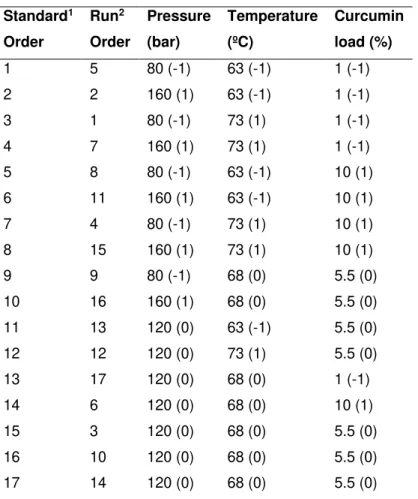
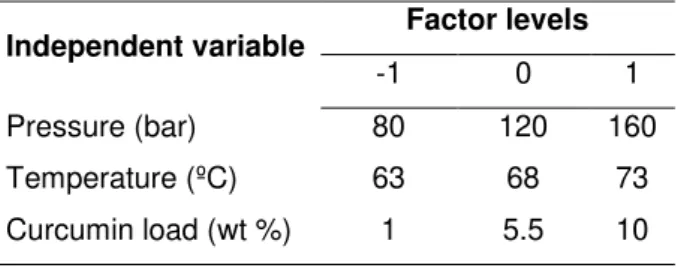
3.1.2.3. Experimental design (Process Optimization)RSM was used to model and optimize encapsulation conditions. RSM consists of a set of mathematical and statistical methods developed for modelling phenomena and finding combinations of several experimental factors (variables) that will lead to optimum responses. With RSM, several variables are tested simultaneously with a minimum number of trials, using special experimental designs that enable to find interactions between the variables which cannot be identified with classical approaches. The encapsulation of curcumin through PGSS® was carried out following a CCFC, as a
function of three factors: pressure, temperature and curcumin load in mixture. The design consisted of seventeen randomized runs with three replicates at the central point described intable 3.1. To normalize parameters, each of the coded variables was forced to range from -1 to 1, so that they all affect the response more evenly (Table 3.2). The repetitions of the centre points are used to determine the experimental error, which is assumed to be constant along the experimental domains. The fit of the models was evaluated by the R2 and Radj2.
Table 3.1| Three-factor, three-level face-centered cube design used for RSM.
1 No randomized
2 Randomized
Independent variables Standard1 Order Run2 Order Pressure (bar) Temperature (ºC) Curcumin load (%)
18
Table 3.2| Real values of the variables for the coded ones.
3.2. Particles’ characterization
3.2.1. DSC measurements
DSC was performed using a TA instruments Q200 (module MDSC) with the aim of studying the thermal behaviour of the particle’s components. An amount of 5-10 mg of curcumin or pure lipid (beeswax) was weighted into an aluminium pan and sealed in the calorimeter. The samples were heated from 0 °C to 220 °C at a rate of 10 °C/min under nitrogen atmosphere. After encapsulation procedure an amount of 0.4-1 mg of lipid particles were tested to in the same conditions.
3.2.2. Encapsulation efficiency
To measure the total curcumin encapsulated in PGSS produced microparticles, curcumin (an amount of 5 and 7 mg of particles) was extracted from the lipidic matrix with 30 mL of methanol in an ultrasound bath during 30 min. The result of this disruption was later analysed by a UV/Vis spectrophotometer (model EPOCH, 219 Bio-Tek, USA) wavelength of 420 nm. To eliminate non-dissolved solids before the quantification the solution was filtered (25 mm - 0.20 µm).
The determined absorbance is proportional to the amount of curcumin in solution. The amount of curcumin in the solution is reported in this work as the percentage of encapsulated curcumin. Standard solutions of curcumin to be used in the calibration was obtained by using standard samples in methanol with concentrations between 1 and 25 µg/mL (R2 =0.9992). For reproducibility purposes
average and standard deviation were calculated from 3-4 replicates. On the other hand, the encapsulation efficiency (Eq. 1) was obtained comparing the final and initial load of curcumin in the experiment.
𝐸𝐸 (%) =𝐼𝑛𝑖𝑐𝑖𝑎𝑙 𝑐𝑢𝑟𝑐𝑢𝑚𝑖𝑛 𝑎𝑚𝑜𝑢𝑛𝑡 × 100 (𝐄𝐪. 𝟏)𝐹𝑖𝑛𝑎𝑙 𝑐𝑢𝑟𝑐𝑢𝑚𝑖𝑛 𝑙𝑜𝑎𝑑
3.2.3. Particle size and morphology
The morphology of the particles was analyzed and imaged by SEM (Jeol, JSM-5310 model, Japan). Samples were prepared for observation by covering with gold/palladium (Au/Pd), approximately
300 ˚A of gold, in a sputter coater (Quorum Technologies, model Q150TES). Micrographs of the
prepared aliquots were taken at an acceleration voltage of 20 kV.
Independent variable Factor levels
19
The particle size of the samples was measured by a LD equipment model Malvern Mastersizer 2000. The particle size measurements are defined as d0.5 (known as the median particle size by volume) being the result the average from 3 measurements. The span value is also reported, that is, the ratio between d0.5 and (d0.9-d0.1); span values near to 1 represent narrow PDI.
3.2.4. Curcumin formulations sterilization
Briefly, microparticles were put directly in contact with UV irradiation during 30 min at room temperature in laminar flux chamber and to further confirm sterility, microparticles were incubated in tryptone soya broth at 37 °C in a humidified atmosphere of 5 % CO2 to ensure the absence of bacterial
contamination (data not shown).
3.3. In Vitro Release of Curcumin
The in vitro curcumin release assays were performed in two different environments: simulated intestinal fluid and simulated gastric fluid, both without the presence of enzymes. This test had three different approaches: solubility in SIF, SGF and SIF plus SGF. For each fluid 5 mg of PGSS microparticles were deposited in a sealed flask with 30 ml of SIF or SGF. The system was kept under constant agitation (50 rpm) at 37 °C during 48 h. In the last-mentioned approach, the compound was subjected to two hours of incubation with SGF to which a volume of SIF (1: 2 respectively) was added over the remaining assay time. At a desired time interval, an aliquot of 1 mL was removed, with replacement with fresh fluid, from each flask to quantify through UV/Vis measurements like described in 3.2.2. The aliquots were filtered (25 mm - 0.20 µm) before the UV/Vis analysis to avoid the interference of microparticles. The same procedure was repeated to free curcumin, where 1 mg of curcumin (95 %) was submitted to 40 mL of digestion fluids.
3.4. In vitro Digestion
An in vitro digestion procedure, as described by Minekus et al. 90 was used to evaluate the
transformation that encapsulated curcumin suffer throughout the gastrointestinal tract. These methods try to mimic physiological conditions in vivo, considering the presence of digestive enzymes and their concentrations, pH, digestion time, and salt concentrations, among other factors, to mimic closely the oral, gastric an intestinal phase property.
3.4.1. Oral Phase
20
3.4.2. Gastric PhaseThe mixture resulting from the oral phase was submitted to a gastric phase by adding 8 mL of Simulated Gastric Fluid and 1 mL of pepsin (2000 U/mL in fluid) from porcine gastric mucosa. In this phase, 1M HCl was added until pH 3 was stablished and Milli-Q water to perform a final volume of 20 mL. The gastric digestion step was conducted for 2 hours at 37°C, under constant agitation.
3.4.3. Small Intestine Phase
To the gastric digestion mixture were added 11 mL of Simulated Intestinal Fluid, 2.5 mL of bile extract porcine and 5 mL pancreatin from porcine pancreas (200 U/mL in the fluid). Finally, the mixture was brought to pH 7 by addition of 6 M NaOH and Milli-Q water added to a final volume of 40 mL. Like gastric phase incubation of intestinal digestion mixture occurred for 2 hours at 37 °C, under constant agitation.
To stop enzymatic action on the intestinal digestion sample proceeded the addition of 10 μL of Pefabloc® per millilitre of digested sample. The mixture was transferred to Amicon® Filter and centrifuged
(Mikro 220R, Hettich, Germany) at the maximal velocity, normally 4000 G, for 40 minutes. All collected samples were stored at -20 °C until further use.
3.5. In vitro cell-based assays
3.5.1. Cell Culture and sub culturing
To determine cellular viability as used a standard trypan blue staining procedure followed by cell counting in hemocytometer. Every time complete medium or trypsin was needed, to cell culturing, were pre-warmed at 37 °C.
3.5.1.1. Adherent cells culture
The human colon adenocarcinoma Caco-2 cells, purchased from Deutsche Sammlung von Microorganismen und Zellkulturen (Braunschweig, Germany) and HT29-MTX-E12 (12040401), European Collection of Authenticated Cell Cultures, were grown in a standard medium Dulbecco's DMEM supplemented with 10 % heat-inactivated FBS, 1 % MEM NEAA and 1 % PST and maintained at 37 °C in a humidified atmosphere of 5 % CO2. The stock cells were maintained as monolayers in 175
cm2 culture flasks regularly sub-cultured when reaching 70-80 % of confluence with medium change
21
3.5.1.2. Non-adherent cells cultureHuman Burkitt's lymphoma Raji B (85011429) from European Collection of Authenticated Cell Cultures were grown in a standard DMEM medium supplemented with 10 % heat-inactivated FBS, 1 % MEM NEAA and 1 % PST and maintained at 37 °C in a humidified atmosphere of 5 % CO2. The stock
cells were maintained as monolayers in 175 cm2 culture flasks with medium change every three days.
3.5.2. In vitro cell models
The techniques employed to obtain the models used for the in vitro experiments were described in Araújo et al. paper 55. Monocultures of Caco-2 cells and a triple co-culture of
Caco-2:HT29-MTX-E12:Raji B with a proportion of 90:10 between Caco-2 and HT29-MTX-E12 cells, respectively, to reach a monolayer with a final density of 1x10⁵ cells/cm2 were grown in the apical compartment of Transwell®
inserts and were maintained for 21 days under a 5 % CO2 humidified atmosphere at 37 °C.
Caco-2 monolayer and a co-culture of Caco-2 cells with HT29-MTX-E12 were maintained under
standard incubation conditions for 14−16 days, with medium change on both, apical (0.5 mL) and
basolateral sides (1.5 mL) every other day. To reach the triple culture model, 1×10⁶ Raji B cells were
added to the basolateral compartment, after the co-culture grew for 16 days, and maintained during 21 days of culture. 62,91 Until the day of the experiment the basolateral medium, containing the non-adherent
cells, was never changed.
3.5.2.1. Cytotoxicity assay - MTS
Toxicity assays were performed using completely differentiated Caco-2 cells. When cultured in special conditions, suffer spontaneous differentiation expressing a morphological and functional resemblance to mature enterocytes monolayer. 66
Cells were seeded in 96-well culture plates at a density of 2 x 10⁴ cells/well, and the medium
changed every 48 hours. The cells were allowed to grow for 5-7 days, until confluence and differentiation were reached. Testing samples were diluted in DMEM culture medium with 0.5 % FBS, and then added to the wells, except to the control cells which contained the solvent alone. The incubation with the different formulations was carried out for 24 hours and in a 5 % CO2 humidified atmosphere at 37 °C.
The experiments were done in triplicates with cells between passages 40 and 45. 92
Cytotoxicity evaluation was performed by colorimetric MTS assay. This assay is based on the conversion of a tetrazolium salt into a coloured, aqueous soluble formazan product by mitochondrial activity of viable cells at 37 °C. Following the incubation period (24 h at 37 °C in a 5 % CO2 humidified
22
quantity of formazan produced was measured in a spectrophotometer (EPOCH, 219 Bio-Tek, USA) at 490 nm and is directly proportional to the number of living cells in culture.
The results were expressed as percentage (%) of cell viability relative to the control. The plates were also examined under the microscope to assess the degree of cell survival.
3.6. Triple co-culture validation and morphology
After 21 days in culture, Transwell® membranes were washed twice in PBS, following primary
fixation with 3 % (v/v) glutaraldehyde (AGAR Scientific, AGAR cientific) during 45 min at room temperature. After the incubation period the glutaraldehyde was discarded, and membranes were washed with 0.1 M sodium-cacodylate buffer, following a dehydration step through a graded series of ethanol. In this step the cells were not long in touch with the different concentrations of ethanol and a solution of 75 % (v/v) HMDS was added to culture during 10 min at room temperature. After this time, inserts were air drier and membrane were analysed and imaged by SEM (Jeol, JSM-5310 model, Japan).
Samples were prepared for observation by covering with gold/palladium (Au/Pd), approximately
300 ˚A of gold, in a sputter coater (Quorum Technologies, model Q150TES) and micrographs of the
prepared membranes were taken at an acceleration voltage of 15 kV. All reagents used in this fixation protocol were purchased from Sigma-Aldrich.
3.6.1. Alcian Blue assay
Alcian Blue 8GX obtained from Sigma-Aldrich (St. Louis, USA) was used to stain mucus secretion on the surface of monolayer of HT29-MTX-E12 cells. The confluent culture was washed twice
with 200 μL PBS and fixed using 10 % (v/v) paraformaldehyde (contains 4 % (w/v) formaldehyde) for 20
minutes. To remove paraformaldehyde cells were rinsed again with PBS and then stained using 100 μL
alcian blue stain (1 % (w/v) alcian blue in 3 % (v/v) acetic acid/water at pH 2.5) for 15 minutes. The inserts were washed several times with PBS to remove the excess marker solution and then air-dried.
3.7. Curcumin transport assay
3.7.1. Cell monolayer integrity
With medium change (every 48 h) the TEER of Caco-2 and co-culture monolayers was measured with EVOM epithelial voltohmmeter equipped with chopstick electrodes (World Precision Instruments, Sarasota, FL, USA to monitor cellular integrity before the permeability studies. To ensure integrity and confluence of entirely differentiated monolayers only inserts with TEER value higher than