November 2012
Host-induced changes in the cell surface
N-
linked
glycoproteins, from
Aspergillus fumigatus.
Search for specific targets with potential for
clinical therapy and/or diagnosis.
Sofia Matos Flores Évora Garcia
This dissertation is presented to obtain a Master degree in Structural and Functional Biochemistry
Supervisor: Sara Monteiro, PhD (ISA)
Co-supervisor: Regina Freitas, PhD (ISA)
Jury:
I
Host-induced changes in the cell surface
N-
linked
glycoproteins, from
Aspergillus fumigatus.
Search for specific targets with potential for clinical
therapy and/or diagnosis.
Sofia Matos Flores Évora Garcia
This dissertation was carried out at the Plant Physiology Laboratory at the Department of Natural Resources, Environment and Territory (DRAT), Instituto Superior de Agronomia of
Universidade Técnica de Lisboa.
November 2012
III
Host-induced changes in the cell surface
N-
linked
glycoproteins, from
Aspergillus fumigatus.
Search for specific
targets with potential for clinical therapy and/or diagnosis.
© Sofia Matos Garcia, FCT/UNL, UNL
V Acknowledgments
I would like to express my sincere thanks to all the people who directly or indirectly helped and supported me during this project.
To my supervisor at the Institute of Agronomy (ISA, UTL), Sara Monteiro (PhD), thank you for having me in your workgroup for the past three years, and for believing in me and in my abilities. For your teaching and scientific rigor that has made this work a promising task. And most important, thank you for your friendship, constant availability and concern about my professional and personal growth.
To my co-supervisor, Regina Freitas (PhD) for her friendship and great support, and for sharing her knowledge with me during this project.
To Professor Ricardo Boavida Ferreira, for his teaching and incredible insights that helped this work to grow.
To Gabriela Almeida (PhD) for accepting me in her laboratory, at the Faculty of Science and Technology (FCT, UNL), and for her willingness to resolve any questions that might arise.
To the Proteomics Department of the Institute of Molecular Pathology and Immunology of the University of Porto, for the analysis and aid in interpretation of the spectra by MALDI-TOF-MS, allowing the development of a significant portion of this work.
To Ana Margarida Pinheiro, Filipe Rollo and Andreia Ferreira, for their friendship, support and patience inside and outside of the laboratory, mostly when things were not going well. For the fun moments that helped me to forget the stress of work.
To all my colleagues, Ricardo, Catarina, João, Alex, Ana and Ana Cristina, at the Plant Physiology laboratory (ISA, UTL). Thank you for your help and support during this project.
To Teresa de Jesus Matos and Mara Valada, for their friendship, great support and for being always available to listen to my emotional outbursts when things were not going well.
To Ana Filipa Abreu, Catarina Mourato, Tiago Mendes and all my closest friends for the support they have given me.
VII Resumo
O fungo Aspergillus fumigatus é responsável por causar aspergilose invasiva pulmonar, uma doença fatal em pacientes imunodeprimidos. O desenvolvimento deste tipo de doenças está, geralmente, associado a uma resposta imunológica deficiente no hospedeiro, mas também, a alterações fenotípicas a nível celular do próprio fungo. A sequenciação do genoma completo do fungo, veio permitir o estudo do proteoma e dos seus constituintes, tornando possível explicar o porquê de tais alterações.
O conjunto das diferentes estruturas de natureza glicídica, os oligossacáridos, que constituem as glicoproteínas e glicolípidos presentes na membrana celular dos organismos, através de ligações N- e O-glicosídicas, são definidos como exoglicoma. Em estudos realizados observou-se que as alterações que ocorrem no exoglicoma celular de A. fumigatus são as principais causas do seu potencial infecioso. Como tal, a sua identificação e caracterização é essencial, de forma a aumentar o conhecimento do comportamento patogénico do fungo. Neste trabalho, foram utilizadas diversas técnicas experimentais, desenvolvidas em proteómica e glicómica, numa tentativa de identificar os principais componentes do proteoma da membrana celular de A. fumigatus, como também a estrutura
dos N-oligossacáridos que constituem o exoglicoma do fungo. Foram utilizados dois métodos
de deteção de glicoproteínas, que têm como base a ligação de lectinas a oligossacáridos específicos e a oxidação dos grupos glicosídicos, seguida da conjugação com um substrato cromogénico ou marcado. Numa tentativa de identificar as glicoproteínas que compõem o proteoma recorreu-se à espectrometria de massa, sendo, no entanto, os resultados inconclusivos. Certos factores, como a ausência de homologia entre os péptidos sequenciados das proteínas de membrana analisadas, com sequências proteicas já descritas, em A. fumigatus, foram avaliados e questionados, mas ainda sem resposta que suporte uma
identificação.
Palavras-chave: Aspergillus fumigatus, exoglicoma, lectina, glicoproteínas, N-oligossacáridos
IX Abstract
The fungus Aspergillus fumigatus is responsible for causing invasive aspergillosis in human lungs, a fatal disease in immunocompromised patients. The development of such diseases is typically associated with a deficient immune response in the host as well as with phenotypic changes at cellular level of the fungus itself. The sequencing of the fungal genome has allowed the study of the proteome and its constituents, making it possible to explain the reason to such changes.
The collection of carbohydrate moieties present in N- and O-linked glycoproteins and glycolipids, which protrude outwards from the cell membrane, has been defined as the exoglycome. Furthermore, studies have demonstrated that changes suffered by the exoglycome of A. fumigatus are the main cause of the fungus infectious potential. Therefore, identification and characterization of the different carbohydrate structures that comprise the fungal exoglycome has become of great importance in order to increase the knowledge of the fungus pathogenicity.
In this study, several experimental techniques developed in proteomics and glycomics areas were used in an attempt to identify the main components of the cell membrane proteome of A.
fumigatus as well as the N-linked oligosaccharides structure that comprise the fungal
exoglycome. Two methods for glycoprotein detection were used that are based in the non-covalent binding of lectins to specific oligosaccharides and the oxidation of carbohydrate groups followed by conjugation with a chromogenic or tagged substrate. In an attempt to identify the glycoproteins that comprise the proteome was performed, mass spectrometry was used, however the results were inconclusive. Certain factors, such as lack of homology between the sequenced peptides from membrane proteins with protein sequences already described in the databases were evaluated and questioned, but with no conclusive answers.
XI List of Contents
Acknowledgments………...…….………...…….. V Resumo……….………....……….. VII Abstract………..………..……….... IX List of Figures………..………..……….. XV List of Tables………..……….……. XIX List of Abbreviations………..…………...…...……….… XXI
1. Introduction……….………..1
1.1 Aspergillus fumigatus……….………….1
1.2 Pathogenesis of Aspergillus fumigatus………...…….…………. 2
1.2.1 Establishment of the disease………...………..….. 3
1.2.2 The fungal cell wall………...………..………... 5
1.2.3 Fungal dissemination………...……….……….… 6
1.3 The fungal plasma membrane………...…………...……….……….…. 6
1.3.1 Membrane proteins……….……… 6
1.3.2 The Exoglycome……..………..………...……….………. 7
1.3.3 Glycoproteins: N- and O-glycans..………...……….….….. 8
1.3.3.1 N- and O-glycans……….……….……….. 9
1.3.3.1.1 N-linked oligosaccharides……….……...…....…….. 10
1.3.3.1.2 O-linked oligosaccharides……….……...……….……. 11 1.3.4 Lectins………..………..………...……….……..……… 11
1.3.4.1 General approach……….…………...……….... 11
1.3.4.2 Physico-chemical properties………..……… 12 1.3.4.3 Lectin classification according to carbohydrate specificity……..…….…………. 12 1.3.4.4 Applications………....……….. 13 1.4 Objectives………..……….……….…….…14
2. Materials and Methods……….……....15
2.1 Strain and cell culture……….…...15
2.2 Membrane extraction and purification………....…... 15
2.3 Extraction of A. fumigatus plasma membrane proteome…...….…………..…….…… 15
2.4 Protein quantification………...………… 15
2.5 Marker enzymes of specific cell membrane systems……….………...…….……. 16 2.5.1 Enzyme activity from the vesicle-vacuole membrane system……….……...16
2.5.2 Enzyme activity from the mitochondrial membrane system………..…….16
2.5.3 Enzyme activity from the endoplasmic reticulum membrane system……....……...…16
2.5.4 Enzyme activity from the Golgi apparatus membrane system…….……..…….…..… 16
2.5.5 Enzyme activity from the plasma membrane system…………...………...…... 17
XII
2.7 Two-dimensional electrophoresis (2DE)………...…………..……….……… 17
2.8 Polypeptide silver staining………..………...……….……… 18
2.9 Polypeptide staining with colloidal blue………...… 19 2.10 Glycopolypeptide gel stain with Pro-Q Emerald 300……...…...…… 19
2.11 Western blotting and Affinoblotting………...………. 20
2.11.1 Western blotting……….………....………. 20
2.11.2 Affinoblotting……….…….……….……….… 20
2.12 A. fumigatus plasma membrane protein isolation by lectin affinity chromatography... 21
2.12.1 Affinity chromatography on concanavalin-A-Sepharose gel………...……....……… 21
2.12.2 Affinity chromatography on Affisep-PNA-Adsorbent gel…….………...……. 21
2.12.3 Affinity chromatography on Affisep-MAL-Adsorbent gel and Affisep-UEA-Adsorbent gel……….………...… 22
2.13 MALDI-TOF-MS analysis and protein identification by peptide mass fingerprinting ………...………...… 23 2.14 Oligosaccharide-chain purification and quantification from A. fumigatus plasma membrane proteome….……….………..…………... 24
2.14.1 Oligosaccharide release and purification………....……..…... 24
2.14.2 Oligosaccharide quantification………...…..……….………… 24
3. Results and Discussion………...……….…..…… 25
3.1 Determination of enzyme activity on different membrane systems………...… 25
3.2 One dimensional electrophoretic (1DE) analysis of the plasma membrane proteome profile of A. fumigatus……….... 26
3.3 Two dimensional electrophoretic (2DE) analysis and evaluation of the plasma membrane profile of A. fumigatus……….…... 27
3.4 A. fumigatus plasma membrane glycoproteome profile through fluorescence methods……….………. 28
3.4.1 One dimensional electrophoresis (1DE) analysis using the Pro-Q Emerald 300 gel stain method……….… 28
3.4.2 One dimensional electrophoresis (1DE) and two dimensional electrophoresis (2DE) analysis by lectin affinoblotting………...……. 29
3.5 Isolation of A. fumigatus plasma membrane protein by lectin affinity chromatography and one dimensional electrophoresis (1DE)………..…… 32
3.5.1 Affinity chromatography on concanavalin-A-Sepharose gel………..……… 32
3.5.2 Affinity chromatography on Affisep-PNA-adsorbent gel………... 34
3.5.3 Affinity chromatography on Affisep-MAL-adsorbent gel………. 36
3.5.4 Affinity chromatography on Affisep-UEA-adsorbent gel……….… 38
3.5.5 1DE comparison of the lectin-bound protein fractions collected from all four affinity chromatography columns………... 40
XIII
3.6 Protein identification by peptide mass fingerprint (PMF)………..…… 43 3.7 Quantification of glycoproteins of oligosaccharide levels present
in an A. fumigatus cell membrane protein sample………...……… 50
4. Conclusion……….… 51
5. References………...….. 53
XV List of Figures
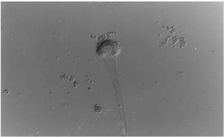
Pages Figure 1.1 Light microscopy of typical A. fumigatus sporulating structures. 1
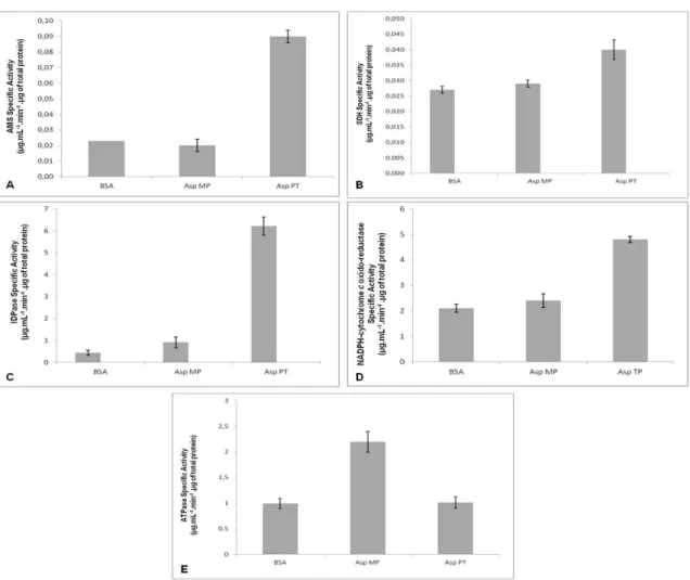
Figure 3.1 Measurement of marker enzyme specific activities from whole cells, total purified cell membrane sand bovine serum albumin (BSA, negative control). Protein extracts were prepared from whole cells (Asp PT) and total cell membrane fraction (Asp MP). The specific activities of AMS (A) (vacuole marker; specific activity was expressed in μg of p-nitrophenol reduced/ mL .min. μg total protein), SDH (B) (mitochondrial marker; specific activity was expressed in μg of DCPIP reduced/ mL .min. μg total protein), IDPase (C) (Golgi apparatus marker; specific activity was expressed in μg of Pi/ mL .min. μg total protein), NADPH-cytochrome c oxido-reductase (D) (endoplasmic reticulum marker; specific activity was expressed in μg of cytochrome c reduced/ mL .min. μg total protein), and ATPase (E)(plasma membrane marker; specific activity was expressed in μg of Pi produced per/ mL .min. μg total protein) were measured.
25
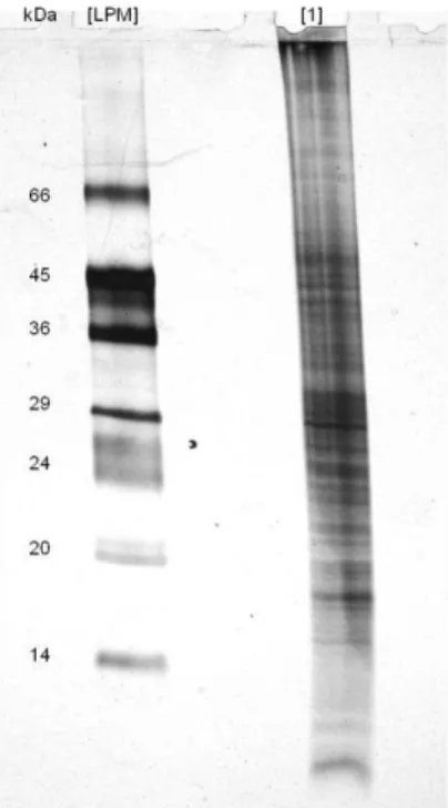
Figure 3.2 Electrophoretic analysis of A. fumigatus plasma membrane proteins, through a 17.5% (w/v) polyacrylamide gel. The lanes contain [LPM] low protein molecular mass markers (14-66 kDa). [1] Plasma membrane protein from A. fumigatus (100 μg). The gel was silver stained.
26
Figure 3.3 2DE electrophoresis of the cell membrane proteome, of A. fumigatus, through a 17.5% (w/v) polyacrylamide gel. Four hundred μg of total cell membrane protein was loaded. The gel was stained for total polypeptides with colloidal blue. In the left side, the molecular mass markers (14-66 kDa) are indicated.
27
Figure 3.4 Detection of glycoproteins in polyacrylamide gel using Pro-Q Emerald 300 dye. [LPM] Low protein molecular mass markers (14-66 kDa). [1] Electrophoretic analysis through a 17.5% (w/v) polyacrylamide gel of A. fumigatus plasma membrane proteins. The gel was stained with colloidal blue and 100 μg of total cell membrane protein was loaded. [2] Glycoprotein detection using Pro-Q Emerald 300 gel stain method after electrophoresis through a 17.5% (w/v) polyacrylamide gel (250 μg of total cell membrane protein was loaded).
29
Figure 3.5 Lectin affinoblot detection of cell membrane oligosaccharides of A. fumigatus, Polypeptides were separated by 1DE (through a 17.5% [w/v] polyacrylamide gel) and blotted onto a PVDF membrane. 150 μg of total cell membrane protein was loaded. [PM] Protein molecular mass markers (10-250 kDa). The affinoblots for oligosaccharide detection were performed with 4 different lectins, namely: [1] Con A; [2] PNA; [3] MAL; and [4] UEA.
30
Figure 3.6 Lectin affinoblot detection of cell membrane oligosaccharides of A. fumigatus, Polypeptides were separated by 2DE (through a 17.5% [w/v] polyacrylamide gel) and blotted onto a PVDF membrane. 500 μg of total cell membrane protein was loaded in each gel. [PM] Protein molecular mass markers (10-250 kDa). The affinoblots for oligosaccharide detection were performed with 4 different lectins, namely: (a) concanavalin A; (b) peanut agglutinin; (c) Maackia amurensis lectin; and (d) Ulex europaeus agglutinin.
31
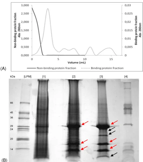
Figure 3.7 Electrophoretic analysis of the cell membrane proteome of A. fumigatus. (A) Affinity chromatography on Con A-Sepharose gel. Elution was performed with 0.5 M methyl-α-D-mannopyranoside, as the inhibiting sugar. One mL fractions of non-bound and bound protein were collected for each absorbance reading at 280 nm. (B) 1DE through a 17.5% (w/v) polyacrylamide gel was performed with the collected fractions of non-bound protein [2] and bound protein [3]. One hundred μg of total cell membrane protein sample [1] and a negative control [4] were also loaded. [LPM] Low protein molecular mass markers (14-66 kDa). The black arrows ( ) indicate the electrophoretic bands that were extracted from the gel for posterior protein identification by PMF. The red arrows ( ) indicate a possible contamination with Con A.
XVI
Figure 3.8 (A) Affinity chromatography of the cell membrane proteome of A. fumigatus on Affisep-PNA-adsorbent gel. Elution was performed with 0.5 M D -galactose, as the inhibiting sugar. One mL fractions of non-bound and bound protein were collected for each absorbance reading at 280 nm. (B) 1DE through a 17.5% (w/v) polyacrylamide gel was performed with the collected fractions of non-bound protein [2] and bound protein [4]. One hundred μg of total cell membrane protein sample [1] and a negative control [3] were also loaded. [LPM] Low protein molecular mass markers (14-66 kDa). The black arrows ( ) indicate the electrophoretic bands that were extracted from the gel for posterior protein identification by PMF.
35
Figure 3.9 (A) Affinity chromatography of the cell membrane proteome of A. fumigatus on Affisep-MAL-adsorbent gel. Elution was performed with 0.5 M α -lactose, as the inhibiting sugar. One mL fractions of non-bound and bound protein were collected for each absorbance reading at 280 nm. (B) 1DE through a 17.5% (w/v) polyacrylamide gel was performed with the collected fractions of non-bound protein [2] and bound protein [4]. One hundred μg of total cell membrane protein sample [1] and a negative control [3] were also loaded. [LPM] Low protein molecular mass markers (14-66 kDa). The black arrows ( ) indicate the electrophoretic bands that can be detected on lane [1] and [4], having the same molecular weight.
37
Figure 3.10 (A) Affinity chromatography of the cell membrane proteome of A. fumigatus on Affisep-UEA-adsorbent gel. (B) 1DE was performed with the collected fractions. (A) Affinity chromatography of the cell membrane proteome of A. fumigatus on Affisep-UEA-adsorbent gel. Elution was performed with 0.5 M L-fucose, as the inhibiting sugar. One mL fractions of non-bound and bound protein were collected for each absorbance reading at 280 nm. (B) 1DE through a 17.5% (w/v) polyacrylamide gel was performed with the collected fractions of non-bound protein [2] and bound protein [4]. One hundred μg of total cell membrane protein sample [1] and a negative control [3] were also loaded. [LPM] Low protein molecular mass marker (14-66 kDa). The black arrows ( ) indicate the electrophoretic bands that were extracted from the gel for posterior protein identification by PMF.
39
Figure 3.11 1DE comparison of the lectin-bound protein fractions collected from all four affinity chromatography columns. [LPM] Low protein molecular mass markers (14-66 kDa). The lanes correspond to the bound protein fractions of UEA [1], MAL [2], PNA [3] and Con A [4].
41
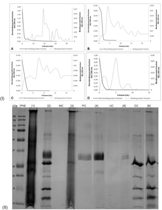
Figure 3.12 Sequential purification of lectin-binding proteins. (I) Affinity chromatography spectra for: (A) MAL, (B) PNA, (C) UEA and (D) Con A. (II) 1DE through a 17.5% (w/v) polyacrylamide gel was performed with the collected fractions of non-bound protein [2] and bound protein ([3] MAL; [4] PNA; [5] UEA; [6] Con A). One hundred μg of total cell membrane protein sample [1] and a negative control of each column ([MC] MAL; [PC] PNA; [UC] UEA; [CC] Con A) were also loaded. [PM] protein molecular mass markers (10-250 kDa).
42
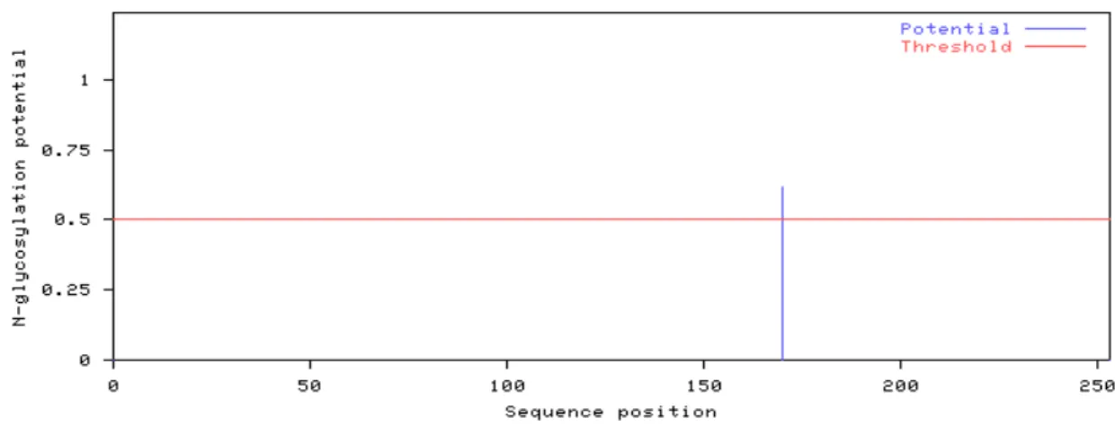
Figure 3.13 Predicted positions of N-glycosylated sites for the GPI anchor related
protein. 44
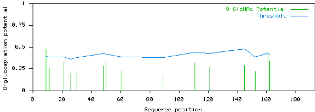
Figure 3.14 Predicted positions of O-glycosylated sites for the GPI anchor related protein.
45
Figure 3.15 Predicted positions of O-glycosylated sites for the 40S ribosomal protein
S9. 45
Figure 3.16 Predicted positions of N-glycosylated sites for the predicted protein. 46
XVII
Figure 3.18 Predicted positions of N-glycosylated sites for the fluconazole resistance protein.
47
Figure 3.19 Predicted positions of O-glycosylated sites for the fluconazole resistance
protein. 47
Figure 3.20 Predicted positions of N-glycosylated sites for the WD repeat protein. 48
Figure 3.21 Predicted positions of O-glycosylated sites for the WD repeat protein. 48
Figure 3.22 Predicted positions of N-glycosylated sites for the C6 transcription factor. 49
Figure 3.23 Predicted positions of O-glycosylated sites for the C6 transcription factor. 49
Figure 6.1 MALDI-TOF mass spectrum of Con A1 polypeptide. 57
Figure 6.2 MALDI-TOF mass spectrum of Con A2 polypeptide. 57
Figure 6.3 MALDI-TOF mass spectrum of Con A3 polypeptide. 58
Figure 6.4 MALDI-TOF mass spectrum of PNA 2 polypeptide. 58
Figure 6.5 MALDI-TOF mass spectrum of PNA 3 polypeptide. 59
XIX List of Tables
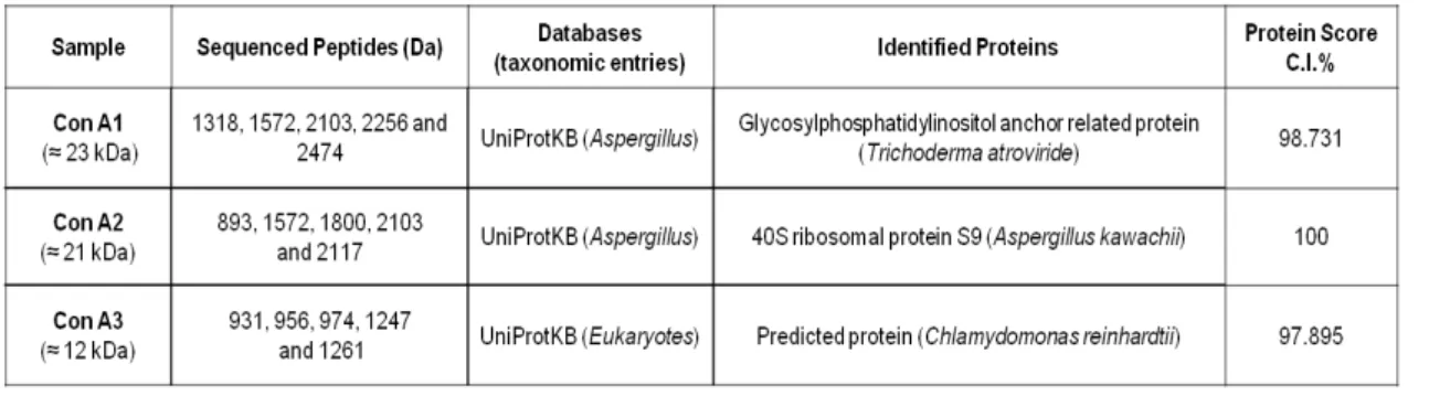
Pages Table 3.1 Protein identification determined by PMF. The protein samples were
extracted from the 1DE gel performed for the Con A affinity chromatography.
44
46 Table 3.2 Protein identification determined by PMF. The protein samples were
XXI List of Abbreviations
1DE One-dimensional electrophoresis
2DE Two-dimensional electrophoresis
AMS α-Mannosidase
Asn Asparagine
ATP Adenosine triphosphate
Con A Concanavalin A
DCPIP Dichlorophenolindophenol
DMF N,N-Dimethylformamide
DTT 1,4-Dithiothreitol
EDTA Ethylenediamine tetraacetic acid
EGTA Ethylene glycol tetraacetic acid
Flu1P Fluconazole resistance protein
G6PT Glucose-6-phosphate transporter
GalNAc N-Acetylgalactosamine
GlcNAc N-Acetylglucosamine
Glut4 Glucose transporter
GPI Glycosylphosphatidylinositol
H1N1 Influenza A virus
H5N1 Influenza A virus subtype
IA Invasive Aspergillosis
IDPase Inosine diphosphatase
IEF Isoelectric focusing
kDa Kilo Dalton
IPATIMUP Institute of Molecular Pathology and Immunology of the University of Porto
MAL Maackia amurensis lectin
MALDI-TOF-MS Matrix-assisted laser desorption/ionization – time-of-flight – mass spectrometer
MFS Major facilitator superfamily
MS Mass spectrometry
XXII mV Mili Volts
NADPH Nicotinamide adenine dinucleotide phosphate
PAMPs Pathogen-associated molecular patterns
pI Isoelectric point
Pi Inorganic phosphate
PMF Peptide Mass Fingerprint
PMSF Phenylmethylsulfonyl fluoride
PNA Peanut agglutinin
PRRs Pattern-recognition receptors
PVDF Polyvinylidene fluoride
ROS Reactive oxygen species
SDH Succinate dehydrogenase
SDS Sodium dodecyl sulfate
SDS-PAGE Reducing polyacrylamide gel electrophoresis
Ser Serine
TCA Trichloroacetic acid
Thr Threonine
TLRs Toll-like receptors
UEA Ulex europaeus agglutinin
UniProtKB UniProt Knowledgbase
v/v Volume/volume
w/v Weight/volume
1 1. Introduction
1.1 Aspergillus fumigatus
The genus Aspergillus, which includes almost 200 species, has a tremendous impact on public health, either beneficial as the workhorse of industrial applications and detrimental as plant and human pathogens (Dagenais and Keller, 2009).
The saprophyte Aspergillus fumigatus (Figure 1.1) has emerged as one of the most critical
fungal pathogens in distinct clinical settings, being a major threat to the immunocompromised individual. A. fumigatus can be found worldwide and plays an important role in recycling carbon
and nitrogen. Its natural ecological niche is the soil, wherein it survives and grows on organic debris (Latgé, 1999). This fungal species has a very simple biological cycle, one characteristic of which is its high sporulating capacity, which results in the ubiquitous presence of large numbers of conidia (1 to 100 conidia/m3) in the air indoors and outdoors (Latgé, 2001).
The conidia released into the atmosphere have a diameter small enough (2 to 3 μm) to reach
the lung alveoli (Latgé, 1999) and their dissemination simply relies on disturbances of the environment and strong air currents. In fact, A. fumigatus conidia are constantly inhaled by humans but rarely cause adverse effects, owing to a very successful response by the innate immunological system of healthy individuals. For most patients, disease occurs predominantly in the lungs, although dissemination to almost any organ may occur in the most severely predisposed. When a conidium overcomes the immune defense mechanisms, it germinates and produces a branched, septate vegetative mycelium that invades the lung tissues. The virulence of A. fumigatus can then be caused either by the production of fungal proteins that promote mycelia growth into the lung parenchyma and/or by structural features of the conidia which
confer resistance to the host’s antifungal mechanisms (Latgé, 2001). A compromised immune system due to immunosuppressive therapies and congenital defects may lead to the
2
development of invasive aspergillosis (IA) in experimental models, as well as in naturally occurring human infections. Under these conditions, the biological characteristics of the fungus (a fast-growing thermophillic species with small sized conidia and no specific nutritional requirements) may be sufficient for infection (Latgé, 2001).
In the past 10 to 20 years, the pathogenicity of this fungus has suffered several alterations due to the increasing number of immunosupressed patients and the severity of immunosupressing therapies. In spite of a pronounced increase in the incidence of IA, the pathogenicity of A. fumigatus is still poorly understood. Due to the unique nature of its infection process,
understanding its molecular basis, in particular the host defense reactions might be the key to develop new therapies effective on immunocompromised hosts.
1.2 Pathogenesis of Aspergillus fumigatus
Of all human pathogenic species of Aspergillus, A. fumigatus is the major causative agent of human infections, followed by A. flavus, A. terreus, A. niger and the model organism, A.
nidulans (Dagenais and Keller, 2009). Aspergillus species are exogenous fungi, which can
colonize the respiratory mucosa of patients with underlying localized bronchopulmonary disorders, such as healed tuberculous cavities, bronchiectasis, and cystic fibrosis, as well as occasionally invading the respiratory mucosa regardless of systemic immunological conditions (Amitani and Kawanami, 2009). The noninvasive aspergilloma may arise from repeated exposure of the host to conidia as well as from pre-existent pulmonary cavities, as healed lesions on patients that have suffered from tuberculosis. IA is the most critical disease caused by this fungus on immunocompromised patients. The most susceptible individuals to this life-threatening disease are individuals with hematological malignancies such as leukemia; solid-organ and hematopoietic stem cell transplant patients; patients on prolonged corticosteroid therapy, which is commonly utilized for the prevention and/or treatment of graft-versus-host disease in transplant patients; individuals with genetic immunodeficiencies such as chronic granulomatous disease; and individuals infected with human immunodeficiency virus (Dagenais and Keller, 2009). Mortality rates range from 40% to 90% in high-risk populations and are dependent on factors such as host immune status, the site of infection and the treatment regimen applied (Lin and Teutsch, 2001).
3
Saccharomyces cerevisiae) fungi. These studies showed that A. fumigatus does not share a
common set of genes with other fungal pathogens (Hohl and Feldmesser, 2007).
1.2.1 Establishment of the disease
The primarily way to a human fungal infection is through conidia inhalation, followed by their deposition on bronchioles or alveolar spaces. On healthy individuals, A. fumigatus conidia that do not endure mucociliary removal encounter epithelial cells or alveolar macrophages, responsible for phagocytosis and elimination of conidia, as well as for primary inflammatory response initiation; this process recruits neutrophils to the site of infection. Fungal attributes, which allow the survival and growth of A. fumigatus within the host, together with a dysfunction
on host defenses, increases the risk to develop IA. As conidia start developing, the factors which influence hyphal growth rate and resistance to infection mechanisms, invasion and dissemination, as well as secondary metabolite production, have a decisive effect upon disease establishment. However, these factors are secondary to the immune status of the host.
As mentioned before, there are two main pulmonary sites for infection: the lung epithelium and the alveola. Several observations (Latgé, 2003) suggest that the epithelium can serve as a focus of infection for IA establishment: (a) epithelial cells can engulf conidia which subsequently remain alive intracellularly; (b) the lung epithelium is damaged following immunosuppressive therapies and graft rejection, facilitating binding of conidia to altered or activated epithelial cells; and (c) corticosteroids, a risk factor for IA, reduce the release or efficacy of antimicrobial peptides and proteins constitutively synthesized at epithelial surface cells. With respect to epithelial cells, the soluble antimicrobial compounds that they secrete play a direct role in airway defense. Members of the defensin family of antimicrobial peptides have broad-spectrum activity against multiple microbes and are produced in vitro following the incubation of epithelial cells with A. fumigatus (Alekseeva et al., 2009; Dagenais and Keller, 2009).
4
associated with this fungus. This observation requires further analysis since limited density
testing of other aspergilla has been performed (Hohl and Feldmesser, 2007). Removal of sialic acid residues diminishes conidial phagocytosis, but not surface binding, by cultured macrophages and type II pneumocytes (Hohl and Feldmesser, 2007). Therefore, immune function may be enhanced by sialylation, which may also provide a protected reservoir for conidia.
Regarding the alveola, conidia that have not been trapped intracellularly at the epithelial level end up in the alveola where they encounter the main lung phagocytic cell, the alveolar macrophage (Latgé, 2003). Alveolar macrophages are the primary resident phagocytic cells of the respiratory tract and a critical component of the host defense against Aspergillus conidia. These cells are able to phagocytize Aspergillus conidia in an actin-dependent manner, a process mediated by the recognition of pathogen-associated molecular patterns (PAMPs) by host cell pattern-recognition receptors (PRRs), including toll-like receptors (TLRs) and dectin-1 (a lectin; Dagenais and Keller, 2009). Conidial engulfment is very quick, lasting from 1 to 2 h, and is not affected by the immune status of the host (Latgé, 2003).
In addition to these pathogenic attributes, A. fumigatus produces a variety of secondary
metabolites, including gliotoxin, which contributes to its pathogenicity, particularly during hyphal growth (Sugui et al., 2007; Dagenais and Keller, 2009). The biological activity of gliotoxin is
based on an internal disulfide bridge that can bind and inactivate proteins via a sulfide:thiol exchange, as well as via reactive oxygen species (ROS) produced by redox cycling between the oxidized and reduced forms of the toxin (Dagenais and Keller, 2009).
All the interactions mentioned above between fungal cells and host innate immune system, as well as interactions with the surrounding environment that may occur, rely upon a functional and permeable fungal cell wall. In fact, the initial sensing by the host innate immune system of fungal pathogens is most likely associated with components of the cell wall (Ferreira et al.,
2012). Moreover, localization of specific polysaccharides in the outermost part of the fungal cell wall is thought to be one of the first steps in the recognition of fungal pathogens by the host innate immune system (Ferreira et al., 2012).
As previously noted, phagocytosis of Aspergillus conidia is believed to be a process mediated by the recognition of PAMPs by host cell PRRs (Dagenais and Keller, 2009) This interaction is crucial to defeat microorganisms as it boosts phagocytosis and cytokine release (Heesemann et al., 2011), triggering a fierce chemical warfare between pathogen and host if pathogenesis is to
5
1.2.2 The fungal cell wall
All fungal cell walls are critical in maintaining cell shape and integrity in environments that range from the surface of grapes to human tissues, and are highly cross-linked structures, which adapt to highly variable growth conditions in a dynamic and flexible way (Cummings and Doering, 2009). The main structural components of the A. fumigatus cell wall are polysaccharides that can be divided in two groups, depending on their solubility in hot alkali (Bernard and Latgé, 2001). The alkali-insoluble fraction is primarily composed by galactomannans, chitin and β-1,3-glucans, and the alkali soluble fraction is composed mainly of
α-(1,3)-glucans with some galactomannan (Bernard and Latgé, 2001). Galactomannans are covalently linked to the non-reducing ends of β-1,3/1,4-glucans, and are mainly coated with glycosylphosphatidylinositol (GPI) proteins, which contain N- and O-glycans derived primarily
from the process of glycosylation (Cheng, 2011). Like all other eukaryotes, filamentous fungi possess three types of protein glycosylation, N-glycosylation of asparagines (Asn) residues, O-glycosylation of threonine (Thr) or serine (Ser) residues, and GPI-anchoring of the C-terminus of some proteins. Glycans are highly efficient vehicles for information storage and their biosynthesis requires a complicated non-template assembly line.
Carbohydrates from fungal cell walls have been used as prime targets to develop new drugs or by the pathogens themselves to their own benefit. For example, the antifungal echinocandins (e.g. caspofungin), target fungal cell wall β-1,3-glucan synthesis and are used clinically to treat yeasts (as Candida albicans) and IA (Ferreira et al., 2012). Nevertheless, they do not completely inhibit in vitro growth of A. fumigatus since they are active only at the tips and
branch points of the filaments, where β-1,3-glucan synthesis is most active (Ferreira et al., 2012). Many of these carbohydrates, more specifically oligosaccharides, comprise both the cell wall and the plasma membrane, which protects the cytoplasm from the extracellular milieu. The plasma membrane of fungal pathogens contains many glycoproteins and glycolipids, whose oligosaccharide side-chains, collectively termed the exoglycome (discussed in section 1.3.2), hence excluding cell wall carbohydrates, are projected towards the cell exterior (Ferreira et al.,
6
1.2.3 Fungal dissemination
A. fumigatus dissemination can occur through the lung and via the bloodstream to other organs.
The growing hyphae that escape host defenses may invade the endothelial cell lining of blood vessels to gain access to the vasculature. Hyphal invasion occurs from the abluminal side to the luminal side of endothelial cells, inducing endothelial cell activation but little cell damage (Kamai et al., 2006; Dagenais and Keller, 2009). Hyphal fragments can break off into the bloodstream
and invade the endothelium at other sites, resulting in hematogenously disseminated disease (Dagenais and Keller, 2009).
1.3 The fungal plasma membrane
The fungal plasma membrane is the target for the largest group of antifungal and antimicrobial proteins (Theis and Stahl, 2004). Over 500 naturally occurring proteins have been reported that are believed to interact with lipid components of the plasma membrane, leading to pore formation, efflux of cellular components, and changes in the membrane potential (Tossi et al., 2000).
The main function of the plasma membrane is to define the permeability barrier of cells and to serve as a matrix for proteins involved in a variety of cell functions such as energy targeting, signal transduction, solute transport, DNA replication, secretion, etc (Theis and Stahl, 2004). The cell membrane is formed by a lipid bilayer composed of sphingolipids, phospholipids, sterols and proteins. The plasma membrane proteins exhibit an enormous diversity of structures but share at least two common features (Ferreira et al., 2012): a positive net charge under physiological conditions, which promotes interaction with negatively charged microbial surfaces; and an amphipathic structure that allows incorporation into pathogen membranes.
The fungal plasma membrane differs from those of higher eukaryotes with respect to embedded sterols (Theis and Stahl, 2004): cholesterol (zoosterols), campesterol, sitosterol, and stigmasterol (considered phytosterols) or ergosterol (present in the cell membrane of fungi). Ergosterol, and the ergosterol pathway, is reported to be the target to most antifungal agents currently used for clinical treatment of fungal infections.
Furthermore, membrane-acting antifungal and antimicrobial proteins have been shown to interact with phospho- and sphingolipids, which make up the major part of the plasma membrane.
1.3.1 Membrane proteins
In A. fumigatus, a large number of proteins that play important roles in cell survival and invasion
are located in plasma membrane, endoplasmic reticulum, Golgi membrane, and other membrane systems (as mitochondria and vesicle-vacuole) (Ouyang et al., 2010).
7
attached to it is a variety of membrane proteins which enable efficient cell performance. Membrane proteins vary according to the type of cell and their subcellular location. Moreover, they can be classified as integral or peripheral, and are organized in different domains. Extramembrane domains are usually involved in the signaling pathway of cells or in cell-cell interactions. On the other hand, intramembrane domains facilitate molecule movement through the membrane, by channel formation. Furthermore, there are domains close to the intracellular membrane layer which display a broad spectrum of functions, such as structural protein anchoring to the membrane to engage intracellular signaling pathways. In many cases, membrane protein functions and their polypeptide chain topology can be predicted based on homology between these proteins and other well characterized membrane proteins.
Integral membrane proteins are characterized by a hydrophobic domain which interacts directly with the hydrophobic core of the lipid bilayer. Non ionic detergents are widely used in solubilization and characterization of these proteins (Bordier, 1981). These non ionic detergents (as Triton X-100, Triton X-114, Brij 35, Brij 58, Tween 20, Tween 80, Nonidet P-40, octyl β -glucoside and Mega 8) have a hydrophilic head, free of electric charge, and are used to shatter lipid-lipid or lipid-protein interactions. They also possess a limited capacity to break interactions between proteins and are usually used to isolate active membrane proteins; also, they are often referred to as non-denaturing detergents. During protein solubilization, these detergents substitute a large amount of lipid molecules that are in direct contact with the hydrophobic domain of membrane proteins, leading to the formation of micelles – globular structures formed by an aggregate of surfactant molecules. In contrast to integral membrane proteins, which possess amphiphilic structures, hydrophilic proteins show little or no interaction with non-ionic detergents and their physico-chemical properties are not influenced by the presence of such detergents in solution (Bordier, 1981).
1.3.2 The Exoglycome
The word exoglycome was proposed by Ferreira et al. (2012) to represent the oligosaccharide
side chains presented at the outer surface of the cell membrane, thus excluding the cell wall carbohydrates, whenever present, which tend to have a passive or “not-so-active” role in mediating cell communication. The exoglycome was then defined as the collection of carbohydrate moieties present in N- and O-linked glycoproteins and glycolipids which protrude outwards from the cell membrane (Ferreira et al., 2012).
8
Specific recognition processes between fungal parasites and their host cell targets may be mediated by the interaction of carbohydrate-binding proteins on the surface of one type of cell with complementary sugars on the surface of another (Ferreira et al., 2012).
1.3.3 Glycoproteins: N- and O-glycans
The glycome, which is the carbohydrate analog to the genome, proteome and metabolome, denote the total collection of carbohydrate species present in a biological unit (i.e. cell, tissue, organ, organism, species, and ecosystem) under any given set of environmental and physiological conditions. More recently, the concept of functional glycomics has emerged after discovering the array of biologically active roles played by certain carbohydrates, the set of which comprise the functional glycome. The exoglycome may therefore be considered as a subset of the functional glycome.
A. fumigatus cellular membrane glycoproteins contain different structures of oligosaccharide residues of great importance not only in cell biological processes but also in cell and cell-molecule recognition, including those relating to pathogenic agents. The extensive stereochemistry, multiple hydroxyl groups and oxygen atoms, and accessible hydrophobic regions make oligosaccharides ideal ligands for precise interactions with recognition sites in proteins (Albersheim et al., 1992). Regarding their role in biological processes, several theories
have been reported (Varki, 1993), such as, for example: an aid in the conformation and stability of proteins; the provision of target structures for microorganisms, toxins and antibodies; the masking of such target structures; control of the half-life of proteins and cells; the modulation of protein functions; and the provision of ligands for specific binding events mediating protein targeting, cell-matrix interactions or cell-cell interactions. Also, the ‘coating’ by oligosaccharides of many apoprotein surfaces on glycoproteins may serve to protect the polypeptide chain from
recognition by proteases or antibodies and the ‘coating’ of glycoconjugates covering a whole cell can present a ‘glycocalyx’ of substantial proportions (Varki, 1993).
The oligosaccharide side chains comprising the A. fumigatus exoglycome exhibit a large
capacity for storing biological information, but also a high antigenic potential. The oligosaccharides are expressed in a cell-type specific and temporally manner to allow cell phenotypes to change dynamically in response to environmental stimuli (Zaia, 2008). They are synthesized in a non-template controlled manner and mature structures arise by the coordinated expression of numerous genes that code for glycosyltransferases, glycosidases, and other enzymes that synthesize and remodel oligosaccharide chains, as well as accessory enzymes involved in the synthesis and transport of nucleotide sugars (Ferreira et al., 2012).
9
importance of this fungus in clinical studies has become quite relevant, it remains unclear whether the A. fumigatus exoglycome changes during a human fungal infection.
It is certainly difficult to predict specific rules for oligosaccharide functions due to the effects of a changing glycosylation pattern, even within a given group of proteins (e.g. cell surface receptors or enzymes), which may be highly variable and quite unpredictable (Varki, 1993). Moreover, the same modification in glycosylation can have a dramatically opposite effect on in vivo function versus that observed under in vitro conditions (Varki, 1993).
Several reasons make carbohydrates complex to analyze and very difficult to synthesize (Ferreira et al., 2012), including structural complexity of their linear or branched, oligo or
polymer structures; the different configurations/conformations monosaccharides can adopt (ring opened or closed, different ring sizes and conformations); isomeric diversity, including the two possible anomeric stereochemical linkages between units (α and β); diversity of secondary modifications of monomers (e.g. methylation, sulphation, acetylation and phosphorylation); different modes of attachment for cell-surface oligosaccharides (including glycolipids and N- or O-linked glycoproteins); their indirect relationship to the genome; the range of molecular
contexts in which the modifications are found and the fact that most carbohydrates lack chromophores or fluorophores, a property that makes their detection difficult.
From the technological point of view, a minimum amount of each oligosaccharide is required for the structural characterization of a fungal exoglycome. The extraction of a suitable amount of isolated fungal cells from the host turns out to be, most of the time, impossible, even from specific organs in which the fungus seems to be encountered in higher amounts (Ferreira et al., 2012).
1.3.3.1 N- and O-glycans
Glycosylation is the most universal and structurally diverse form of posttranslational modification of proteins, and occurs by the attachment of a glycan to a protein either at an Asn residue, termed N-glycosylation, or at a Ser, Thr, hydroxylysine or hydroxyproline residue, called O-glycosylation (Deshpande et al., 2008). Glycosylation can contribute to protein secretion, stability, and immunogenicity, and in the case of membrane glycoproteins and glycolipids, these
oligosaccharides may mediate a cell’s communication with the outside world (Deshpande et al., 2008).
10 1.3.3.1.1 N-Linked oligosaccharides
All N-linked oligosaccharides are derived from the precursor Glc3Man9GlcNAc2, which is
attached to the protein during translation and subjected to various modifications in the Golgi and in the endoplasmic reticulum (An and Lebrilla, 2011). In addition, N-glycans share a common pentasaccharide core region (Varki and Sharon, 2009) and can be generally divided into three main classes (Stanley et al., 2009): (i) high-mannose-type, in which only mannose residues are attached to the core; (ii) complex-type, in which “antennae” initiated by N-acetylglucosaminyltransferases (GlcNAcTs) are attached to the core; (iii) and hybrid-type, in which only mannose residues are attached to the Man-α-1,6 arm of the core and one or two antennae are on the Man-α-1,6 arm. These oligosaccharides are generally larger than O-linked
oligosaccharides (typically 10 to 20 monosaccharide residues) with a single common core (An and Lebrilla, 2011).
Maturation of N-glycans comprises several biosynthetic processes that can be divided into three components: sugar additions to the core, mostly occurring in the trans-Golgi network (a highly dynamic series of interconnected tubules and vesicles at the trans face of the Golgi stack); elongation of branching N-acetylglucosamine (GlcNAc) residues by sugar additions; and
“capping” or “decoration” of elongated branches (Stanley et al., 2009). The most important
“capping” or “decoration” reactions involve the addition of sialic acid, fucose, galactose, N-acetylgalactosamine (GalNAc), and sulfate (Stanley et al., 2009).
Yeasts and other fungi typically produce high-mannose-type N-glycans by adding up to 100 mannose units, including β-linked mannoses, whereas formation of mammalian oligosaccharides generally involves the removal of mannose, followed by the addition of GlcNAc, galactose, fucose, and sialic acid (Kainz et al., 2008). Filamentous fungi are known to carry small, high-mannose N-linked oligosaccharides (Deshpande et al., 2008).
Understanding N-linked oligosaccharides pathways is important because they affect many
properties of glycoproteins including their conformation, solubility, antigenicity, and recognition by oligosaccharide-binding proteins (Stanley et al., 2009). For example, defects in N-linked oligosaccharide synthesis lead to a variety of human diseases (Stanley et al., 2009).
Of all different types of glycosylation, the N-asparagine linked sugar chains are the easiest to manipulate in experimental systems (Varki, 1993), such as enzymatic or chemical removal of complete sugar chains, producing an aldehyde reducing end (An and Lebrilla, 2011); inhibition of initial glycosylation; alteration of oligosaccharide processing; elimination of specific glycosylation sites; and the study of natural variants and genetic mutants in glycosylation.
11
chromatography on specific oligosaccharide-binding proteins called lectins (Stanley et al.,
2009).
1.3.3.1.2 O-Linked oligosaccharides
An O-linked oligosaccharide (or O-(Ser/Thr)-linked oligosaccharide) is typically attached to the apoprotein moiety of a O-glycoprotein by linking a GalNAc residue to a Ser or Thr residue, and can be extended into a variety of different structural core classes (Varki and Sharon, 2009). O-Glycosylation is diversified according to the origins of organisms with respect to sugar components and the linkage modes among sugars (Goto, 2007).
Regarding protein O-glycosylation in fungi, the structures of the O-linked oligosaccharides are highly dependent on the fungi. In filamentous fungi, O-linked oligosaccharides containing branched structures of carbohydrate chains and sugars, except for mannose, are more variable than those of yeasts (Goto, 2007). O-Mannosylation is commonly found in glycoproteins of many higher eukaryotes as well as in most fungi, including the filamentous fungi (Deshpande et al., 2008).
A number of proteins localized in the cell wall, plasma membrane and organelles membranes are O-glycosylated and are related to proper cell morphology maintenance. However, very little
is known about the glycosylated proteins and their function in filamentous fungi (Goto, 2007).
1.3.4 Lectins
1.3.4.1 General approach
Lectins are proteins of non-immune origin (thus excluding immunoglobulins) which bind in a stable manner (thus excluding enzymes and carbohydrate sensor/transport proteins) to carbohydrates (Ribeiro et al., 2012). Lectins are a specific class of carbohydrate-binding
proteins or receptors with a role in decoding the oligosaccharide-encoded messages.
12
Lectins exhibit diversity of specificity towards simple sugars, although they regularly display a much higher binding affinity for oligosaccharides and glycoproteins, having the capacity to precipitate glycoproteins and branched oligosaccharides due to their usual multivalency (Ferreira et al., 2012). This specific recognition between lectins and carbohydrates is of great importance as they have been used as reliable biochemical, cytochemical and histochemical probes in the study of subtle differences between the cell surface glycoconjugates of malignant and non-malignant cells that are otherwise undetectable with the available monoclonal antibodies. Recognition of specific oligosaccharides by lectins may explain, for example, human infection by flu viruses, how swine H1N1 and avian H5N1 viruses cross the species barrier and infect the human population, and many other exoglycome changes involved in dissemination of fungal diseases such as IA.
Screening large numbers of lectins for the capacity to target such oligosaccharides alterations could provide ways for an early detection/control of such ailments (Ferreira et al., 2012). Nowadays, many different plant lectins have been detected, isolated and identified by conventional, laboratory biochemical techniques.
1.3.4.2 Physico-chemical properties
Although there are noticeable differences on carbohydrate specificity, lectins share common molecular properties as being usually comprised of two or four 25 to 30 kDa subunits (Lis and Sharon, 1986), each with a single, small carbohydrate combining site with the same specificity. Many are glycoproteins or metalloproteins containing a tightly bound Ca2+ and a transition metal ion, predominantly Mn2+, per subunit, which are required for carbohydrate binding (Lis and Sharon, 1998). Lectins combine with carbohydrates by a network of hydrogen bonds and hydrophobic interactions, and coordination with metal ions (Lis and Sharon, 1998). The hydrogen bonds are formed between carbohydrate hydroxyl groups and amino groups, hydroxyl groups, and oxygen atoms of the protein.
1.3.4.3 Lectin classification according to carbohydrate specificity
According to the monosaccharide ligand towards which lectins exhibit the highest affinity, they are classified in seven main groups: mannose and glucose, galactose/N-acetylgalactosamine (GalNAc), N-acetylglucosamine (GlcNAc), fucose, N-acetylneuraminic acid (sialic acid), mannose-6-phosphate, and N-acetylgalactose-4-sulfate (Ambrosi et al., 2005). All these sugars are in the D-configuration, except for fucose (Ambrosi et al., 2005).
Although lectins are generally classified in seven groups, in this dissertation, affinoblotting and affinity chromatography (lectin column chromatography) assays were developed with lectins belonging to only four groups, namely concanavalin A (Con A), peanut agglutinin (PNA),
Maackia amurensis lectin (MAL), and Ulex europaeus agglutinin (UEA). It has been reported
13
others, exhibit N-acetyl-D-glucosamine and methyl-α-D-mannoside residues (as shown by lectin-binding assays; Leal et al., 2011), which are recognized by MAL and Con A, respectively.
Moreover, it has been described that lectins may be of value as important tools in mycological diagnostics, as well as in identification and typing fungal agents of isolates in culture, tissue samples and fixed histopathologic specimens (Leal et al., 2011).
Con A (jack bean protein) belongs to the mannose/glucose group, which is the second major group. The ability of Con A to bind with high affinity to certain N-linked carbohydrates has made
it a widely used tool to investigate the properties of normal and transformed cells, as well as to isolate carbohydrates, glyconjugates, and cells on Con A-affinity matrixes (Mandal et al., 1994). As a tetramer, at neutral and alkaline pH, Con A consists of four identical subunits (Gunther et al., 1973), and each subunit contains a binding site which binds ligands with unmodified hydroxyls at positions 3, 4, and 6 of an α-glucopyranose or α-mannopyranose unit or residue (Weatherman et al., 1996).
PNA is also known as Arachis hypogaea lectin. PNA belongs to the galactose/ GalNAc group as well as the vast majority of lectins. It binds the carbohydrate sequence galactosyl-β(1,3) -N-acetylgalactosamine, and its activity is inhibited by lactose and galactose, which compete for the binding site of PNA (Lotan et al.,1975). A fairly stable and non-glycosylated lectin, PNA has
proven as a potential structure-specific probe in glycobiology, especially as it sharply discriminates between sialylated and non-sialylated forms of its most powerful inhibitor carbohydrate group, the galactosyl-β(1,3)-N-acetylgalactosamine, unlike the jack fruit seed lectin, jacalin (Chacko and Appukuttan, 2001).
MAL belongs to the N-acetylneuraminic acid group, and is commonly used as a glycoanalytical
tool to probe biological targets for α-2,3-linked sialic acids (Geisler and Jarvis, 2011). Immobilized MAL interacts with high affinity with complex-type Asn-linked oligosaccharides containing terminal sialic acid in α-2,3-linkage to galactose (Wang and Cummings, 1988). It has been referred that MAL binds most preferably to terminal α-2,3-Gal-β-1,4-Glc(NAc) in N-linked
oligosaccharides (Geisler and Jarvis, 2011).
UEA is associated to the fucose residue owing to its capacity to bind to many glycoproteins and glycolipids containing L-Fuc α1-2-D-Galβ1-4-D-GlcNAc residues, which is the highest affinity ligand known to UEA (Loris et al., 1998). Thus the inhibitory carbohydrate is α-L-fucose (fuc) (Blonski et al., 2007).
1.3.4.4 Applications
14
glycoconjugates and their preceding intracellular assembly and routing in normal and genetically engineered cells, analysis of mechanisms involved in correct glycosylation by lectin-resistant cell variants, detection of disease-related alterations of glycan synthesis, blood group typing and definition of secretor status, and cell marker for diagnostic purposes including infectious agents (viruses, bacteria, fungi, parasites).
1.4 Objectives
In order to achieve a better understanding of the host-induced changes upon the Aspergillus fumigatus cell membrane N-linked glycoproteins, this workplan aims, in a first stage, to identify
the main components of A. fumigatus cell membrane proteome as well as to detect and identify the glycoproteins present in the cell membrane. The identification of the N-linked oligosaccharides (herein simply referred to as the A. fumigatus exoglycome), that are projected into the external milieu from the fungal cell membrane surface, will also be performed.
15 2. Materials and Methods
2.1 Strain and cell culture
In this study the Asp1-56/03 strain (provided by the Instituto de Higiene e Medicina Tropical, New University of Lisbon) of Aspergillus fumigatus was inoculated in GYP medium (0.5% [w/v] yeast extract, 0.5% [w/v] peptone and 2% [w/v] glucose), for 2 to 3 days, at 37 °C, in a 3D culture system.
2.2 Membrane extraction and purification (Burghoom et al., 2002)
A. fumigatus cells were collected by filtering through cheesecloth, maintained at 0 to 4 °C and
resuspended in homogenization buffer (50 mM Tris-HCl pH 7.5, 0.3 M sucrose, 1% [w/v] glucose, 1 mM ethylenediamine tetraacetic acid [EDTA], 2 mM 1,4-dithiothreitol [DTT]) containing 1 mM phenylmethylsulfonyl fluoride (PMSF). The cells were broken in a French pressure cell at 20.000 lb/in2. After cell disruption, the cells were centrifuged in an Eppendorf F34-6-38 rotor for 20 min, at 3,024 g, recovering the supernatant. The crude cell membrane
fraction was pelleted from the supernatant with a second centrifugation in a Beckman SW48 rotor, for 1 h, at 66,000 g and 4 °C. Crude membranes were washed in membrane wash buffer
(10 mM Tris-HCl pH 7.5, 1 mM ethylene glycol tetraacetic acid (EGTA), 1 mM DTT, 20% [v/v] glycerol) containing 0.5 mM PMSF and resuspended in membrane wash buffer. Purified plasma membranes were collected at the 42%-62% (w/w) sucrose interface of a step gradient containing 1 mM EDTA, 1 mM DTT and 10 mM Tris-HCl (pH 7.0) after centrifugation for 3 h at 66,000 g in a Beckman SW48 rotor, at 4 °C. The membranes were washed in membrane wash buffer for 1 h at 66,000 g in the SW48 rotor and resuspended in membrane wash buffer. For subsequent use, the membrane sample was stored at -80 °C.
2.3 Extraction of A. fumigatus plasma membrane proteome
For A. fumigatus plasma membrane total protein extraction enough solid urea, ammonium
bicarbonate and sodium dodecyl sulfate (SDS) stock solution were added directly to the previously purified plasma membrane sample to prepare a denaturing buffer containing 8 M urea, 0.4 M NH4HCO3 and 0.1% (w/v) SDS. Afterwards, the sample was sonicated for 6 min at 4
°C with ultrasounds for homogenization.
2.4 Protein quantification
16
2.5 Marker enzymes of specific cell membrane systems
The catalytic activities of specific membrane marker enzymes were examined to confirm that the proteins extracted from Aspergillus fumigatus were indeed from the plasma membrane. The membrane markers used were from the vesicle-vacuole fraction (Chanda et al., 2009), mitochondrial fraction (Chanda et al., 2009), endoplasmic reticulum (Ugalde et al., 1992), Golgi apparatus (Ugalde et al., 1992) and plasma membrane (Perzov et al., 2000).
2.5.1 Enzyme activity from the vesicle-vacuole membrane system
For the vesicle-vacuole membrane system, the catalytic activity of α-mannosidase (AMS) was assayed according to the method described by Chanda et al. (2009). Ten μL of the A. fumigatus
purified plasma membrane sample (total protein range from 3 to 10 μg) was added to a reaction mix containing 0.5 mL of sodium succinate buffer (50 μM Na-succinate, pH 5.0); then, 3 μL of 0.1 M p-nitrophenyl substrate was added. The reaction was stopped by addition of 0.8 mL of 1 M Na2CO3 and the absorbance determined at 405 nm. The specific activity was expressed in
nmol of p-nitrophenol produced per min per μg total protein.
2.5.2 Enzyme activity from the mitochondrial membrane system
For the mitochondrial membrane system, the catalytic activity of succinate dehydrogenase was measured according to Chanda et al. (2009). Ten μL of the A. fumigatus purified plasma
membrane sample (total protein range from 3 to 10 μg) was added to a reaction mixture consisting of 0.3 M KH2PO4, 8.5 mM KCN and 50 μg of dichlorophenolindophenol (DCPIP, Na
salt). Sodium succinate buffer (50 μM Na-succinate, pH 5.0) was then added to this reaction mixture and the absorbance was recorded at 600 nm. Enzyme specific activity was expressed
in μmol DCPIP reduced per minute per μg total protein.
2.5.3 Enzyme activity from the endoplasmic reticulum membrane system
NADPH-cytochrome c oxido-reductase was used as an endoplasmic reticulum membrane marker according to Ugalde et al. (1992). The reduction of cytochrome c was measured at 550 nm in the A. fumigatus purified plasma membrane sample. The 0.6 mL reaction mixture contained 0.45 mM cytochrome c, 50 mM KH2PO4 pH 7.2, and 3 mM NADPH, plus 50 mM NaCN and 0.4 μM antimycin A, which were included to inhibit cytochrome oxidase activity. The
reaction was started with 20 to 50 μg protein.
2.5.4 Enzyme activity from the Golgi apparatus membrane system
Inosine diphosphatase (IDPase) activity, a commonly used marker of the Golgi body membrane system, was measured by the liberation of inorganic phosphate (Pi), according to Ugalde et al.
(1992), associated with IDP hydrolysis after incubation for 10 min at 30 °C in a 0.5 mL reaction mixture containing 5 mM IDP, 5 mM MgCl2, 10 mM piperazine-N,N-bis(2-ethane sulfonic acid
17
membrane protein. The reaction was stopped by addition of 0.1 mL 50 % (w/v) trichloroacetic acid (TCA). The liberation of inorganic phosphate (Pi) was measured at 700 nm.
2.5.5 Enzyme activity from the plasma membrane system
ATPase activity was used as a plasma membrane marker. According to the method described by Perzov et al. (2000), ATP hydrolysis associated to the A. fumigatus plasma membrane was assayed in 0.5 mL of a reaction medium consisting of 5 mM ATP (vanadate-free), 5 mM KCN, 1 mM NaN3, and 10 mM 2-(N-morpholino)ethanesulfonic acid (MES)-KOH (pH 6.0) in the presence of 100 μM NaVO3. The reaction was initiated by the addition of 0.1 mL from A.
fumigatus plasma membrane gradient fractions, and allowed to proceed for 10 to 20 min at 30
°C. The ATPase activity was measured by the release of Pi associated to ATP hydrolysis according to the method described by Ames (1966), and the change in absorbance recorded at 700 nm.
2.6 Reducing, denaturing polyacrylamide gel electrophoresis (SDS-PAGE)
For one-dimensional electrophoresis (1DE), the A. fumigatus plasma membrane proteome was washed with 10% (w/v) TCA to remove contaminants, and centrifuged at 15,800 g, for 10 min.
The pelleted fraction was resuspended in 1 mL of acetone, and kept at -20 °C for 30 min. After protein precipitation, the sample was again centrifuged at 15,800 g, for 10 min. The pellet was
finally resuspended in sample buffer (2% [w/v] SDS, 0.08 M Tris-HCl pH 6.8, 0.1 M β -mercaptoethanol (ME),15% glycerol and 0.006% m-cresol purple) for electrophoresis. To achieve full protein denaturation, the sample was heated at 100 °C for 3 min in a water bath. Protein separation by SDS-PAGE was performed in a discontinuous buffer system according to the method proposed by Weber and Osborn (1969) and Laemmli (1970):
- Separation gel: 12.5% (w/v) acrylamide, 0.1% (w/v) bis-acrylamide, 375 mM Tris-HCl pH 8.8, 0.03% (v/v) tetramethylethylenediamine (TEMED) and 0.03% (w/v) ammonium persulfate (PSA);
- Concentration gel: 5% (w/v) acrylamide, 0.13% (w/v) bis-acrylamide, 125 mM Tris-HCl pH 6.8, 0.05% (v/v) TEMED and 0,1% (w/v) PSA.
Electrophoresis took place under constant current intensity and voltage (70 mA and 220 V, respectively) conditions and 220 V.
2.7 Two-dimensional electrophoresis (2DE)