Surface photochemistry: benzophenone as a probe for the study of silica and
reversed-phase silica surfaces†
L. F. Vieira Ferreira,*
aI. Ferreira Machado,
aJ. P. Da Silva
a,band T. J. F. Branco
a Received 11th January 2006, Accepted 27th March 2006First published as an Advance Article on the web 7th April 2006 DOI: 10.1039/b600384b
This work reports the use of benzophenone, a very well characterized probe, to study new hosts: two reversed-phase silicas. Laser-induced room temperature luminescence of argon purged solid powdered samples of benzophenone adsorbed onto the two different reversed-phase silicas, RP-18 and RP-8, revealed the existence of a low energy emission band in contrast with the benzophenone adsorbed on 60 A˚ pore silica, where only triplet benzophenone emits. This low energy emission band was identified as the fluorescence of the ketyl radical of benzophenone, which is formed as the result of a hydrogen atom abstraction reaction of the triplet excited benzophenone from the alkyl groups of the surface of the reversed silicas. Such emission does not exist for benzophenone adsorbed onto 60 A˚ pore silica. Room temperature phosphorescence was obtained in argon purged samples for all the surfaces under use. The decay times of the benzophenone emission vary greatly with the alkylation of the silica surface when compared with “normal” silica surface. A lifetime distributions analysis has shown that the shortest lifetimes for the benzophenone emission exist in the former case. Triplet–triplet absorption of benzophenone was detected in all cases and is the predominant absorption in the case of 60 A˚ pore silica, while benzophenone ketyl radical formation occurs in the case of the reversed silicas. Diffuse reflectance laser flash photolysis and gas chromatography-mass spectrometry techniques provided complementary information, the former about transient species and the latter regarding the final products formed after laser irradiation, both at 266 nm or 355 nm. Product analysis and identification show that the degradation photoproducts are dependent on the excitation wavelength, the
photochemistry being much more rich and complex in the 266 nm excitation case, where an a-cleavage reaction occurs. A detailed mechanistic analysis is proposed.
1.
Introduction
Time resolved laser induced luminescence, diffuse reflectance laser flash-photolysis and ground state diffuse reflectance absorption spectroscopy are relatively new techniques that can be applied
to study opaque and crystalline systems.1–8 These solid state
photochemical methods have been recently applied by our group to study several organic compounds adsorbed onto different hosts such as p-tert-butylcalix[4], [6] and [8]arenes and derivatives,9–11
mi-crocrystalline cellulose,12–14silicalite and cyclodextrins,15–17silica18
and resorcinarene frameworks,19among others.
Benzophenone (BZP) is an extremely useful molecule for prob-ing new hosts. The n→ p* absorption transition was found to be very sensitive to the environment characteristics and also exhibits a photochemistry which depends on the host properties.9–10,12,20
In a very recent paper,20 we reported a comparative study of
the luminescent properties of BZP adsorbed onto a mesoporous channel-type solid support material called MCM-41 with other nano-porous materials, namely silicalite (a de-aluminated zeolite)
aCentro de Qu´ımica-F´ısica Molecular-Complexo Interdisciplinar, Instituto
Superior T´ecnico, Av. Rovisco Pais, 1049-001, Lisboa, Portugal. E-mail: [email protected]
bFCT, Universidade do Algarve, Campus de Gambelas, 8005-139, Faro,
Portugal
† Electronic supplementary information (ESI) available: Fig. S1 and S2. See DOI: 10.1039/b600384b
and H-ZSM-5 (with an acidic internal surface). Triplet state luminescence was observed in all cases, hydrogen bonded excited benzophenone was detected on MCM-41 while protonated excited BZP luminescence was observed only on H-ZSM-5. Time resolved diffuse reflectance absorption technique was also applied to the powdered solid samples of these compounds proving undoubtedly the inclusion of BZP within the cavities of these three hosts, and the existence of triplet state of BZP in air equilibrated samples in all cases, following laser excitation. BZP•OH radical was also detected and is one of the important intermediates.
Reversed-phase silicas are used in reversed-phase chromatogra-phy, where the stationary phase is non-polar (often a hydrocarbon) and the mobile phase is a relatively polar or polar solvents such as methanol or acetonitrile and water, or a mixture of them.21
These silicas have octyl- or octadecyl siloxane groups on the silica surface (the silanol groups of the “normal” silica surface ≡SiOH react with Cl(CH3)2Si–R in acidic hot media, forming the
organosiloxane,≡SiO–(CH3)2Si–R and HCl, where R=
octyl-or octadecyl-groups). These long hydrocarbon groups are aligned parallel to one anther and perpendicular to the surface of the particle, forming a brush-like, non-polar silica surface.21 Many
successful partition chromatography separations were achieved with the use of these materials.
This work reports the use of benzophenone, a very well char-acterized probe, to study new hosts (two reversed-phase silicas). In these hosts all surface silanols were replaced by alkylsiloxane
groups with alkyl chains with eighteen or eight carbon atoms, therefore transforming the polar surface of the normal silicas into surfaces with a non-polar character. A comparison of the
photochemical behaviour of BZP was made with 60 A˚ pore size
silica.
Experimental evidence will be presented for the first time, showing the occurrence of a photochemical reaction of benzophe-none on the surface of the reversed-phase silica, resulting in the formation of a new species, the diphenylketyl radical, which is also excited within a single laser pulse and emits its own lumine-scence.
A detailed chromatographic study of the laser (and lamp) irradiated samples was made, which provided experimental results pointing to a clear dependence of the photochemistry on the excitation wavelength under use.
2.
Experimental
MaterialsSilicalite (Union Carbide) and 60 A˚ pore silica gel (Merck, 70– 230 mesh) were used as powdered solid supports as received. Benzophenone (Koch-Light, Scintillation grade) and the solvents hexane, dichloromethane, iso-octane, methanol and acetonitrile were from Merck, LiChrosolv grade. These compounds were first analysed by chromatography before use.
RP-18 and RP-8 were purchased from Merck (LiChrosorb grade) and used as received. The specific surface areas indicated by the producer are 150 and 250 m2g−1respectively.
Sample preparation
The samples used in this work were prepared using the solvent evaporation method. This method consists in the addition of a solution containing the probe to the previously dried powdered solid substrate, followed by solvent evaporation from the stirred slurry in a fume cupboard. Hexane or dichloromethane were used for sample preparation in the case of the 60 A˚ silica and for the reversed-phase silicas because of the high solubility of BZP in these solvents and also because these solvents evaporate easily at room temperature in the fume cupboard. In the case of the BZP/silicalite samples iso-octane was used for BZP inclusion into the channels of the adsorbent, because the solvent molecule is not able to penetrate into the channels due to its molecular dimensions. The final solvent removal was performed overnight in an acrylic chamber with an electrically heated shelf (Heto, Model FD
1.0-110) with temperature control (30± 1◦C) and under moderate
vacuum at a pressure of ca. 10−3Torr.
Solvent evaporation in the fume cupboard was done in the dark. All samples are kept in the dark before the experiments are performed. The experiments are also performed in dark conditions so that only the light arising from the excitation sources (lamps and lasers) reaches the samples under study.
Methods
1. Ground state diffuse reflectance absorption spectra (GSDR).
Ground state absorption spectra for the solid samples were
recorded using an OLIS 14 spectrophotometer with a diffuse reflectance attachment. Further details are given elsewhere.1,12–14
2. Laser induced luminescence (LIL) and diffuse reflectance laser flash photolysis (DRLFP) systems. Schematic diagrams of the LIL and of the DRLFP systems are presented in ref. 1 and 16. Laser flash photolysis experiments were carried out with the third or the fourth harmonic of a Nd:YAG laser (355 and 266 nm,
ca. 6 ns FWHM, ∼10–30 mJ pulse−1) from B. M. Industries
(Thomson-CSF, model Saga 12-10), in the diffuse reflectance mode. The light arising from the irradiation of solid samples by the laser pulse is collected by a collimating beam probe coupled to an optical fiber (fused silica) and is detected by a gated intensified charge coupled device Oriel model Instaspec V, (Andor ICCD, based on the Hamamatsu S57 69-0907). The ICCD is coupled to a fixed imaging compact spectrograph (Oriel, model FICS 77441). The system can be used either by capturing all light emitted by the sample or in a time-resolved mode by using a delay box (Stanford Research Systems, model D6535). The ICCD has high speed gating electronics (2.2 ns) and intensifier and covers the 200–900 nm wavelength range. Time-resolved absorption and emission spectra are available in the nanosecond to second time range. Transient absorption data are reported as percentage of absorption (% Abs.), defined as 100DJt/Jo= (1 − Jt/Jo)100, where Joand Jtare diffuse reflected light from sample before exposure to the exciting laser pulse and at time t after excitation, respectively. For laser-induced
luminescence experiments with a N2laser (PTI model 2000, ca.
600 ps FWHM,∼1.1 mJ pulse−1), was also used. In this case
the excitation wavelength is 337 nm. With these set-ups, both fluorescence and phosphorescence spectra are easily available (by the use of the variable time gate width and start delay facilities of the ICCD).
3. Irradiation and product analysis. Photodegradation stud-ies under lamp irradiation were conducted in a reactor previously used to study the photochemistry of several compounds at the solid/gas interface.22,23Lamp irradiation was performed at 254 nm
using a 16 W low-pressure mercury lamp (Applied Photophysics) without filters and without refrigeration and a xenon lamp with a glass filter. The samples were irradiated in a quartz cell placed at 1 cm from the lamp surface, in air equilibrated conditions, during 3.5 h. Laser irradiation at 355 nm and 266 nm, in argon atmosphere, was also performed. In this case the samples were irradiated in a quartz cell during 0.5 h at five pulses (30 mJ) per second. The samples were mixed every 5 min during the laser irradiation process. Non-irradiated and irradiated samples were analysed after extraction with acetonitrile (a known weight of sample in a known volume of solvent) followed by centrifugation. Photolysis was followed by HPLC using a Merck-Hitachi 655A-11 chromatograph equipped with detectors 655A-22 UV and Shimadzu SPD-M6A photodiode array. A column LiChroCART 125 (RP-18, 5 lm) Merck was used and the runs were performed using mixtures water–acetonitrile. The extracts were also analysed by GC-MS using a Hewlett Packard 5890 Series II gas chromato-graph with a 5971 series mass selective detector (E.I. 70 eV). A Restek RTX-20 capillary column with 20 m and 0.18 mm id was used. The initial temperature 70◦C was maintained during 5 min and then a rate of 5◦C min−1was used up to 250◦C.
3.
Results and discussion
Ground state diffuse reflectance absorption spectra
Ground state diffuse reflectance absorption spectra for BZP adsorbed onto RP-18, RP-8, 60 A˚ pore silica and silicalite, were obtained with the use of an integrating sphere.1,12–14
The BZP ground state absorption S0 → S1 transition (n →
p*) has an absorption maximum at about 347 nm for silicalite as host material and a clear vibrational structure characteristic of the carbonyl group in the excited state24can be observed when BZP
is included into the rigid channel structure of this adsorbent (see Fig. S1, in the electronic supplementary information, ESI).†
Silicalite is a de-aluminated analogue of ZSM-5 zeolite, with straight and zig-zag channels interconnected. Due to the lack of substitutional aluminium, this adsorbent has no catalytic or exchange properties when compared to the ZSM-5 zeolites. Silicalite is the only known hydrophobic form of silica and is capable of adsorbing organic molecules up to about 6 A˚ of kinetic diameter, even removing them from water. It provides a very rigid and hydrophobic environment for the BZP adsorption inside both channels25and this is reflected in the vibrational structure of both
absorption and emission spectra.15
In the case of the other three surfaces, RP-18, RP-8 and
60 A˚ silica (all curves were normalized at 315 nm and the BZP
concentration was 250 lmol g−1), the absorption curves appear
now more and more as broad bands, shifted to the blue in that order. These hypsochromic shifts are quite characteristic of the n→ p* transition with increasing polarity of the surface.12,15,20The
broadening of the spectra is probably related to both heterogeneity of the adsorbent and a much smaller rigidity of the adsorbed probe. Solution absorption spectra of BZP, for instance in cyclohexane and ethanol, also exhibit this type of influence of polarity, characteristic of the n→ p* transition.24
Clearly, going from RP-18 to RP-8 and to 60 A˚ silica, an increase in the surface polarity is observed, quite consistent with the surface characteristics: long alkyl chains with 18 carbons in RP-18, only 8 carbons in RP-8 and contact with the hydroxyl groups of the surface silanols in the case of 60 A˚ silica.
Room temperature laser induced phosphorescence
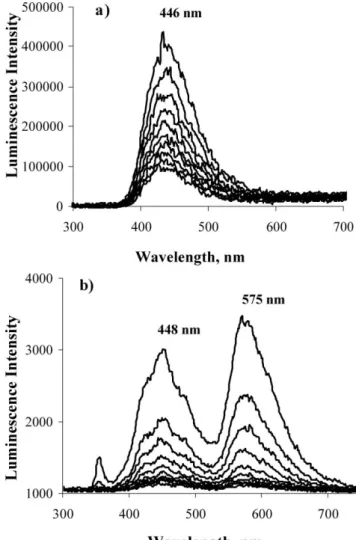
Fig. 1 presents the room temperature phosphorescence spectra of BZP onto the surfaces of the 60 A˚ pore silica gel (part a) and of the RP-18 reversed-phase silica (part b).
Similar luminescence data for BZP adsorbed on silicalite and included into the cavities of calixarenes are presented in ref. 15 and 9, 10 respectively. Those time resolved spectra were obtained with air equilibrated conditions and were identical to the ones obtained with argon purged samples. Lifetimes of about 80 ls were determined for the calixarene inclusion9–11and 3.1 ms for
inclusion into the narrower channels of silicalite,25as compared to
about 50 ls for benzophenone microcrystals, all determined at the
maximum emission wavelength (about 448 nm).12
The luminescence spectrum of benzophenone adsorbed onto reversed-phase silica surface does not present a rise time in a nanosecond timescale, as Fig. 1a shows. Other time resolved spectra in a 1–10 ns timescale were recorded (data not presented in the paper) and confirm this behaviour.
Fig. 1 a) Laser induced phosphorescence spectra for argon purged
samples of benzophenone adsorbed onto 60 A˚ pore silica. The excitation wavelength was 337 nm and curves were recorded every 1 ms after the laser pulse (t0 = 1 ms). b) Laser induced phosphorescence spectra for
argon purged samples of benzophenone adsorbed onto reversed-phase silica (RP-18). The excitation wavelength was 355 nm and curves were recorded every 50 ns after the laser pulse (t0= 0 ns).
It is important to note that the emission of BZP on the silica gel surface peaks at 446 nm and that this emission is very broad when compared to the emission into the silicalite channels or within the calixarenes cavity, in accordance with the very broad absorption spectrum. The heterogeneity of the adsorbent is also reflected in the wide range of lifetimes determined for this phosphorescence emission of BZP as we will show now.
Recently we have developed a new tool for the lifetime distribu-tions analysis of emissions of probes adsorbed onto heterogeneous
surfaces.25 This new methodology allows for asymmetric
distri-butions and uses Voigt profiles (Gaussian–Lorentzian product) instead of pure Gaussian or Lorentzian distributions. A very simple and widely available tool for fitting has been used, the Microsoft Excel Solver. This is a very convenient way to treat the emission or transient absorption decay data because it reflects the multiplicity of sites available for the probe onto the specific surface under study.
A detailed study of the luminescence decay curves of pyrene in-cluded within p-tert-butylcalix[4]arene cavities and benzophenone into silicalite channels has been reported recently.25
The use of the sum of a few exponentials to analyse the decay of probes onto heterogeneous surfaces is a description without physical meaning,25,26 and the best tool to study decays
of a probe onto heterogeneous surfaces is a lifetime distribution analysis (as Scaiano and co-workers did recently, e.g. ref. 26– 28). The validation of its conclusions being sustained by other spectroscopic studies.
There was no need here for the use of convolution with the excitation pulse because decay times analysed are in the millisecond or hundred of nanoseconds time domain and the excitation pulse width was 0.6 ns. Modelling studies have shown that for decays with lifetimes greater than 3 ns the decay curve is not affected by this excitation pulse.
When applied to the BZP/60 A˚ silica case (Fig. 2a), and
after recording the decay traces within various instrumental timescales (experimental information was obtained starting in the nanosecond timescale and also in the 0.01 ls or longer timescales), the lifetimes distribution analysis evidences a broad band centred at 0.3 ls, which we assigned to different conformations of adsorbed BZP due to different adsorption sites of the silica gel surface (the 60 A˚ size reported for the pore average diameter means that the
maximum of the distribution of the pore sizes is at 60 A˚ but,
of course, larger and smaller pores exist, as is clearly indicated for several silica gels in ref. 29), therefore resulting in different conformations for BZP and different emissions. However Fig. 2a also shows a peak at 4.2 ms, showing that some fraction of adsorbed benzophenone molecules is very rigidly adsorbed in
Fig. 2 Lifetime distributions recovered from luminescence decays of
argon purged samples of a) benzophenone adsorbed onto 60 A˚ pore silica, observed at 450 nm; b) benzophenone adsorbed onto reversed-phase silica (RP-18), observed at 450 nm or 575 nm. The insets show the fitting of the recovered decay superimposed on the experimental data.
Scheme 1 Schematic representation of the surfaces of a small pore silica
and also of RP-18 and RP-8.
some cases and emits with a long lifetime, comparable to the one of silicalite adsorption into the narrower channels.25 Some
of the pores are particularly suitable for BZP, as the schematic representation presented in Scheme 1 suggests, showing different possibilities for hydrogen bond formation to the silanols of the surface.
Two different time gates were used for the BZP/60 A˚ silica
case. Data presented in Fig. 1a were obtained with the use of a very large time gate (20 ms width) to record all room temperature phosphorescence. A small time gate (50 or 100 ns width) was also used to show the short time behaviour immediately after the laser excitation pulse. A special emission of BZP in the nanosecond timescale was detected, very similar to the one reported in Fig. 3b of ref. 20 for BZP on MCM-41. This emission peaks at ca. 430 nm, and for these reasons, we assign again this emission to hydrogen bonded BZP emission (data not shown).
In the case of the BZP/RP-18 sample, the use of a small time gate width or large gate width enabled us to observe only one time resolved luminescence spectrum and this means that there is no hydrogen bonded BZP emission in the nanosecond timescale in this case: superimposed on the normal benzophenone phosphorescence emission we detected the appearance of a low energy emission band, which peaks at 575 nm immediately after the laser pulse. This low energy emission band is shown in Fig. 1b. Very similar results (not shown) were obtained for the BZP/RP-8 samples.
All experimental luminescence results presented until now for
BZP/60 A˚ silica, BZP/RP-18 and BZP/RP-8 samples were
obtained with the use of argon purged samples. Air equilibrated surfaces do not provide detectable emissions or only provide small emissions, showing the oxygen quenching of the excited species is very effective for these powdered solid silica surfaces.
All results at timescales shorter than 6 ns (laser pulse width) are simply a reflection of the forced shape of the distribution.
In what regards the assignment of the low energy emission band for the BZP/RP-18 or BZP/RP-8 samples two main hypotheses were assumed as starting points: emission from transient species derived from benzophenone alone or from new species resulting from benzophenone photochemical reaction with the host. The second hypothesis seems to be the most logical one. The simple fact that the low energy emission band peaks at 575 nm (and its lifetime) points to be an emission from the ketyl radical of
benzophenone which emits at this wavelength range, as the result of a two photon excitation process (fluorescence emission).30–32It also
excludes emissions from aryl alkyl ketones which phosphoresces around 420 nm.16,33,34
Previous work indicates that calixarene,9–11cellulose,12–14or
b-cyclodextrin27are good hydrogen atom donors towards BZP (in
solid powdered samples), so it sounds reasonable to assume that efficient ketyl radical formation of benzophenone may also occur here.
The lifetime emission distribution for this case is presented in Fig. 2b: only one emissive species is detected (a single distribution of lifetimes peaking at 110 ns), with a much shorter lifetime when compared with the BZP emission onto the porous silica. Other important experimental observation is that the same lifetime distribution curve was obtained at 450 nm or 575 nm, indicating that the lifetime of the ketyl radical emission is the same as the
3BZP* emission. From solution data in the literature, we know
that the lifetime of the excited diphenylketyl radical is ca. 4 ns in solvents such as toluene or acetonitrile at room temperature.30
How is it possible that on the RP-18 surface this species lives hundreds of nanoseconds?
Two species are emitting on the RP-18 surface,3BZP* and the
excited diphenylketyl radical. The hydrogen atom abstraction from the alkyl chain is competing with the phosphorescence emission and a remarkable quenching effect exists, therefore reducing the lifetime emission from millisecond to about one hundred nanoseconds. This is a much longer lifetime when compared with the excited diphenylketyl radical and this means that this species only has a pseudo lifetime of 110 ns. The emission of the
excited diphenylketyl radical is the same as the one of3BZP*
because the former excited species is formed from an energy
transfer process from 3BZP* and emits in an “instantaneous”
manner when compared with the energy donor, therefore in a time resolved spectra, it exhibits the same lifetime of the species which is in its origin. The ketyl radical emission exists with one single laser pulse and could be recorded immediately after that laser pulse. In this case, and taking into account the pulse duration (about 6–7 ns for 355 nm excitation), the ketyl radical can be formed, excited and also emit during one laser pulse. At later times it fluoresces as soon as it is formed and excited by energy transfer.
This argument for the excitation of the ketyl radical at times larger than the excitation laser pulse, by an energy transfer mechanism (triplet–doublet energy transfer) was reported before for solution studies by Naqvi and Wild31and Netto-Ferreira and
Scaiano.30In the solution case a rise time was detected for the ketyl
emission.31When BZP is adsorbed onto the reversed-phase silica
surface no rise time was detected in the nanosecond timescale. This is due to the confinement of the donor and acceptor species (3BZP* and BZPH•radical are closely entrapped within the long alkyl chains of the reversed-phase silica surface).
The high intense fluorescence band of the diphenylketyl radical is easily detected due to its relatively high fluorescence quantum yield, which is 0.11± 0.02 in benzene, as reported in ref. 35.
For low loadings (∼10 to 50 lmol of BZP per gram of the RPS) the ketyl emission is hardly detectable and this is consistent with the fact that the energy transfer mechanism responsible for the observed emission of the excited diphenylketyl radical is concentration dependent.
By keeping the samples in the dark we prevent the possibility of the room light being absorbed, therefore producing ketyl radicals before DRLFP experiments were performed. Also the possibility that the analysing light from the DRLFP set-up produced ketyls that stayed entrapped was excluded by performing ground state diffuse reflectance absorption spectra with our diffuse reflectance laser flash photolysis set-up. The ketyl radicals were only detected after laser pulse excitation.
In order to perform the LDA analysis, several decay curves in different instrumental timescales were recorded. A superposition of those decay traces was made by normalization of each decay curve at a time range where they overlap, in order to produce a composite decay with closely spaced data at short times and larger spaced values at long times. This procedure was adopted before (e.g. ref. 26,36,37) and is necessary because the abscissa is log t, therefore a very large time range had to be used.
Diffuse reflectance laser flash photolysis
Time-resolved absorption spectra of samples of BZP/60 A˚ silica, BZP/RP-18 and BZP/RP-8 complexes were obtained by the use of diffuse reflectance laser flash photolysis technique, developed by Wilkinson and co-workers.2–4In this study the use of an intensified
charged coupled device as detector allowed us to obtain time-resolved absorption spectra with nanometer spectral spacing.1,9–11
Fig. 3a, b and c show the time-resolved absorption spectra of 250 lmol of BZP per gram of the solid powdered substrate. Spectra 3a and 3b were obtained for argon purged samples, exciting at 355 nm, while spectra 3c were obtained after 266 nm excitation.
Diffuse reflectance laser flash photolysis provided us further experimental information of relevance in order to solve the problem of the identification of the species responsible for the low energy emission band peaking at 575 nm for the reverse phase silicas as adsorbents, as we will show now.
Transient absorption spectra of the BZP/60 A˚ silica sample
have shown the simultaneous formation of triplet
benzophe-none and also of hydroxylbenzophebenzophe-none radical (BZP•OH),20
as Fig. 3a shows. The triplet–triplet absorption spectra of mi-crocrystalline BZP on a silica surface has been published a long time ago,4,29,37 but usually in a smaller wavelength range
(400–700 nm or even smaller). The reason why we repeat the spectrum here is to show the importance of obtaining the transient absorption in a large spectral domain: at about
395 nm the dominant absorption on this surface is the BZP•OH
radical absorption which remains even in the 20 ms timescale,
i.e., after the complete disappearance of the BZP transient
absorption.
The triplet–triplet absorption spectra of benzophenone was easily identified from comparison with the one published by
Wilkinson and Willsher.4 The transient absorption which peaks
at 395 nm can be assigned to BZP•OH radical by comparison with
previously reported spectra in solution,38,39 or on the MCM-41
surface.20
In part b of Fig. 3, which shows the transient absorption spectra obtained after 355 nm excitation of BZP on the RP-18 surface, the BZP•OH radical absorption can be seen at about 395 nm, but the dominant absorption is now peaking at 555 and 320 nm, i.e. we have the ketyl radical from BZP transient absorption which lives
Fig. 3 Diffuse reflectance transient absorption spectra for argon purged samples of a) benzophenone adsorbed onto 60 A˚ pore silica, 250 lmol g−1.
(kexc = 355 nm). Curves 1 and 2 refer to 1 and 5 ls after laser pulse
while curve 3 was obtained at 20 ms. b) Benzophenone adsorbed onto reversed-phase silica (RP-18), 500 lmol g−1. (k
exc= 355 nm). Curves 1, 2
and 3 refer to 1, 5 and 20 ls after laser pulse while curve 4 was obtained at 20 ms. c) Benzophenone adsorbed onto reversed-phase silica (RP-8), 250 lmol g−1. (k
exc= 266 nm). Curves 1, 2 and 3 refer to 0, 1 and 5 ls after
laser pulse while curve 4 was obtained at 20 ms.
longer than the triplet BZP, as shown in this Fig. 3. So now we have a double experimental evidence for the diphenylketyl radical: its fluorescence and its transient absorption spectra, both time resolved.
The interesting fact in Fig. 3c when compared to Fig. 3b, where 266 nm radiation was used to excite BZP adsorbed on RP-8 (similar results were obtained for RP-18, but those data are not shown) is that the benzoyl radical transient absorption11
is now well visible in the shorter wavelength range and at longer timescales. The identification could be made, again by comparison
with previously published work,11 and this means that the BZP
photochemistry is wavelength dependent, i.e. when using 266 nm laser excitation a much more rich photochemistry was obtained, which results, as we will see now in detail with the help of
chromatographic separation and identification of products studies (GC-MS).
Photodegradation products studies
Lamp irradiation at 254 nm. The photoproducts were first studied under lamp irradiation (254 nm), in argon atmosphere and air equilibrated conditions, in all solid supports. As
ob-served in previous photodegradation studies of BZP,9–11,20
2-hydroxybenzophenone (2-OHBZP) is the main photodegrada-tion product for BZP on silica. The other isomers of
hydrox-ybenzophenone (m/z = 198) and traces of compounds with
m/z = 258, assigned to phenylbenzophenones (PhBZP), were
also found. In air equilibrated conditions benzoic acid is also one of the major degradation products, suggesting the involvement of molecular oxygen on its formation. 2-OHBZP and benzoic acid were also detected after lamp irradiation (254 nm) of
BZP/calixarene complexes9–11and MCM-41 surface.20The same
photodegradation products were obtained on RP-18. Benzhydrol was also found on this support but is a minor degradation product in these conditions. The presences of PhBZP(s) indicate that BZP can undergo a-cleavage. Since the C–CO bond energy of benzophenone is higher than the excitation energy of the lowest
triplet state, benzophenone in the T1 state cannot dissociate.
Therefore, as already reported in solution,40 this reaction must
occur from a higher excited state and a wavelength depen-dence of the photoproduct distribution should in principle be found.
Laser irradiation at 355 and 266 nm. A study of the photo-product distribution was also made under 355 nm and 266 nm laser irradiation, in argon atmosphere. Fig. S2 of ESI† shows the photoproduct distribution on silica surface at 266 nm (part a) and 355 nm irradiation (part b). In what regards 266 nm irradiation, while under lamp irradiation 2-OHBZP is the main degradation
product, in these conditions other isomers (m/z = 198) are
formed in a significant amount. Products resulting from the
a-cleavage of BZP, namely biphenyl and PhBZP(s) (m/z = 258),
are formed in larger quantities than those obtained under lamp irradiation conditions. This result confirmed that this reaction pathway only takes place from a state higher in energy than that of np* state. The photoproduct distribution also indicates that the a-cleavage pathway is more important under laser than under lamp irradiation conditions. This result seems to indicate the importance of obtaining higher excited states of BZP, allowing therefore for a very efficient a-cleavage reaction. It is also important to emphasise that benzhydrol was not detected on the silica surface.
Under 355 nm irradiation, 2-OHBZP and one of its isomers are the main degradation products on silica (see Fig. S2b of ESI).† All products derived from the a-cleavage were not found in these irradiation conditions, confirming that the excitation to electronic states with higher energy than the np* states are required for this a-cleavage reaction to occur. Benzhydrol was also not detected in these irradiation conditions.
Fig. 4a shows the photoproduct distribution on RP-18 after 266 nm irradiation. As expected, 2-OHBZP and products resulting from the a-cleavage are also present in this solid support. Given the reductive nature of RP-18, benzhydrol was also expected in this solid support. The used conditions did not allow for a good
Fig. 4 Chromatographic results (GC-MS) of the extracts of benzophe-none adsorbed onto reversed-phase silica (RP-18). With a) 266 nm excitation, b) 266 nm excitation with detection of the ion with m/z= 184 and c) 355 nm excitation. Similar results were obtained for RP-8.
chromatographic separation of benzhydrol form BZP because the latter compound is present in much large concentration (the products were analysed at conversions lower than 5%) and
they show similar retention times. The single m/z = 184 ion
chromatogram (the molecular ion of benzhydrol) showed that this compound is one of the major degradation products in this solid support (see Fig. 4b).
The results obtained on RP-18 in 355 nm indicated that 2-OHBZP and benzhydrol, the latter detected in the same way as for 266 nm irradiation are the main photodegradation products (see Fig. 4c). Products showing mass spectra compatible with compounds with aliphatic structure were also detected in RP-18 at 266 nm (see Fig. 4a, products with retention time 33.29 and
33.62 min.) and at 355 nm (see Fig. 4c), product with retention time 32.90 min. These products result from the reaction of the probe with the solid support, resulting namely from hydrogen abstraction and ketyl radical formation.
Reaction pathways
The obtained transient absorption and photoproduct data for BZP on silica and reversed-phase silica in the absence of oxygen can be explained considering three main reaction pathways: hydrogen atom abstraction from the adsorbent, which gives benzhydrol through the ketyl radical; a-cleavage, which gives ben-zaldehyde, biphenyl and phenyl benzophenone isomers through benzoyl and phenyl radicals and photo hydrolysis, which gives
2-hydroxybenzophenone through BZP•OH radical formation (see
Fig. 5).
Hydrogen abstraction is expected to be the main reaction pathway on RP-18 due to the reductive nature of this support. In fact, on this solid support the ketyl radical was detected by both time resolved luminescence and laser flash photolysis and benzhydrol was the main photodegradation product under all the studied conditions. Upon 266 nm irradiation the a-cleavage of BZP emerges as another important degradation pathway. This primary photoreaction process is in agreement with the formation of benzaldehyde, biphenyl and phenyl benzophenone isomers, and explains the broadening of the transient absorption below 400 nm observed after 266 nm excitation. In fact the benzoyl radical was detected in the transient absorption studies and absorbs in this spectral region for all three adsorbents. This reaction was
already reported20 and only occurs when a highly excited state
of BZP is formed. This is in agreement with our results since both transient absorption and photodegradation products are compatible with this degradation pathway, which is only observed
under 266 nm excitation, that promotes the p→ p* transition.
Photo hydrolysis also occurs on the RP-18 surface. Both 2-OHBZP and its precursor (BZP•OH) were detected, suggesting that in spite of the expected hydrophobic character of this surface, some water molecules must be present in order to allow this reaction. This OH adduct can result from the BZP radical cation, after reaction with water.41,42 The formation of the radical cation, according to ref.
7,41 and 42, is due to an electron transfer process from the excited
benzophenone to, in this case, the silica surface.7 However no
absorption assignable to the radical cation transient was detected between 300 nm and 800 nm, probably due to the low absorption in this spectral range and/or to its fast reaction,20,41in a time-range
which is not detectable in our set-up. In all three solids under study, we were not able to confirm this pathway for the OH adduct formation. However, the participation of water in this reaction pathway is a necessity.
On silica no data compatible with hydrogen abstraction were found. This result is in agreement with the low hydrogen donor ability of the silica surface. On this surface all data suggest that photo hydrolysis is the main degradation pathway. The detection of photoproducts and transient absorption compatible with the a-cleavage was also observed on this solid support, under 266 nm irradiation. This is in agreement with the results obtained on RP-18 and RP-8 and confirms that the photochemical behaviour of BZP is wavelength dependent.
Fig. 5 Reaction mechanisms: benzophenone adsorbed onto 60 A˚ pore silica or reversed-phase silicas (RP-18 and RP-8).
Conclusions
BZP proved to be a very useful probe for the study of the reversed silica surfaces as compared to a simple porous silica surface, because it provided information regarding the polarity of these surfaces and also regarding the surface photochemical characteristics of these hosts.
The photochemistry of BZP onto porous silicas and reversed-phase silicas is determined by the nature of the surface (silanols
versus long alkyl chains). BZP phosphorescence, BZP ketyl
radical fluorescence and hydrogen bonded BZP luminescence were detected, depending on the nature of the surface.
A lifetime distributions analysis (LDA) provided important information regarding the adsorption sites of BZP on 60 A˚ pore silica and also revealed an important quenching effect on the case of reversed silica.
Diffuse reflectance transient absorption spectra revealed the presence of the triplet state of BZP in all supports under study, but also of the diphenylketyl radical, BZP•OH radical, benzoyl radical and other transient species, depending on the surface and on the energy of the excitation radiation.
The photochemistry of BZP on surfaces is in fact wavelength dependent: with 266 nm excitation a-cleavage reaction predomi-nates and a variety of photoproducts was obtained and identified by GC-MS, while with 355 nm excitation only BZP ketyl radical and/or OHBZP formation was detected.
Acknowledgements
T.J.F.B. thanks FCT for Doctoral fellowship SFRH/BD/ 8143/2002. J.P.S. thanks FCT for a Post-Doctoral fellowship
SFRH/BPD/15589/2001 and I.F.M. thanks FSE for financial support.
References
1 A. M. Botelho do Rego and L. F. Vieira Ferreira, in Handbook of
Surfaces and Interfaces of Materials, ed. H. S. Nalwa, Academic Press,
San Diego, 2001, vol. 2, ch. 7, pp. 275–313.
2 F. Wilkinson and G. P. Kelly, in Photochemistry on Solid Surfaces, ed. M. Anpo and T. Matsuara, Elsevier, Amsterdam, 1989, pp. 31–47. 3 F. Wilkinson and G. P. Kelly, in Handbook of Organic Photochemistry,
ed. J. C. Scaiano, CRC Press, Boca Raton, 1989, vol. 1, ch. 12, pp. 293–314.
4 F. Wilkinson and C. J. Willsher, Detection of triplet–triplet absorption of microcrystalline benzophenone by diffuse-reflectance laser flash photolysis, Chem. Phys. Lett., 1984, 104, 272–276.
5 J. C. Scaiano and H. Garcia, Intrazeolite Photochemistry: toward supramolecular control of molecular photochemistry, Acc. Chem. Res., 1999, 32, 783–793.
6 S. Hashimoto, Zeolite photochemistry: impact of zeolites on pho-tochemistry and feedback from phopho-tochemistry to zeolite science,
J. Photochem. Photobiol., C: Photochem. Rev., 2003, 4, 19–49.
7 J. K. Thomas, Physical Aspects of Radiation-Induced Processes on SiO2, c-Al2O3, Zeolites and Clays, Chem. Rev., 2005, 105, 1683–1734.
8 J. K. Thomas, Effect of SiO2and zeolite surfaces on the excited triplet
state of benzophenone: a spectroscopic and kinetic study, Photochem.
Photobiol. Sci., 2004, 3, 483–488.
9 L. F. Vieira Ferreira, M. R. Vieira Ferreira, J. P. Da Silva, I. Ferreira Machado, A. S. Oliveira and J. V. Prata, A novel laser-induced luminescence resulting from benzophenone/O-propylated
p-tert-butylcalix[4]arenes complexes. A diffuse reflectance study, Pho-tochem. Photobiol. Sci., 2003, 2, 1002–1010.
10 L. F. Vieira Ferreira, M. R. Vieira Ferreira, A. S. Oliveira, T. J. F. Branco, J. V. Prata and J. C. Moreira, Diffuse reflectance studies of b-phenylpropiophenone and benzophenone inclusion complexes with calix[4], [6] and [8] arenes, Phys. Chem. Chem. Phys., 2002, 4, 204–210. 11 L. F. Vieira Ferreira, I. Ferreira Machado, A. S. Oliveira, M. R. Vieira Ferreira, J. P. Da Silva and J. C. Moreira, A diffuse reflectance
comparative study of benzil inclusion within p-tert-butylcalix[n]arenes (n= 4, 6 and 8) and silicalite, J. Phys. Chem. B, 2002, 106, 12584– 12593.
12 L. F. Vieira Ferreira, J. C. Netto-Ferreira, I. Khmelinskii, A. R. Garcia and S. M. B. Costa, Photochemistry on surfaces: matrix isolation mechanisms for study of interactions of benzophenone adsorbed on microcrystalline cellulose investigated by diffuse re-flectance and luminescence techniques, Langmuir, 1995, 11, 231– 236.
13 L. F. Vieira Ferreira, M. R. Freixo, A. R. Garcia and F. Wilkinson, Photochemistry on surfaces: fluorescence quantum yield determination of dyes adsorbed on microcrystalline cellulose, J. Chem. Soc., Faraday
Trans., 1992, 88, 15–22.
14 L. F. Vieira Ferreira, A. R. Garcia, M. R. Freixo and S. M. B. Costa, Photochemistry on surfaces: the solvent-matrix effect on the swelling of cellulose. An emission and absorption study of adsorbed auramine-O,
J. Chem. Soc., Faraday Trans., 1993, 89, 1937–1944.
15 L. F. Vieira Ferreira, M. R. Vieira Ferreira, A. S. Oliveira and J. C. Moreira, Potentialities of diffuse reflectance laser-induced techniques in solid phase: a comparative study of benzophenone inclusion within p-tert-butylcalixarenes, silicalite and microcrystalline cellulose,
J. Photochem. Photobiol., A, 2002, 153, 11–18.
16 L. F. Vieira Ferreira, A. S. Oliveira and J. C. Netto-Ferreira, in
Fluorescence Microscopy and Fluorescent Probes 3, ed. A. Kotyc, Espero
Publishing, Prague, 1999, pp. 199–208.
17 J. P. Da Silva, L. F. Vieira Ferreira, A. M. Da Silva and A. S. Oliveira, A comparative study of the photophysics and photochemistry of 4-chlorophenol adsorbed on silicalite and b-cyclodextrin, J. Photochem.
Photobiol., A, 2002, 151, 157–164.
18 L. F. Vieira Ferreira, M. J. Lemos, M. J. Reis and A. M. Botelho do Rego, UV/Vis absorption and luminescence and X-ray photoelectron spectroscopic studies of rhodamine dyes adsorbed onto different pore size silicas, Langmuir, 2000, 16, 5673–5680.
19 B. Q. Ma, L. F. Vieira Ferreira and P. Coppens, Structure and photolu-minescence of a benzil nanocolumn in a C-methylcalix[4]resorcinarene-based framework, Org. Lett., 2004, 6, 1087–1090.
20 J. P. Da Silva, I. Ferreira Machado, J. P. Lourenc¸o and L. F. Vieira Ferreira, Photochemistry of benzophenone adsorbed on MCM-41 surface, Microporous Mesoporous Mater., 2005, 84, 1–10.
21 D. A. Skoog and D. M. West, in Analytical Chemistry: An introduction, Saunders College Pub., Philadelphia, 4th edn, 1986, ch. 18.
22 J. P. Da Silva, A. M. Da Silva, I. V. Khmelinskii, J. M. G. Martinho and L. F. Vieira Ferreira, Photophysics and Photochemistry of Azole Fungicides Triadimefon and triadimenol, J. Photochem. Photobiol., A, 2001, 142, 31–37.
23 J. P. Da Silva and L. F. Vieira Ferreira, Surface photochemistry of pesticides: an approach using diffuse reflectance and chromatographic techniques, Environ. Sci. Technol., 2004, 38, 2849–2856.
24 N. J. Turro, Modern Molecular Photochemistry, Benjamin Cummings, Menlo Park, CA, 1978.
25 T. J. F. Branco, A. M. Botelho do Rego, I. Ferreira Machado and L. F. Vieira Ferreira, A luminescence lifetime distributions analysis in heterogeneous systems by the use of Excel’s Solver, J. Phys. Chem. B, 2005, 109, 15958–15967.
26 J. C. Scaiano, M. Kaila and S. Corrent, Intrazeolite Photochemistry. 19. Effect of the “spectator” pyridine on the behavior of the carbonyl triplet states in the zeolite NaY, J. Phys. Chem. B, 1997, 101, 8564–8568. 27 M. Barra and J. C. Scaiano, Photoinduced transient phenomena in cyclodextrin solid complexes: photochemistry of aromatic ketones,
Photochem. Photobiol., 1995, 62, 60–64.
28 M. Barra and K. A. Agha, Application of diffuse laser-flash photolysis to the study of aromatic probes in b-cyclodextrin solid complexes,
J. Photochem. Photobiol., A, 2002, 109, 293–298.
29 N. J. Turro, Photochemistry of Ketones adsorbed on Porous Silica,
Tetrahedron, 1987, 43, 1589–1616.
30 J. C. Netto-Ferreira and J. C. Scaiano, Photochemistry and photo-physics from the excited states of diaryl ketyl radicals, Res. Chem.
Intermed., 1989, 12, 187–201.
31 K. R. Naqvi and U. P. Wild, Room temperature fluorescence of the diphenyl ketyl radical, Chem. Phys. Lett., 1976, 41, 570–574. 32 M. R. Topp, Biphotonic excitation of the fluorescence of benzophenone
ketyl, Chem. Phys. Lett., 1976, 39, 423–426.
33 H. L. Casal and J. C. Scaiano, Intrazeolite photochemistry. I. Phos-phorescence enhancement of aromatic ketones included in silicalite,
Can. J. Chem., 1984, 62, 628–629.
34 G. Scharf and J. D. Winefordner, Phosphorescence characteristics of acetophenone, bemzophenone, p-aminobenzophenone and Michler’s ketone in various environments, Talanta, 1986, 33, 17–25.
35 R. W. Redmond and J. C. Scaiano, A two-laser technique for the evaluation of the quantum yields of fluorescence from excited reaction intermediates. The case of diphenylketyl radicals, Chem. Phys. Lett., 1990, 166, 20–25.
36 G. Kelly, C. J. Willsher, F. Wilkinson, J. C. Netto-Ferreira, A. Olea, D. Weir, L. J. Johnston and J. C. Scaiano, Intrazeolite photochemistry. VI. Diffuse reflectance laser flash photolysis and product studies of diphenylmethyl radicals on solid supports, Can. J. Chem., 1990, 68, 812–819.
37 S. Kazanis, A. Azarani and L. J. Johnston, Diffuse reflectance laser flash photolysis studies of reactions of triplet benzophenone with hydrogen donors on silica, J. Phys. Chem., 1991, 95, 4430–4435.
38 M. B. Ledger and G. Porter, The photochemistry of aromatic carbonyl compounds in aqueous solution, J. Chem. Soc., Faraday Trans., 1972,
68, 539–553.
39 S. B. Sharma, M. Mudaliar, B. S. M. Rao, H. Mohan and J. P. Mittal, Radiation chemistry oxidation of benzaldehyde acetophenone and benzophenone, J. Phys. Chem. A, 1997, 101, 8402–8408.
40 Y. Kajii, T. Suzuki, Y. Takatori, K. Shibuya and K. Obi, Photodis-sociation of Higly-Excited Triplet State of Benzophenone Studied by a Time-Resolved Thermal Lensing Technique, Bull. Chem. Soc. Jpn., 1992, 65, 1349–1355.
41 S. Baral-Tosh, S. K. Chattopadhyay and P. K. Das, A laser flash photolysis study of paraquat reduction by photogenerated aromatic ketyl radicals and carbonyl triplets, J. Phys. Chem., 1984, 88, 1404– 1408.
42 F. Elisei, G. Favaro and H. Gorner, Ion-forming processes on 248 nm laser excitation of benzophenone in aqueous solution: a time-resolved absorption and conductivity study, J. Photochem. Photobiol., A, 1991,
59, 243–253.