http://dx.doi.org/10.1590/s2175-97902017000215185
A
r
* C o r re s p o n d e n c e : N . V. D h a n d a p a n i . J S S C o l l e g e o f P h a r-macy, Ootacamund. JSS University, Mysore - 643001 – India. E-mail: [email protected]
Ameliorating the antitumor activity of lenalidomide using PLGA
nanoparticles for the treatment of multiple myeloma
Veera Venkata Satyanarayana Reddy Karri
1, Nagasamy Venkatesh Dhandapani
1, Sai Sandeep
Mannemala
2,3, Kollipara Radhakrishna
1, Shashank Mulukutla
4, Dedeepya Sudunagunta
21Department of Pharmaceutics, JSS College of Pharmacy, Ootacamund, JSS University, Mysore, India, 2Department of Pharmaceutical Analysis, JSS College of Pharmacy, Ootacamund, JSS University, Mysore, India, 3Department of Pharmacy,
Annamalai University, Annamalai Nagar, Tamil Nadu, India, 4Department of Pharmacology, JSS College of Pharmacy, Ootacamund, JSS University, Mysore, India
Lenalidomide (LND) is an anti-cancer drug and an efective derivative of thalidomide used for multiple
myeloma therapy. Because of its poor solubility in water, LND is known to cause low oral bioavailability (below 33%), and as a direct consequence of this, the dosing frequency is extended thus increasing risk of toxicity. To improve its bioavailability and sustained release, the present study aims to formulate polymeric nanoparticles (NPs) for LND using [Poly (lactic-co-glycolic acid)] (PLGA) as a polymer. The
polymeric NPs were evaluated for particle size, SEM, XRD, drug content, entrapment eiciency (EE), in vitro release studies and in vivo bioavailability studies in rats. The formulated NPs possessed a size of 179±0.9 nm and a zeta potential of -24.4 ± 0.2 mV. The drug loading and EE of the optimized formulation was 32 ± 0.37 % and 78 ± 0.92% respectively. After oral administration of LND PLGA-NPs, the relative bioavailability was enhanced about 3.67-fold compared to LND. This study demonstrates the novel drug
delivery for LND with PLGA-NPs as efective drug delivery system for sustained delivery of LND.
Uniterms: Multiple myeloma/treatment. Lenalidomide/uses. PLGA/sustained release. Polymeric nanoparticles/development. Solubility.
INTRODUCTION
Classified as a plasma cell malignancy, multiple myeloma (MM) is identified by the bone marrow accumulation of terminally differentiated B cells. Irrespective of therapeutic progression, MM remains without a cure till date. (Morgenroth et al., 2011). Lenalidomide (LND), a thalidomide analogue is an immunomodulatory agent with antineoplastic and antiangiogenic properties which has been cleared for clinical application for the treatment of MM and transfusion dependent anemia (Kastritis, Dimopoulos, 2007; Richardson et al., 2006). LND is of-white to pale yellow powder marketed with the trade name of Revlimid. Revlimid hemihydrate (commercial form) has poor oral bioavailability (< 33%) because of its inadequate solubility in water. LND has a shorter half-life of 3 hours
(Song et al., 2014). Novel therapeutic approaches are, therefore, urgently needed. Hence, the main target of this study was to formulate a nanoparticulated drug delivery system for LND which can enhance the solubility of LND, consequently improving its bioavailability.
the solubility of hydrophobic compounds. Therefore, we expect that PLGA-NPs drug delivery system can improve poor oral bioavailability of LND along with sustained release to achieve intended therapeutic efect on level with the marketed products, while keeping its anti-neoplastic activity constant. The ultimate goal of this delivery system is to ensure the applicability to a wider population of cancer patients.
MATERIAL AND METHODS
Material
Lenalidomide (98.7%),and thalidomide (99.1%) (internal standard, IS) were obtained as a gift sample from NatcoPharmaLtd., (Hyderabad, India). PLGA, tetrahydrofuran, acetonitrile and acetone of HPLC grade was purchased from Sigma Aldrich, (Mumbai, India). The HPLC grade water was obtained by using Milli-Q Academic system, Millipore (Bangalore, India). All other chemicals used in this study were of analytical grade.
Methods
Preparation of lenalidomide nanoparticles (Venkatesh et al., 2015)
The nanoprecipation technique was the method of choice for the preparation of the nanoparticles as previously described (Venkatesh et al., 2015). Briely, LND (20 mg) and PLGA (100 mg) were dissolved in dichlromethane (5 mL; Solution A) and were subjected to sonication for 5 min to dissolve all substances.125 mg of Poloxamer F-127 was dissolved in 50 mL of deionized water (Solution B). Solution A was then incorporated into solution B under magnetic stirring at varying rpm using a syringe with a flow rate of 1 mL/10 min. The schematic representation of fabrication of LND-PLGA-NPs is shown in Figure 1. The obtained nanosuspension was centrifuged, and lyophilized with cryoprotectant (2% sucrose) and tested for various characterization parameters.
Drug loading and entrapment efficiency
Drug loading and entrapment eiciency are assessed after freeze-drying and adding 10 mL of acetonitrile (a common solvent for drug and PLGA) to facilitate the coat of the NPs to get dissolved. The obtained suspension was diluted appropriately with mobile phase to determine drug content and entrapment efficiency. Samples were measured at an absorbance of 220 nm using high pressure liquid chromatography (Figure 2). The following equations
were utilized for the determination of drug loading and entrapment eiciency of LND in NPs.
Characterization of nanoparticles
Particle size and zetapotential (Karri et al., 2015) The prepared nanoparticles were washed with double distilled water (iltered through 0.22µm) several times before particle size analysis. The average particle size and zeta potential of the LND-PLGA-NPs were determined by Particle Size Analyzer (Malvern Instruments Ltd, UK) which allows sample measurement in the range of 0.020-2000µm.
Scanning electron microscopy (SEM)
samples were observed under a SEM (JEOL Ltd, Japan) operated at 15-keV pulse under diferent resolutions.
Differential scanning calorimetry
DSC analysis was performed using DSC Q200. (TA instruments, U.S.A). The samples sealed in aluminum pans and heated at a rate of 10 °C per/min within a temperature range of 30 to 300 °C with constant nitrogen gas supply at a rate of 40 mL/min. DSC analysis was performed for PLGA, LND and LND-PLGA nanoparticles.
X-Ray diffraction (XRD) studies
An XRD peak mainly depends on the crystal size as they indicate the crystalline nature at particular value at 2θ range. Molecular arrangements of LND alone and in nanoparticulate formulations were performed on an X-ray difractometer (PANalytical X’Pert Pro, The Netherlands)
by applying CuKα radiation. The data was collected with an angular range from 3° to 50°2θ in continuous mode using a step size of 0.0202θ and step time of 5 sec.
ANALYTICAL CONDITIONS
Quantiication of LND in NPs and plasma samples was achieved using Shimadzu HPLC (LC 20 AD) (Kyoto, Japan) connected to a PDA detector (SPD-M20A). The chromatographic separation was performed on a Luna C18 column (150 × 4.6 mm) (Phenomenex, USA)with a mobile phase containing 20 mM KH2PO4: acetonitrile (86:14 v/v) at a pH of 4.2, supplied at a flow rate of 1 mL/min. Sample detection was carried out at 220 nm. Data collection and instrumental control was achieved by means of LC Solutions software (SP 1.1). Thalidomide was used as internal standard (IS), as it belongs to same class of LND and offered acceptable resolution with LND peak. Retention times of LND and IS were 4.5and 6.7 min respectively. The calibration curves were linear in the range of 0.5–30 µg/mL(r = 0.997) in NPs, and 20-600 ng/mL(r=0.998) in rat plasma, suggesting that the method was linear over the selected range (Mannemala, Nagarajan, 2015b). The intra and inter-day accuracy and precision were within CV%≤ 6%, indicating the method meets the acceptance criteria (FDA, 2001). The extraction eficiency in case of spiked plasma samples was 98.7±3.1%, suggesting that the procedure was consistent and robust (Mannemala, Nagarajan, 2015a).
In vitro release studies (Gomathiet al.,2014)
The dialysis bag difusion technique was incorporated to analyze the in vitro drug release of LND from NPs. The drug loaded NPs were placed in the dialysis bag (3 mL) and immersed into 250 mL of HCl bufer (pH 1.2) for a period of 48 h. The receptor phase was stirred and the temperature was maintained at 37 °C. At predetermined intervals (0, 0.5, 1, 1.5, 2, 3, 4, 6, 8, 10, 12, 24 and 48 h), samples were drawn from the receptor compartment and equal volumes of fresh media were replaced to maintain sink equilibrium. The amount of drug dissolved was quantitated by high pressure liquid chromatography at 220 nm.
Cytotoxicity studies
Human multiple myeloma U266 cells were obtained from National center for cellular sciences (NCCS), Pune, India. Minimal essential medium (MEM) was used to culture the cells which was further supplemented with 10% fetal bovine serum (FBS), 3% L-glutamine, FIGURE 2 -Representative chromatograms corresponding to
100 U/mL pencillin-G and 100 µg/mL streptomycin (Himedia) in a humidiied atmosphere of 5% CO2:95% air at 37±2oC. A 96 well microtiter plate was seeded with 1lakh cells per milliliter of the U266 cell suspension for performing the cytotoxicity assay. After 24 h of seeding, fresh medium containing different concentrations of LND-PLGA-NPs suspension was added to the plate. A 10 mg/mL working concentration was freshly obtained by dissolving 0.5 mg of nanoparticles in 4.5 mL of DMSO and was iltered through a 0.22 µ ilter prior to each assay. Blank cells (cells without test samples) were incubated with DMSO whose negligible presence in the wells was not found to be afecting the interfering with experiments.
In vivo bioavailability studies (Maet al., 2012)
All the animal investigations were performed as per the requisite protocol approved by the Institutional Animal Ethical Committee of JSS College of Pharmacy, Ooty, India. Approval letter no (JSSCP/IAEC/M.PHARM/ PH.ANALYSIS/02/2012-13). In vivo studies were carried out in healthymale Wistar rats of 150–200g.The animals were housed in individual cages in the animal house for 10 days prior to the initiation of the study to facilitate environmental acclimatization and had access to feed and water ad libitum.12 h circadian rhythms were maintained and the temperature was kept constant throughout the study period. The animals were divided into 3 groups containing three animals in each group. The animal dose (30 mg/kg) was chosen based on the surface area ratio from human to rat. Group 1 pertained to the control, group 2 were treated with free drug and group 3 were treated with LNDPLGA-NPs.Blood samples, each not more than 500 µL were withdrawn from the tail vein at 0, 0.5, 1, 2, 3, 4, 6, 8, 10, 12, 24 and 48 h period using a sterilized syringe. Fasting blood samples (0th hour) were withdrawn early in the morning. The blood samples were collected in ria-vials containing anticoagulant (100 µL of 11% sodium citrate) and centrifuged at 4000 rpm for 15 min to separate the plasma and was stored at -20oC. The plasma samples were deproteinized by mixing the samples with equal volumes of 10% perchloric acid and vortexed for 2 min followed by centrifugation at 4000 rpm for 15 min. The supernatant liquid was separated and analyzed. The amount of LND in plasma samples by was estimated using HPLC at optimized chromatographic conditions.
Statistical analysis
Statistical data were analyzed using GraphPad Prism® 6 program (Graph pad Inc., USA).
In vitro
The experiments were replicated at least three times. The amount of each drug in the receptor compartment was estimated through HPLC. Data were shown as mean±S.D (n = 3). The differences between two samples were determined by student’s t-test (p < 0.05).
In vivo
The experiments were replicated at least six times. The amount of drug in plasma was estimated through HPLC. Data was analyzed by one-way analysis of variance (ANOVA), and the mean diferences between groups were considered to be signiicant at p<0.05. Data were shown as mean±S.D (n = 6). The relative oral bioavailability of NFV was calculated according to the equation:
AUCA and AUCB represent the area under the blood concentration time curve of LND-PLGA-NPs and LND suspension, and doseA and doseB mean the dose of LND-PLGA-NPs and LND suspension following oral administration.
RESULTS AND DISCUSSION
Preparation
Particle size and zeta potential analysis
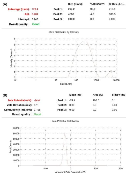
The mean particle size of LND NPs was found to be179.4±0.9nm (Figure 3a) and the zeta potential were found to be -24.4±0.2mV (Figure 3b). The experiment was performed in triplicate (n=3) in order to ensure reproducibility and minimize the error. PDI values were found to be 0.464, which indicates that the system has a relatively narrow distribution. This narrow distribution ensures good stability of PLGA-NPs suspension by avoiding problems such as Ostwald ripening. Zeta potential reports had showed that the prepared PLGA
nanoparticles had typical negative zeta potential attributed to PLGA preparations due to carboxyl groups present in their structure. The Zeta potential of the prepared NPs is also a prominent factor to ensure stability. Highly charged NPs are capable to remain stable as colloidal suspension. Since the prepared NPs have shown high zeta potential which ensure the stability of the formulation. The morphological evaluation (size, shape and morphology) of the LND-PLGA-NPs was performed using SEM (Figure 4). SEM studies conirm that nanoparticles are in the range of 50-300 nm with smooth surface and spherical shape which ensures the drug release in sustained manner.
DSC and XRD studies
The DSC thermograms of LND, PLGA, and NPs are shown in Figure 5. The DSC curve of LND exhibit an exothermic peak at peak temperature of 265 oC corresponding to its melting point. The polymer, PLGA had shown peak at a temperature of 65.12. As per the DSC graph of LND-PLGA-NPs, the characteristic exothermic peak of LND was observed with minimal intensity and shifted to lower temperature range. This indicates the conversion of crystalline nature of LND to amorphous form which ensures the better stability. Furthermore, it also conirms that LND was entrapped into PLGA-NPs. Reported indings have suggested that with an increase in the amorphous nature of the therapeutic system, a corresponding increase in the efficiency of delivery is observed (Abdelwahed et al., 2006).
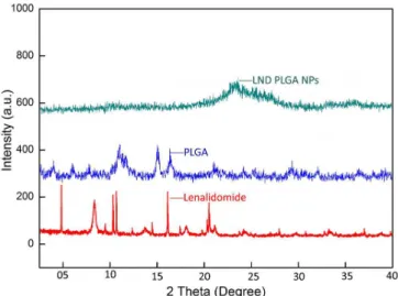
The physical nature of pure LND, PLGA-NPs and LND loaded PLGA-NPs were observed using X-ray diffraction analysis (Figure 6). Pure LND possessed a crystal structure when compared to LND-PLGA-NPs, which were in amorphous form. Pure LND has shown multiple peaks at diferent 2θ values indicating crystalline form of pure LND. However, the intensity of LND peaks were greatly reduced in PLGA-NPs signifying the conversion of crystalline form LND to amorphous form and LND loading in to PLGA-NPs.
In vitro drug release
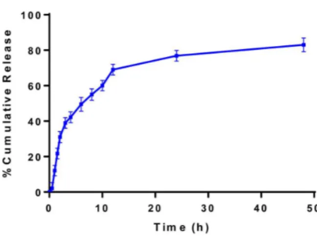
In vitro release studies of LND loaded PLGA-NPs were studied up to 48 h in HCl bufer (pH 1.2) for a period of 48 h. The summarized in vitro release of LND-PLGA-NPs is shown in Figure 7, as cumulative percentage drug release. The NPs has shown a biphasic pattern of drug release. Initially, a burst release of drug was observed (39%) followed by sustained release. 40% of LND was released within 6 hours and about 76.89 ± 2.99% and 83 ± 3.90% was released in 24 and 48 h respectively. This clearly depicts the pronounced time prolongation of the drug release. The initial burst release of drug may be due to drug desorption of surface entrapped or adhered LND from the particle surface, and the sustained release can be characterized by the drug difusion through the polymeric matrix and subsequent dissolution of drug or erosion of the polymeric matrix. In dissolution media, the release of LND from PLGA-NPs occurred either by the formation of holes in the particles or by their disintegration. A constant rate of release of the drug from the NPs will facilitate better drug control in vivo.
FIGURE 4 - SEM photograph of lenalidomide -PLGA-NPs.
FIGURE 5 -DSC curves of lenalidomide, PLGA, and lenalidomide -PLGA-NPs.
Cytotoxicity studies
The extent of cell viability due to LND-PLGA-NPs was examined by MTT assay in U266 cells. Eight concentrations (1-100 µg/mL) of LND-PLGA-NPs were prepared and tested yielding proof that the NPs exhibited significant toxicity against U266 cells. Cytotoxicity concentration (CTC50) value for NPs was calculated from the concentration and subsequent response; it was found to be34.09 µg/mL (Figure 8). This proves the cytotoxic potential of the PLGA-NPs and lends further credence to the viability if this concept.
In vivo bioavailability studies
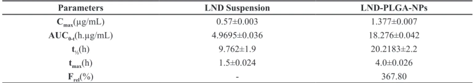
The formulations (LND aqueous suspension, dispersion of the LND-loaded PLGA NPs) were administered by oral gavage with a single dose of 30 mg/kg. The nanoparticles were dispersed in ultra-pure water. Table I and Figure 9 shows the pharmacokinetic
data of the drug suspension and nanoparticles. Figure 9 depicts a remarkable difference in bioavailability between LND PLGA NPs and LND suspension. After oral administration of LND suspension, the drug was rapidly absorbed and a Cmax of 0.57±0.003 µg/ mL was reached in 1.5 h. Consequently, the plasma concentration decreased abruptly, as the drug was rapidly distributed and metabolized and was detected up to 48 h after administration, resulting in low AUC0-t (4.9695 ± 0.036 h.µg/mL) and low t½ (9.762±1.9 h). Figure 9 shows that peak plasma concentration for LND PLGA NPs (Cmax) of approximately1.37±0.007 µg/mL was achieved at 4 h.
Table I depicts the pharmacokinetic parameters Cmax, AUC0-t, t½and tmax after oral administration of LND loaded PLGA-NPs and suspension. From Table I, it can be perceived that, the Cmax value for LND PLGA NPs (1.377±0.007) was signiicantly higher than LND suspension (0.57±0.003). In the same way, the t½ was rapid for LND-PLGA-NPs (20.2183±2.2), when compared to LND suspension (9.762 ±1.9). From the data in Table I, it is apparent that, the tmax was also rapid for LND PLGA NPs (4.0±0.026), when compared to LND suspension (1.5±0.024). This may be due to the primarily related prolonged absorption phase and sustained release of PLGA-NPs. Likewise, the area under curve AUC for LND PLGA NPs was 3-fold higher than the LND suspension. Finally, the relative bioavailability for LND PLGA NPs was computed and found to be 367.80%, suggesting better absorption of LND-PLGA-NPs compared to the suspension. On the contrary, particles belonging to nanoscale are easily absorbed into the intestinal folds, while larger particle surface area to volume ofers faster FIGURE 8 - MTT assay of lenalidomide - PLGA-NPs.
FIGURE 7 -Release rates of lenalidomide from PLGA-NPs in vitro in HCl bufer pH 1.2 (mean±SD, n = 3).
TABLE I - Pharmacokinetic parameters of lenalidomide after oral administration of LND loaded PLGA-NPs and suspension at the dose of 30 mg/kg
Parameters LND Suspension LND-PLGA-NPs
Cmax(µg/mL) 0.57±0.003 1.377±0.007
AUC0-t(h.µg/mL) 4.9695±0.036 18.276±0.042
t½(h) 9.762±1.9 20.2183±2.2
tmax(h) 1.5±0.024 4.0±0.026
Frel(%) - 367.80
drug dissolution. In current study, there was a signiicant improvement in bioavailability with LND-PLGA-NPs in contrast to LND suspension. The reasons may be many, but it is assumed that nano based drug delivery system enhances the bioavailability by modulating the physiochemical properties of poorly soluble drugs. Overall, our results suggest that LND-PLGA-NPs are most likely to have great potential as a therapeutic, by enhanced pharmacokinetic proiles.
CONCLUSION
Nanoparticles of a poorly soluble drug LND were successfully formulated by nano-precipitation method using PLGA as polymer. The particle size, zeta and SEM reports conirmed that the size of LND-PLGA-NPs was below 200 nm with spherical shape and uniform size distribution. DSC and XRD analysis of NPs conirmed the conversion of the crystalline nature of LND to amorphous form and drug entrapment within the NPs. Cytotoxic studies suggested that LND-PLGA-NPs were toxic towards U266 cancer cells. In vitro and in vivo studies confirmed that LND-PLGA-NPs demonstrate increased bioavailability with a sustained release up to 48 h. Due to their hydrophobic nature, conventional chemotherapeutics of LND suffer from poor solubility and an inability to penetrate the tumors (poor bioavailability). This results in grave side efects that include immune system depletion and metastasis to neighboring organs. Hence, the present PLGA nanoparticles can efectively solve the solubility problem and enhance the bioavailability of LND.
ACKNOWLEDGEMENT
The authors sincerely thank Natco Pharma Ltd, Hyderabad, India for providing lenalidomide and thalidomide as gift samples.
REFERENCES
ABDELWAHED, W.; DEGOBERT, G.; STAINMESSE, S.; FESSI, H. Freeze-drying of nanoparticles: formulation, process and storage considerations. Adv. Drug Deliv. Rev.,
v.58, n.15, p.1688-1713, 2006.
EGUSQUIAGUIRRE, S.; IGARTUA, M.; HERNÁNDEZ, R.; PEDRAZ, J., Nanoparticle delivery systems for cancer therapy: advances in clinical and preclinical research. Clin. Transl. Oncol., v.14, n.2, p.83-93, 2012.
FOOD AND DRUG ADMINISTRATION. FDA. Guidance for
industry: bioanalytical method validation. US Department of Health and Human Services. Silver Spring, MD: Food and Drug Administration, Center for Drug Evaluation and Research (CDER), Center for Veterinary Medicine (CV), 2001. p.1-22.
GOMATHI, T.; GOVINDARAJAN, C.; ROSE, H.R, M.H.; SUDHA, P.N.; IMRAN, P.K.M.; VENKATESAN, J.; KIM, S.-K. Studies on drug-polymer interaction, in vitro release and cytotoxicity from chitosan particles excipient. Int. J. Pharm., v.468, n.2, p.214-222, 2014.
K A R R I , V. V. S . N . ; R A M A N , S . ; K U P P U S A M Y, G . ; MULUKUTLA, S.; RAMASWAMY, S.; MALAYANDI,
R. Terbinaine hydrochloride loaded nanoemulsion based
gel for topical application. J. Pharm. Invest., v.45, n.1, p.79-89, 2015.
KASTRITIS, E.; DIMOPOULOS, M.A. The evolving role of lenalidomide in the treatment of hematologic malignancies.
Expert Opin Pharmacother., v.8, n.4, p.497-509, 2007.
MA, Y.; ZHAO, X.; LI, J.; SHEN, Q. The comparison of
diferent daidzein-PLGA nanoparticles in increasing its oral
MAEDA, H.; WU, J.; SAWA, T.; MATSUMURA, Y.; HORI, K. Tumor vascular permeability and the EPR effect in macromolecular therapeutics: a review. J. Control. Rel.,
v.65, n.2, p.271-284, 2000.
MANNEMALA, S.S.; NAGARAJAN, J.S.K. Development and validation of a generic liquid chromatographic method for the simultaneous determination of five commonly used antimalarial drugs: application to pharmaceutical formulations and human plasma. J. Sep. Sci., v.38, n.9, p.1521-1528, 2015a.
MANNEMALA, S.S.; NAGARAJAN, J.S.K. Development and validation of a HPLC-PDA bioanalytical method for the simultaneous estimation of Aliskiren and Amlodipine in human plasma. Biomed. Chromatogr., v.29, n.3, p.346-352, 2015b.
MORGENROTH, A.; DINGER, C.; ZLATOPOLSKIY, B.D.; AL-MOMANI, E.; GLATTING, G.; MOTTAGHY, F.M.; RESKE, S.N. Auger electron emitter against multiple myeloma targeted endo-radio-therapy with 125I-labeled
thymidine analogue 5-iodo-4′-thio-2′-deoxyuridine. Nucl. Med. Biol., v.38, n.7, p.1067-1077, 2011.
NOORI KOOPAEI, M.; KHOSHAYAND, M.R.; MOSTAFAVI, S.H.; AMINI, M.; KHORRAMIZADEH, M.R.; JEDDI TEHRANI, M.; ATYABI, F.; DINARVAND, R. Docetaxel loaded PEG-PLGA nanoparticles: optimized drug loading,
in-vitro cytotoxicity and in-vivo antitumor efect. Iranian J. Pharm. Res., v.13, n.3, p.819-833, 2014.
RICHARDSON, P.G.; MITSIADES, C.; HIDESHIMA, T.; ANDERSON, K.C. Lenalidomide in multiple myeloma.
Expert Rev. Anticancer Ther., v.6, n.8, p.1165-1173, 2006.
SONG, J.-X.; YAN, Y.; YAO, J.; CHEN, J.-M.; LU, T.-B. Improving the solubility of lenalidomide via cocrystals.
Cryst. Growth Des., v.14, n.6, p.3069-3077, 2014.
VEGA, E.; GAMISANS, F.; GARCÍA, M.L.; CHAUVET, A.; LACOULONCHE, F.; EGEA, M.A. PLGA nanospheres for the ocular delivery of flurbiprofen: drug release and interactions. J. Pharm. Sci., v.97, n.12, p.5306-5317, 2008.
VENKATESH, D.N.; BASKARAN, M.; KARRI, V.V.S.R.; MANNEMALA, S.S.; RADHAKRISHNA, K.; GOTI,
S. Fabrication and in vivo evaluation of Nelinavir loaded
PLGA nanoparticles for enhancing oral bioavailability and
therapeutic efect. Saudi Pharm. J., v.23, n.6, p.667-674, 2015.