©Revista Brasileira de Fisioterapia
POSTURAL CHARACTERIZATION OF YOUNG FEMALE OLYMPIC
GYMNASTS
G
UIMARÃESMMB
1, S
ACCOICN
2& J
OÃOSMA
31 Physical Therapy, Speech Therapy and Occupational Therapy Department, Medicine School, University of São Paulo - USP, São Paulo, SP - Brazil
2 Laboratory of Movement Biomechanics and Human Posture, Physical Therapy, Speech Therapy and Occupational Therapy Department, Medicine School, University of São Paulo - USP
3 Laboratory of Musculoskeletal Assessment, Physical Therapy, Speech Therapy and Occupational Therapy Department, Medicine School, University of São Paulo - USP
Correspondence to: Silvia Maria Amado João, Cipotânea Street, nº 51, Cidade Universitária, Butantã, CEP 05360-160, São Paulo, SP – Brazil, e-mail: [email protected]; [email protected]
Received: 27/09/2006 - Revised: 08/03/2007 - Accepted: 22/05/2007
ABSTRACT
Objective: To identify the postural alterations among children aged 8 to 12 years who perform Olympic gymnastics. Method: Eighty-four students aged 8 to 12 years were assessed: 38 who were participating in Olympic gymnastics and 46 who were not. Each child was photographed in the anterior and posterior frontal planes and in the sagittal plane. The photos were analyzed using the CorelDraw v. 11.0 software. The line-guide and dimension tools (angular, vertical and horizontal dimensions) were used to determine the parameters of the nineteen qualitative and five quantitative variables. Results: Comparison between the test group subjects (TG) and control group subjects (CG) showed significant differences in the variables of medial rotation of the femur (CG 56.52%; TG 39.47%), antepulsion of the pelvis (CG 43.48%; TG 76.32%) and trunk rotation (CG 67.39%; TG 23.68%). There were also found significant differences in the measurements of knee valgism (CG 4.06 ± 2.32 cm; TG 3.14 ± 1.49 cm), pelvic inclination (CG 0.69 ± 0.39 cm; TG 0.53 ± 0.33 cm) and tibiotarsal angle (CG 86.93 ± 2.90 cm; TG 87.11 ± 4.17 cm). Conclusion: Analysis of the results showed a tendency towards better alignment of the lower limbs in the test group than in the control group. There was greater anterior pelvic inclination and a tendency towards lumbar hyperlordosis in the test group. These factors may predispose individuals participating in Olympic gymnastics to misalignment of skeletal structures, thereby leading to painful conditions that may limit their sports activities.
Key words: Posture; physical therapy; child, sport; gymnastics.
INTRODUCTION
Good posture may be defined as the ability to maintain the body center of mass in such a relation to the support base that falls are avoided and correct execution of movements is allowed1. Other authors define good posture as the alignment in which the center of mass of each body segment is located vertically over the next segment2.
Postural evaluation is extremely complex and should take in account intrinsic and extrinsic factors that may influence the posture of an individual, such as the physical characteristics of the environment where the subject lives, emotional and socio-cultural status, physical activity, obesity, physiological alterations from growth and human development such as the growth spurt, sexual maturation, sex, race, and hereditariness3-7. Sports, as well as exercise programs, may also influence posture, and can cause adaptations that become permanent8,9. Sports’ training is based on the constant repetition
of certain movements. Such repetition may lead to unbalances in the musculoskeletal system, causing alterations in strength, flexibility, balance, and motor coordination, and also directly influencing bone growth. Such influences may predispose the individual to develop postural alterations which, in turn, may predispose the athlete to injure10,11.
Several studies in the literature associate sports training to postural alterations in athletes9,12,13; others relate postural alterations to injuries; however, few examine the association between postural alterations and the practice of Olympic Gymnastics.
The objective of this study was to characterize the posture of children that practiced Olympic Gymnastics compared to non-athlete children of the same age, in order to inform athletes and professionals about postural alignment of children that practiced this sports modality. This study was performed with the cooperation of the CEPEUSP.
METHODS
Eighty-seven female students between 8 and 12 years were evaluated. Participants composed two groups: a control group with 48 children that did not practice gymnastics, and a test group of 39 children that practiced Olympic Gymnastics. Three subjects were excluded from the sample because they had already had the first menstruation.
Children that practiced Olympic Gymnastics more than two times/week or three hours/week composed the Olympic gymnasts group. The control group was composed of children that did not practice any sport modality or physical activity except for the regular physical activity (physical education) practiced at school14.
Adults responsible for the children filled in a questionnaire including information on duration and frequency of sports practice, practice of other sport modality, presence of chronic diseases and/or injuries related to the sports practice, and maturational age15.
Exclusion criteria were: school children with neuro-muscular or traumatic pathologies, or with any type of pain causing alteration of habitual posture. To take part in the study, children needed to the have the body mass index (BMI)3 defined as weight (kg)/stature (m2), equal or inferior to 19 kg/m2, according to the reference value of normality during childhood16.
Initially, parents of the students were informed about the project through a letter containing information about the postural examination. After explanation of procedures, the legal representative of the school child signed a consent form allowing the child to take part in the study. The project was approved by the Ethics Committee of the HCFMUSP (nº 064/04).
Data from the test group were obtained at the center for sports practice of the University of São Paulo. Data from the control group were obtained from students from Public Schools run by the governments of the City of São Paulo and State of São Paulo. The project received assistance and academic support from the Musculoskeletal Ambulatory and Laboratory of Musculoskeletal Evaluation of the Physical Therapy Course at the USP Medicine School.
For postural analyses, photographs of the subject at the frontal (anterior-posterior) and sagittal (right side) planes were taken. Subjects wore bathing suits or shorts and tops and stood on a 20cm high wood bench in orthostatic position. A board with vertical and horizontal lines (2.00m high; 0.72m wide, with 10cm x 10cm squares) was placed at the background of the subject for reference. The photographic
camera was placed at a height of 78.5cm from the subject and at a distance of 2.56m from the squared board fixed at the wall. The subject stood on the bench, the closest possible to the squared board without touching it.
Subjects had the anatomical landmarks corresponding to the lateral and medial maleolus, head of the fibula, great throcanter of the femur, anterior-superior iliac spines, posterior-superior iliac spines, ulna styloid process, humerus lateral epicondyle, coracoid process, inferior angle of the scapula and spinal processes of the cervical (C5 and C7), thoracic (T2, T7) and lumbar (L1 and L5) vertebrae marked with 13 mm adhesive tags4,5. The protocols of qualitative evaluation of posture by Kendall et al.4and Penha et al. 14 were used. All pictures were aligned to the reference lines of the CorelDraw program version 11.0 to ensure that the horizontal was correctly positioned. The pictures were analyzed by means of the guidelines and dimension (angular, vertical and horizontal dimension) tools.
DATA ANALYSIS
Quantitative variables
Quantitative measures (angles and distances from bone prominences) were obtained through the CorelDraw v. 11.0 software. All measures were converted to real values in order to avoid possible errors of measurement. Measures of the squared board placed behind the subjects in the photographs were used for calibration.
Knee valgus and varus measures were obtained by using the distances between medial malleoli and medial condyles3, respectively, with the horizontal dimension CorelDraw tool. The distance between the inferior angle of the scapula and the spinal processes of the thoracic vertebrae were also obtained by means of the horizontal dimension tool based on the anatomical landmarks. The difference between the distances from the left and right scapulae to the spine axis was used for analysis.
Shoulder and pelvis asymmetry measures were obtained with the vertical dimension tool. Measures were based on the landmarks identified with adhesive tags (anterior-superior iliac spines and coracoid processes). Tibiotarsal angle (TTA) was obtained by means of the angular dimension tool by taking the head of the fibula and lateral malleolus as references. The horizontal axis was determined by the direction of the fifth metatarsal, parallel to the horizontal axis. An TTA angle of 90º was considered to be normal alignment. TTA angles inferior to 90º were classified as closed and TTA angles superior to 90º were classified as open4.
Qualitative variables
In the qualitative analysis 19 variables were observed: tibiotarsal angle (TTA) (open, closed and aligned); frontal knee (valgus, varus or aligned); lateral knee (hyper-extended, semi-flexed, or aligned); hip rotation (medial, lateral or aligned); hip (retroversion, anteversion, or aligned); pelvis (anterior tilt of the pelvis, posterior tilt or aligned); lumbar spine (hyperlordosis, rectified, or aligned); thoracic spine (hyperkyphosis, rectified or aligned); cervical spine (hyperlordosis, rectified or aligned); ankle (valgus, varus or aligned); scapula (adduction, abduction or aligned); pelvic asymmetry (present or absent); shoulder asymmetry (present or absent); trunk rotation (present or absent); shoulder protraction (present or absent); shoulder medial rotation (present or absent); head protraction (present or absent); scoliosis (present or absent); winged scapula (present or absent).
Statistical analysis
The softwares Statistica V. 5.1, and Excel 2000 were used for statistical analyses. Chi-square and Fischer’s exact tests were used for non-parametrical dichotomic variables (categorized as “present or absent” only), the t-test was used for variables with normal distribution, the Mann-Whitney test and Kruskal-Wallis were used for comparisons between groups.
Descriptive analyses included means and standard-deviation. The percentage of children with specific postural characteristics was also described for each group. Significance level was established as p
≤
0.05.RESULTS
Sample characterization
Children were divided into groups according to age. Since there was no significant difference between postural varibles between different age groups, statistical tests were performed considering the entire test and control groups. Mean age of the test group subjects (38) was 10 ± 1 years, height was 1.22 ± 0.26 m and body mass index was 17.26 ± 1.12 Kg/ m2. Children of the test group had been practicing gymnastics for a mean time of 2.26 ± 1.18 years, at a frequency of 2.00 ± 0.23 times a week, for 1.89 ± 0.69 hours a day. Mean age of the control group (46) was 10 ± 1 years, height was 1.29 ± 0.32 m and body mass index was 18.17 ± 0.72 Kg/m2.
Quantitative variables
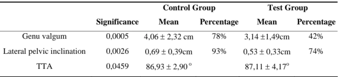
Regarding the quantitative variables, statistically significant differences were found between groups for knee valgus (KV), p= 0.0005, pelvic assymmetry (PA), p= 0.0026, and tibiotarsal angle (TTA), p= 0.0459. Table 1 includes the percentage of children with specific postural characteristics in the control and test groups as well as results of the quantitative analysis for each group.
Qualitative variables
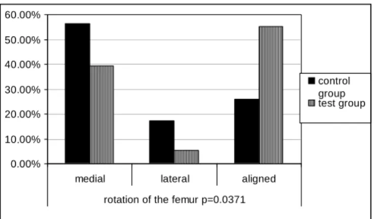
Regarding qualitative variables, a statistically significant difference between groups was observed (p= 0.03) for rotation of the femur, with the test group demonstrating a decreased proportion of children with medial rotation (TG 39.47% and CG 56.52%) and an increased proportion of children with aligned femur (TG 55.27% and CG 26.09%). Figure 1 compares the proportions of medial and lateral rotation of the femur between the test and control groups.
Regarding pelvic and trunk alterations (Figure 2), the test group had a significantly (p= 0.022) greater proportion of children with anterior tilt of the pelvis (TG 76.32% and CG 43.48%). The proportion of trunk rotation was significantly (p= 0.0001) lower in the test group (TG 23.68% and CG 67.39%). No statistically significant difference was found for lumbar hyperlordosis between groups (p= 0.0991). However, test group hand a greater prevalence of hyperlordosis compared to the control group (TG 60.53% and CG 45.65%). Regarding variables related to the upper limbs and shoulder girdle (Figure 3), the percentage of children with abducted scapula was lower in the test group compared to the control group (TG 26.32% and CG 52.17%). The same was observed for winged scapula (TG 34.21% and CG 52.17%) and shoulder medial rotation (TG 42.11% and CG 60.87%). However, none of the differences were statistically significant (abducted scapula: p= 0.0717; winged scapula: p= 0.0988; medial rotation of the shoulder: p= 0.0866).
DISCUSSION
The main objective of this study was to characterize the posture of children that practice Olympic Gymnastics. A trend was found for decreased prevalence (qualitative analysis) of postural alterations in the test group, such as knee valgus, trunk rotation and medial rotation of the femur. The
Table 1. Comparison of the quantitative analysis between test group and control group.
Control Group Test Group
Significance Mean Percentage Mean Percentage
Genu valgum 0,0005 4,06 ± 2,32 cm 78% 3,14 ±1,49cm 42%
Lateral pelvic inclination 0,0026 0,69 ± 0,39cm 93% 0,53 ± 0,33cm 74%
0,00% 10,00% 20,00% 30,00% 40,00% 50,00% 60,00%
medial lateral aligned
rotation of the femur p=0.0371
control group test group
Figure 1. Distribution of lateral and medial rotation of the femur in
the test group and control group.
0,00% 10,00% 20,00% 30,00% 40,00% 50,00% 60,00% 70,00% 80,00% 90,00%
anterior tilt posterior tilt aligned p=0.0001
pelvic = 0.022 trunk
rotation
control group
test group
Figure 2. Distribuition of pelvic and trunk variables in the test group
and control group.
0,00% 10,00% 20,00% 30,00% 40,00% 50,00% 60,00% 70,00% a bduc ted aduc ted a ligned wi n ged sca p u la m e di al ro ta ti on of s h ou ld er s
scapula p=0.0717 p=0.0988 p=0.0866
control group
test group
Figure 3. Distribuition of shoulder and scapula alignament in the test
group and control group.
test group also demonstrated increased prevalence of pelvic anterior tilt and lumbar hyperlordosis, although the difference in this last variable was not statistically significant.
Developmental phases that constitute pre-adolescence and adolescence are characteristic for involving many adaptations and adjustments in the subject’s posture. Between 7 and 12 years of age, the child’s posture is largely transformed as new body proportions originate the need to
seek a new balance14,17. Growth and development of children included in this study are compatible with modifications in knee alignment. Several authors7,18-21 report variation in knee alignment during growth. From birth throughout the first year the knee assumes a varus posture. Thereafter a phase of physiological valgus is initiated and persists until 6 – 7 years of age, when stabilization begins. In this study, it was observed that the in prevalence of knee valgus as well as the distance between melleoli were significantly decreased in the TG (39.47% and 3.14 ± 1.49 cm), when compared to the CG (65.22% and4.06 ± 2.32 cm). The test group also had a decreased proportion of children with medial rotation of the femur (39.47%) compared to the control group (56.52%).
The Olympic Gymnastics athlete child develops static and dynamic balance significantly22. It is known that balance is intimately related to good function of the intrinsic musculature of the foot and ankle and to better alignment of the lower limb (knee valgus and medial rotation of the femur)4,5.
At the age of children included in this study the valgus or varus posture of the knee should be changing towards alignment3,4. However, a high prevalence of knee valgus in the CG children (65.22%) was noted. In the literature, great variability is reported for the prevalence of knee valgus. In a study with 132 girls aged between 7 and 10 years a high prevalence of knee valgus was observed in 7 year-old children (64%). However the prevalence of valgus was lower in other ages14.
In another study with 111 girls, a high prevalence of knee valgus was observed (30%) among children aged 7 and 8 years23. On the other hand, a study performed with children aged 7 to 12 years demonstrated a low prevalence of valgus (11.6%) in a total of 791 subjects24. At the present study the variables were analyzed grouping all ages. Therefore it is not possible to determine if the high prevalence of knee valgus observed was originated in a specific age subgroup or if valgus was equally distributed in all ages. A study with a greater number of individuals in each age group would be necessary to determine if the observed variation is physiological or if it requires specialized intervention. A longitudinal study could analyze variations in postural alignment of athletes throughout development.
support very high loads during falls, jumps, and other similar activities25.
Twenty two juvenile athletes of Olympic Gymnastics, aged 11 to 14 years were followed for five years. The group was compared to a control group composed of girls at similar age that did not practice Olympic Gymnastics. Results demonstrated that the gymnasts had a delay in the first menstruation and a high incidence of injuries, particularly of the lumbar spine, when compared to the control group26. Lumbar hyperlordosis and anterior pelvic tilt result from an attempt to maintain the anterior-posterior balance. Some authors attribute this unbalance to the weakness of the abdominal musculature23, 24. The abdominal musculature acts simultaneously with the extensors of the hip and lumbar spine and is responsible for stabilization of the pelvis and alignment of the lumbar spine5. The abdominal musculature becomes effective during development, especially between the age of 10 to12 years, when the waist becomes relatively narrower and abdominal protrusion decreases4. The posture used during walking and at the end of exercises and exit of the gymnastics apparatuses may also contribute to the presence of hyperlordosis and to the anterior tilt of the pelvis in the children from the test group.
Regarding TTA, children from the test group had a value a little closer from alignment (90º) than the control group, although the mean of the test group was still lower than 90º. The attempt to maintain the anterior-posterior balance by means of lumbar hyperlordosis and the pelvic anterior tilt may also be responsible for the decreased TTA.
Some limitations inherent to the method of analysis (photographs) should be highlighted at the present study. Despite the fact that analysis was performed by a previously trained evaluator, some variables are difficult to observe, such as foot alignment, hip and trunk rotation. Some studies demonstrate the validity of photograph analyses; however low reliability is reported 27.
The present study may contribute significantly to the training methods of this sport modality. The study demonstrated increased anterior pelvic tilt and a trend towards an increase in the prevalence of lumbar hyperlordosis, and these alterations may predispose gymnasts to future injuries and/or painful conditions, as described by other authors25,26. In future studies data from analysis of static posture can be used in comparisons with data from dynamic posture analyses.
CONCLUSION
The present study demonstrated that the young female Olympic Gymnastics athletes that composed the analyzed group had a better alignment of the lower limb (decreased knee valgus, medial rotation of the femur and pelvic
asymmetry). However, there was an increased prevalence of anterior pelvic tilt, as well as a trend for increased lumbar hyperlordosis in the test group. These factors may have relevant clinical implications in the future of these athletes.
Acknowlegments: FAPESP - process nº 03/12100-8.
REFERENCES
1. Westcott SL, Lowes LP, Richardson PK. Evaluation of postural stability in children: current theories and assessment tolls. Phys Ther. 1997;77(6):629-45.
2. Watson AWS, Mac Donncha C. A reliable technique for the assessment of posture: assessment criteria for aspects of posture. J Sports Med Phys Fitness. 2000;40(3):260-70.
3. Ascher C. Variações da postura na criança. São Paulo: Manole; 1976.
4. Kendall FP, McCreary EK, Provance PG. Músculos provas e funções. 4ª ed. São Paulo: Manole; 1995.
5. Tanaka C, Farah EA. Anatomia funcional das cadeias musculares. São Paulo: Editora Ícone; 1997.
6. Teixeira LR. Educação física escolar: alterações posturais e respiratórias na infância e adolescência. São Paulo: Eefusp/dg; 1991.
7. Tolo VT. The lower extremity. In: Morrisy RT, Weinstein SL. Pediatric orthopaedics. 3ª ed. Philadelphia: Lippincott-Raven Publishers; 1996. p. 1047-81.
8. Rodrigues RL, Barbanti VJ. Atividade esportiva e a criança. Principais lesões do aparelho locomotor. In: Conceição JAN. Saúde escolar: a criança, a vida e a escola. São Paulo: Sarvier; 1994. p.170-80.
9. Watson AWS. Sports injuries in footballers related to defects of posture and body mechanics. J Sports Med Phys Fitness. 1995;35(4):289-94.
10. Silva CC, Teixeira AS, Goldberg TBL. O esporte e suas implicações na saúde óssea de atletas adolescentes. Rev Bras Med Esporte. 2003;l9(6):426-32.
11. Ribeiro CZR, Akashi PMH, Sacco ICN, Pedrinelli A. Relação entre alteração postural e lesões do aparelho locomotor em atletas de futebol de salão. Rev Bras Med Esporte. 2003; 9(2):91-7.
12. Beynnon BD, Restrom PA, Alosa DM, Baumhauer JF, Vacek PM. Ankle ligament injury risk factors: a prospective study of college athletes. J Orthop Res. 2001;19(2):213-20.
13. Wojtys EM, Ashton-Miller JA, Huston LJ, Moja PS. The association between athletic training time and the sagittal curvature of the immature spine. Am J Sports Med. 2000;28(4):490-8.
14. Penha PJ, João SMA, Casarotto RA, Amino CJ, Penteado DC. Postural assessment of girls between 7 and 10 years of age. Clinics. 2005;60(1):9-16.
16. Damiani D, de Carvalho DP, de Oliveira RG. Obesidade. In: Nuvarte S. Endocrinologia pediátrica: Aspectos físicos e metabólicos do recém nascido ao adolescente. 2ª ed. São Paulo: Sarvier; 2002. p. 567-75.
17. Bankoff ADP, Brighetti V. Levantamento da incidência de cifose postural e ombros caídos em alunos de 1ª à 4ª séries escolar. Rev Bras Ciênc Esporte. 1986;7(3):93-7.
18. Forlin E, Andújar ALF, Alessi S. Padrões de normalidade do exame físico dos membros inferiores em crianças de idade escolar. Rev Bras Ortop. 1994;29(8):601-6.
19. Health CH, Staheli LT. Normal limits of Knee angle in white children. J Pediatric Orthop. 1993;13(2):259-62.
20. Salenius P, Vanka E. The development of tibiofemoral angle in children. J Bone Joint Surg. 1975;57(2):259-61.
21. Volpon JB. Modificações fisiológicas e patológicas do joelho durante o crescimento. Rev Bras Ortop. 1995;30(12):53-6.
22. Nunomura M, Nista-Piccolo VL. Compreendendo a ginástica artística. São Paulo: Phorte; 2005.
23. Pinho RA, Duarte MFS. Análise postural em escolares de Florianópolis, SC. Rev. bras. ativ. fís. saúde. 1995;1(2):49-58.
24. Rosa Neto FN: Avaliação postural em escolares de 1ª à 4ª série do 1º grau. Rev. bras. ciênc. mov. 1991;5(2):7-10.
25. Gonçalves JLC. Contribuição para o estudo do comportamento da coluna lombo-sagrada em praticantes de ginástica de competição [dissertação]. Porto: Universidade do Porto; 2000.
26. Lindholm C, Hagenfeldt K, Ringertz BM. Pubertal development in elite juvenile gymnasts. Acta Obstet Gynecol Scand. 1994;73(3):269-73.