©Revista Brasileira de Fisioterapia
ASSESSMENT OF MAXIMUM INSPIRATORY PRESSURE IN
NON-COOPERATIVE CRITICAL PATIENTS: COMPARISON BETWEEN TWO
METHODS
G
UIMARÃESFS
1,2, A
LVESFF
3, C
ONSTANTINOSS
4, D
IASCM
1& M
ENEZESSLS
11 Physical Therapy School, Centro Universitário Augusto Motta, Rio de Janeiro, RJ - Brazil
2 Physical Therapy School, Federal University of Rio de Janeiro-UFRJ, Rio de Janeiro, RJ - Brazil
3 Clementino Fraga Filho University Hospital, UFRJ, Rio de Janeiro, RJ - Brazil
4 Military Police Central Hospital of Rio de Janeiro State, Rio de Janeiro, RJ - Brazil
Correspondence to: Fernando Silva Guimarães, Centro Universitário Augusto Motta, Pró-Reitoria de Pesquisa e Pós-Graduação, Praça das Nações, nº 34, Bonsucesso, CEP 21041-010, Rio de Janeiro, RJ - Brasil,
e-mail: [email protected]
Received: 29/11/2006 - Revised: 21/05/2007 - Accepted: 22/05/2007
ABSTRACT
Background: Although mechanical ventilation is necessary for treating acute respiratory insufficiency, it may be associated with deconditioning and respiratory muscle dysfunction. Maximal inspiratory pressure (MIP) evaluation is used to estimate inspiratory muscle strength in artificially ventilated patients, but there is no definition as to the best way to make this measurement. Objective: To compare two methods for MIP evaluation, using four different protocols, among non-cooperative artificially ventilated patients. Method: Thirty non-cooperative patients undergoing the process of weaning off mechanical ventilation were evaluated. In accordance with block randomization, the simple occlusion method (OM) or the unidirectional valve method (UV) was applied to each patient for time periods of 20 and 40 seconds. Additionally, during the 40s measurements, the MIP value at 30s was recorded. Results: The MIP values were higher at 40s than at 20s, both from OM (48.2 ± 21.7 vs. 36 ± 18.7 cmH2O; p< 0.001) and from UV (56.6 ± 23.3 vs. 43.4 ± 24 cmH2O; p< 0.001). The MaxIP values were higher from UV at 40s (UV40) than from OM at 40s (OM40) (56.6 ± 23.3 vs. 48.2 ± 21.7 cmH2O; p< 0.001). There was a difference between UV at 30 and 40s (51.5 ± 20.8 vs. 56.6 ± 23.3 cmH2O; p< 0.001). Conclusion: Among non-cooperative patients, higher MIP values were obtained from the unidirectional valve method with 40s of occlusion than from the other protocols evaluated.
Key words: respiratory muscles; evaluation; weaning from respirator; physical therapy.
INTRODUCTION
It is well established that mechanical ventilation is an essential therapy for patients with acute respiratory insufficiency. In such a context, ventilation support is necessary when ventilation demand becomes superior to the capacity of respiratory muscles as a consequence of several clinical conditions1. Mechanical ventilation reduces or eliminates overload of respiratory muscles. However, it can also be associated with deconditioning and respiratory muscle dysfunction, along with other factors, such as polyneuropathy of the critical patient, sepsis and dysfunction of multiple organs and systems2,3. When problems that produced increased ventilation demand are solved, weaning must be initiated. However, in many patients respiratory muscle weakness can impede withdrawal from mechanical ventilation4. Assessment of patients before weaning is important to avoid respiratory muscle fatigue and to define
presented good prognosis for withdrawal from mechanical ventilation, while patients with MaxIP values greater than – 20 cmH2O failed to wean off. Due to several factors such as study design (prospective or retrospective) weaning method and definition of success and failure, the accurateness of MaxIP measures varies considerably between different studies. In general, studies report sensibility over 80% for cutoff values of – 30 cmH2O9,10,11, - 20 cmH
2O
10,11,12,13 and even - 15 cmH2O11. Because success of withdrawal depends on other factors, specificity is low in all studies. The low specificity indices indicate that patients who fail weaning from mechanical ventilation do not necessarily present reduced MaxIP.
In an attempt to make MaxIP measures reliable, Marini et al.14 described the unidirectional valve method after the assessment of 20 artificially ventilated patients. After a short period of spontaneous ventilation, a device that allowed exhalation only was attached to the airway opening (unidirectional valve). This way, patients were forced to make progressively increasing effort while approaching residual volume. At residual volume MaxIP corresponded to maximum the capability of the patient. The maximum value was reached in 15 or 20 seconds of occlusion. In spite of this result, MaxPI values measured in critical patients are usually underestimated and have poor reliability15,16. The identification of reliable methods of assessment of respiratory muscle strength can contribute to better monitoring of withdrawal from mechanical ventilation as well as to better definition of treatment strategies. The objective of the present study was to compare two methods of assessment of maximum inspiratory pressure implemented trough four different protocols, in non-cooperative patients receiving artificial ventilation.
MATERIALS AND METHODS
Sample
Thirty patients admitted to the Intensive Care Unit of Rio de Janeiro Military Police Central Hospital (Table 1) were assessed in a cross-over randomized trial. Patients included
in the study were receiving invasive ventilatory assistance and were in process of weaning from mechanical ventilation. Individuals with hemodynamic instability, PaO2/FiO2 ratio inferior to 200, intracranial hypertension (ICP > 20 mmHg), deep sedation (Ramsay ≥5), curarization, abdominal surgeries with risk of evisceration, coronary arterial disease or severe cardiac insufficiency were not included in the study. Sample size was calculated with the software Sigmastat 3.1, according to results of Caruso et al.17, considering the statistical test ANOVA, power of 80%, significance level of 5%, expected difference of 28% and standard deviation of 30%. The estimated sample size was 23 individuals.
The research project was approved by the Augusto Motta University Centre Ethics Committee (approval report number 04/06). Relatives responsible for all patients included in this study signed the informed consent form, according to the CONEP 196/96 resolution.
Procedures
Patients were positioned in dorsal decubitus with the head of the bed elevated at 45o for the measurement of maximum inspiratory pressure with the Simple Occlusion Method (OM). The cuff was hyperinsufflated to avoid escape during measurement. After tracheal aspiration, patients rested for five minutes while connected to the mechanical ventilator. To perform the measurement, the mechanical ventilator was disconnected and after 10 seconds a connector attached to the artificial airway was manually occluded at the end of a normal expiration (functional residual capacity level). MaxIP was measured with an Instrumentation Industries analogical manometer, with a measurement range of ± 120 cmH2O at 2 cmH2O steps. Occlusion was maintained for 20 or 40 seconds, according to randomization. The maximum value of each measurement was used for analysis. Three measurements with 2- minute intervals were performed (during intervals the patient was connected to the mechanical ventilator to rest). In order to measure MaxIP with the Unidirectional Valve Method (UV) the same procedure was adopted. At the moment of measurement, however, a low
Subjects Characteristics
Sex 16 M e 14 F
Age (years) 62.6 ± 17.7
PaO2/FiO2 334.4 ± 87.8
APACHE II 16 (10-26)
Admission Cause Clinical Conditions (congestive heart failure - 2,
community-acquired pneumonia - 5, isquemic stroke - 3, bronchoaspiration pneumonia, anoxic encephalopathy, hemorrhagic stroke - 2, acute atrial fibrillation): total: 15. Chirurgical Conditions (exploratory laparotomy - 9, retosigmoidectomy, subarachnoid hemorrhage drainage - 3, total hip arthroplasty, hernioplasty): total: 15.
Table 1. Data related to age and PaO2/FiO2 are presented as mean and standard deviation. The severity score (APACHE II) is presented as median
resistance unidirectional valve (that allowed exhalation only) was connected to the airway opening.
Procedures were performed according to randomization in blocks of 10, considering two techniques of measurement of maximum inspiratory pressure and measurement times (20 or 40 seconds). After the order of measurement techniques (OM or UV) was determined, the respective measurement times were randomized (20 or 40 seconds). The order of measurement times was used for the two measurement techniques for the same patient. Therefore, all patients were assessed with all techniques and times, but in different order according to randomization. Additionally, during the 40-second measurements, maximum inspiratory pressure values at 30 seconds were recorded, without interruption of the test. The two forms of assessment were separated by an interval of thirty minutes, during which patients remained connected to the mechanical ventilator. Ventilatory parameters were not modified during the assessment protocol, and patients were being monitored for cardiac function and oxygen saturation. All assessments were performed by the same examiner.
Statistical analyses were performed with the software SigmaStat 3.1. The Friedmand test was used to compare MaxIP values between the different techniques and measurement times. The Paired t Test was used for the post hoc analysis to compare results of the UV method at 30 and 40 seconds. Differences were considered significant at p< 0.05. Pearson or Spearman tests were used to test for correlations, depending on the type of variable analyzed and results of normality tests.
RESULTS
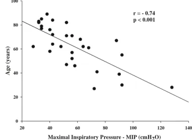
Data regarding patients included in this study are available in Table 1. No differences were found for MaxPi values between OM at 20s (OM20) and UV at 20s (UV20) (36 ± 18.7 and 43.1 ± 24 cmH2O; p> 0.05). Values at 40s were greater than at 20s for both the OM (48.2 ± 21.7 and 36 ± 18.7 cmH2O; p< 0.001) and UV (56.6 ± 23.3 and 43.4 ± 24 cmH2O; p< 0.001) methods (Figure 1). MaxPI values were greater with the UV at 40s (UV40) than with the OM at 40s (OM40) (56.6 ± 23.3 vs 48.2 ± 21.7 cmH2O; p< 0.001) (Figure1). Considering the three measurements performed to obtain the final value of each patient, the mean coefficient of variation was calculated for each protocol: OM20= 0.2; OM40= 0.1; UV20= 0.16; UV= 0.13. The pos hoc analysis revealed a significant difference between UV at 30 and UV at 40s (51.5 ± 20.8 and 56.6 ± 23.3 cmH2O; p< 0.001) (Figure 1). A significant correlation between patient age and MaxIP value measured with UV40 was found (r= -0.74; p< 0.001) (Figure 2). However, no correlation was found between protocols and severity scores (APACHE). MaxIP mean was lower in females (42.3 cmH2O) than in males (69.1 cmH2O). Considering the Black & Hyatt18 equation for the calculus of predicted MaxIP for each individual of the sample,
mean percents of predicted values were calculated for men (60.4 %) and women (62.5 %), with no significant differences between sexes (p= 0.36). Additionally, no significant differences were found between mean percents of predicted values for patients admitted for clinical or surgical reasons (60.9 % and 61.8 %; p= 0.44).
DISCUSSION
Figure 1. Comparison between the methods and protocols for Maximal
inspiratory pressure measurement. * values significantly different from the correspondent method at 20s (p< 0.001). ** values significantly different from the Simple Occlusion Method at 40s (p< 0.001). # values significantly different from Unidirectional Valve Method at 30s (p< 0.001) (post-hoc analysis – Paired t-test).
Maximal Inspiratory Pressure - MIP (cmH2O)
20 40 60 80 100 120 140
0 20 40 60 80 100
r = - 0.74 p < 0.001
A
g
e (y
ea
rs
)
A
g
e (
y
ea
rs
)
Figure 2. Correlation between age and maximal inspiratory pressure
recorded by the Unidirectional Valve Method with an occlusion time of 40s.
and similar to what was found by other authors15,17,2, except for the measure at OM20 for which the mean coefficient of variation was 20%. This result indicates the need for more verbal stimulation in order to improve reliability, especially in patients with difficulty to cooperate.
According to Vitacca et al.19, despite the efforts of the European and American Societies to standardize MaxIP measurements22, more adequate standardization is still needed, since there are a wide variety of methods, results and clinical circumstances in which the measurement is made. Considering that the measurement may be necessary in patients with
reduced ventilatory drive, the increase in inspiratory time in order to increase effort may be an alternative to verbal stimulation, especially for patients that do not interact adequately with the examiner for any reason. Such effect is illustrated by the more negative MaxIP values obtained with the occlusion method at 40s. Some authors used other strategies to increase the ventilatory drive, such as the unidirectional valve method itself 14 and the increase in the dead space20. Similarly to the study by Marini et al.14, a short period of spontaneous ventilation (10s) was implemented before all measurements made in the present study.
MaxIP value at 30s was registered during the measurement at 40s. This data were treated as complimentary information, as the design of the study did not include this assessment as a criterion for randomization. In a pos-hoc analysis comparing values obtained at 30 and 40s a significant difference was found. This result suggests that the best procedure to measure MaxIP is the unidirectional valve with an occlusion of 40s. More studies are necessary to confirm this result, with specific designs to compare different occlusion times for the unidirectional method. Cooperative patients generally have a normal ventilatory drive and do not, as mentioned before, present higher MaxIP values with verbal stimulation. Therefore, prolonged occlusion can be a universal procedure in the application of the method in order to produce more reliable MaxIP values for cooperative or non-cooperative patients.
The use of corticosteroids and other drugs that may influence function of respiratory muscles as well as nutritional status of patients and time since mechanical ventilation was initiated were not controlled during the study. Probably because of heterogeneity of the sample in relation to these factors no significant difference was found for MaxIp percent value predicted by the Black and Hyatt18 equations between patients admitted to the hospital for clinical or surgical reasons. Despite the multifactorial character of the determination of MaxIP, a significant correlation between age and MaxIp was found. Differences between sexes were also found (values were 38.8% lower for females). These results are in accordance with the classical study by Black and Hyatt18 in which authors described an equation to estimate MaxIP according to age and sex. Despite the differences between absolute values of MaxIP, when the equation was used no significant differences were found for the mean percentage of predicted values between sexes.
In face of results it is concluded that, when compared to the other protocols, the unidirectional valve method with occlusion during 40 seconds registers greater values of MaxIp for non-cooperative patients.
REFERENCES
1. McIntyre NR. Respiratory mechanics in the patient who is weaning from the ventilator. Respir Care. 2005;50:275-84.
2. Gayan-Ramirez G, Decramer M. Effects of mechanical venti-lation on diaphragm function and biology. Eur Respir J. 2002; 20:1579-86.
3. Moxham J, Goldstone J. Assessment of respiratory muscle strength in the intensive care unit. Eur Respir J. 1994;7: 2057-61.
4. McIntyre NR, Cook DJ, Ely EW, Epstein SK, Fink JB, Heffner JE, et al. Evidence-based guidelines for weaning and discontinuing ventilatory support. Chest. 2001;120 Suppl 6:S 375-95.
5. Clanton TL, Diaz PT. Clinical assessment of the respiratory muscles. Phys Ther. 1995;75:983-95.
6. Tobin MJ, Brochard L, Rossi A. Assessment of respiratory muscle function in the intensive care unit. Am J Respir Crit Care Med. 2002;166:610-23.
7. Bendixin HH, Bunker JP. Measurements of inspiratory force in anesthetized dogs. Anesthesiology. 1962;23:315-23.
8. Wescott DA, Bendixin HH. Neostigmine as a curare antagonist: a clinical study. Anesthesiology. 1962;23:324-32.
9. Sahn SA, Lakshminarayan S. Bedside criteria for discontinuation of mechanical ventilation. Chest. 1973;63:1002-5.
10. Fiastro JF, Habib MP, Shon BY, Campbell SC. Comparison of standard weaning parameters and mechanical work of breathing in mechanically ventilated patients. Chest. 1988;94:232-8.
11. Yang K, Tobin MJ. A prospective study of indexes predicting outcome of trials of weaning from mechanical ventilation. N Engl J Med. 1991;324:1445-50.
12. Sasson CSH, Mahutte CK. Airway occlusion pressure and breathing pattern as predictors of weaning outcome. Am Rev Respir Dis. 1993;148:860-6.
13. Chatilla W, Jacob B, Guaglionone D, Mantous CA. The unassisted respiratory rate-to-tidal volume ratio accurately predicts weaning outcome. Am J Med. 1996;101:61-7.
14. Marini JJ, Smith TC, Lamb V. Estimation of inspiratory muscle strength in mechanically ventilated patients: the measurement of maximal inspiratory pressure. J Crit Care. 1986;1:32-8.
15. Multz AS, Aldrich TK, Prezand DJ, Karpel JP, Hendler JM. Maximal inspiratory pressure is not a reliable test of inspiratory muscle strength in mechanically ventilated patients. Am Rev Respir Dis. 1990;142:529-32.
16. Polese G, Serra A, Rossi A. Respiratory mechanics in the intensive care unit. Eur Respir Mon. 2005;31:195-206.
17. Caruso P, Friedrich C, Denari SDC, Ruiz SAL, Deheinzelin D. The unidirectional valve is the best method to determine maximal inspiratory pressure during weaning. Chest. 1999;115: 1096-101.
18. Black LF, Hyatt RE. Maximal respiratory pressures: normal values and relationship to age and sex. Am Rev Resp Dis. 1969;99(5):697-702.
19. Vitacca M, Paneroni M, Bianchi E, Clini E, Vianello A, Ceriana P, et al. Maximal inspiratory and expiratory pressure measurement in tracheotomised patients. Eur Respir J. 2006;27:343-9.
20. Truwit JD, Marini JJ. Validation of a technique to assess maximal inspiratory pressure in poorly cooperative patients. Chest. 1992;102:1216-9.
21. Aldrich TK, Spiro P. Maximal inspiratory pressure: does reproducibility indicate full effort? Thorax. 1995;50:40-3.