ISSN 0104-6632 Printed in Brazil
www.abeq.org.br/bjche
Vol. 30, No. 02, pp. 245 - 256, April - June, 2013
*To whom correspondence should be addressed
Brazilian Journal
of Chemical
Engineering
RECOMBINANT
Erwinia carotovora
L-ASPARAGINASE II PRODUCTION IN
Escherichia coli
FED-BATCH CULTURES
G. Roth
1,3, J. E. S. Nunes
1,3, L. A. Rosado
1,2, C. V. Bizarro
2, G.Volpato
3,4, C. P. Nunes
3,
G. Renard
3, L. A. Basso
1,2,3, D. S. Santos
1,2,3and J. M. Chies
3*1
Programa de Pós-Graduação em Biologia Celular e Molecular, Pontifícia Universidade Católica do Rio Grande do Sul, Av. Ipiranga 6690, Partenon, ZC 90610000, Porto Alegre - RS, Brazil.
E-mail: [email protected]; [email protected]; [email protected]; [email protected]; [email protected]
2
Centro de Pesquisas em Biologia Molecular e Funcional, Instituto de Pesquisas Biomédicas, Pontifícia Universidade Católica do Rio Grande do Sul, Av. Ipiranga 6681, Tecnopuc,
Prédio 92A, Partenon, ZC 90619900, Porto Alegre - RS, Brazil. E-mail: [email protected]
3
Quatro G Pesquisa e Desenvolvimento Ltda., Av. Ipiranga 6681, TecnoPUC, Prédio 92A, Partenon, ZC 90619900, Porto Alegre - RS, Brazil. E-mail: [email protected]; [email protected];
[email protected]; *[email protected]
4Instituto Federal de Educação, Ciência e Tecnologia do Rio Grande do Sul,
Rua Ramiro Barcelos 2777, Santana, ZC 90035007, Porto Alegre - RS, Brazil.
(Submitted: January 24, 2012 ; Revised: June 11, 2012 ; Accepted: July 10, 2012)
Abstract - Asparaginases are the cornerstone therapy of many successful combination regimens for the treatment of acute lymphoblastic leukemia (ALL), the most common malignancy in children and adolescents. The aim of this work was to produce recombinant Erwinia carotovora L-asparaginase II in Escherichia coli fed-batch cultures. Using a robust fed-batch technique with pre-determined exponential feeding rates, our bioreactor culture system yielded 30.7 grams of dry cell weight and 0.9 grams of soluble rErAII protein per liter of culture broth. The homogeneous rErAII activity was determined by isothermal titration calorimetry (ITC). The enzyme Km values for the main substrates L-Asn and L-Gln were 33x10-6 M and 10x10-3 M,
respectively. Our work shows that it is possible to produce an active homogeneous rErAII enzyme in the soluble cell fraction through IPTG-induced E. coli fed-batch cultivation.
Keywords: Acute lymphoblastic leukemia; Asparaginase; Erwinia carotovora; Bioreactor; Isothermal titration calorimetry; Escherichia coli.
INTRODUCTION
L-Asparaginase enzymes (L-asparagine amidohy-drolase, E.C. 3.5.1.1) have been successfully used as therapeutic agents for treating acute lymphoblastic leukemia (ALL) for more than 40 years (Broome, 1963; Schwartz et al., 1966). Moreover, T-cell lymphomas, NK tumors and certain subtypes of
myeloid leukemia also respond to asparaginase treatment (Covini et al., 2011).
Khushoo et al., 2005; Ferrara et al., 2006; Onishi
et al., 2010), but the enzymes currently in clinical use are derived solely from Escherichia coli and Erwinia chrysanthemi species, due to their enhanced L-Asn specificity (Schwartz et al., 1966; Wink et al., 2010). Type II L-asparaginase from Erwinia carotovora
should be of interest for therapeutic purposes because it has similar asparaginase but lower glutaminase activity than its E. coli and E. chrysanthemi asparaginase counterparts (Krasotkina
et al., 2004). Low affinity for L-Gln is preferable during anti-cancer therapy (Hawkins et al., 2004). Asparaginase has minimal bone marrow toxicity (Duval, 2002) and its major side effects are anaphylaxis, pancreatitis, diabetes, and coagulation abnormalities that may lead to intracranial thrombosis or hemorrhage (Pui et al., 1981; Evans
et al., 1982; Priest et al., 1982; Sahu et al., 1998). Several asparaginase preparations currently on the market are derived from E. chrysanthemi
(Erwinase) or from E. coli, available in its native form (Asparaginase Medac, Kidrolase, Paronal, Leunase, Elspar) and as a pegylated enzyme (Oncaspar). The available native E. coli asparaginase preparations were developed 20 to 30 years ago, making their production process outdated regarding advances in the understanding of protein folding and translocation (Lee, 1996). Inefficient production methods and the presence of aggregate contaminants have prevented many countries from approving current formulations, leading to the inclusion of asparaginase in the pediatric need-list of the European Medicines Agency (European Medicines Agency, 2006). Moreover, a number of commer-cially available asparaginase preparations have several posttranslational modifications with unclear consequences (Bae et al., 2011).
The combination of recombinant DNA technology and large-scale culture processes has enabled enzymes to be produced in much larger quantities than what can be obtained from natural sources. Applying this technology to therapeutic asparaginase production would mitigate the aforementioned problems. In recombinant DNA technology, the most common host used for protein production is E. coli, mainly due to its well-known molecular genetics and physiology (Riesenberg and Guthke, 1999). Fed-batch techniques for culturing
E. coli have improved productivity, thereby reducing the formation of toxic by-products, enhancing the downstream processing, lowering overall production costs and reducing the technical effort required (Johnston et al., 2002). Interestingly, to the best of our knowledge, there are no reports of recombinant
production of soluble, high-yielding E. carotovora
L-asparaginase II production.
Our research group has published results on recombinant E. carotovora L-asparaginase II with and without signal-peptide (Wink et al., 2010). In this paper, we extend the former work and propose an efficient and simplified production process, where gram-level quantities of high-purity recombinant soluble E. carotovora-derived L-asparaginase II (rErAII) are obtained. For this purpose, we employed
E. coli as the host in a fed-batch cultivation procedure. A one-step high-yield purification scheme was established to ensure homogeneous enzyme production. Finally, the enzyme kinetics of the re-combinant protein that we produced were measured using a calorimetric technique, and its activity was compared to the commercially available Asparaginase Elspar®. The new calorimetric continuous assay displayed advantages over the current published spectrophotometric and colorimetric discontinuous assays. The combination of different experimental strategies presented in this study provides an efficient protocol that may be useful in the industrial production of rErAII.
MATERIAL AND METHODS Strain Selection and Shaker Cultivation
The experiments in this work were performed using the nucleotide sequence [GenBank: AY560097] that encodes for L-asparaginase II from Erwinia carotovora (rErAII). The gene was previously amplified and cloned by Wink and coworkers (Wink
et al., 2010) without the signal peptide encoding region.
The influences of temperature, E. coli strain and culture media were investigated with the one-factor-at-a-time (OFAT) method to observe the optimum level of recombinant protein expression. rErAII expression was assessed in the following E. coli
strains: BL21 (DE3), BL21 (DE3) Star, BL21 (DE3) NH, C41 (DE3), Rosetta (DE3) and C43 (DE3). Temperature was varied after inoculation in two levels (30 and 37 ºC) and three culture media compositions were prepared (Terrific Broth, Luria-Bertani and semi-defined medium). The strains were transformed by electroporation with a recombinant plasmid, pET30-asp, containing the gene encoding for the mature asparaginase II protein region from
Brazilian Journal of Chemical Engineering Vol. 30, No. 02, pp. 245 - 256, April - June, 2013
Transformed cells were selected on Luria-Bertani (LB) agar plates containing the appropriate antibiotics for each E. coli strain. The colonies were grown overnight in 10 mL LB medium (pre-inoculum) containing the appropriate antibiotics at 37 °C. An aliquot of these cultures was used to inoculate flasks (final OD600 nm = 0.1) containing 50 mL of either one
of the two complex media (TB or LB) or the semi-defined medium (0.5 g L-1 NaCl, 1 g L-1 NH4Cl, 20 g L-1
yeast extract, 6 g L-1 Na2HPO4, 3 g L-1 KH2PO4,
30 μg mL-1 kanamycin, 5 g L-1 glucose, 1 mM MgSO4,
1 μg mL-1 thiamine, 1 mL L-1 trace element solution (FeSO4 2.8 g L-1, MnCl2 2 g L-1, CaCl2 2 g L-1, CuCl2
0.26 g L-1, ZnSO4 0.3 g L-1)) in a shaker at 180 rpm.
Recombinant protein expression was induced by adding 1 mM IPTG when the OD600 nm reached 0.4–
0.6. Control cultures were left uninduced. Samples were harvested at several different times after induc-tion. The cells were disrupted, and the expression of rErAII was analyzed in the soluble and insoluble fractions on a 12% SDS-PAGE gel stained with Coomassie Brilliant Blue.
Bioreactor Cultivation
A MCB (Master Cell Bank) with the selected strain was made in 30% glycerol and stored at -20 °C. The pre-inoculum was prepared with 250 mL of LB, 30 µg mL-1 kanamycin and 150 µL of MCB. The culture was grown overnight in a shaker at 180 rpm at 37 °C. The bioreactor was inoculated at an OD 600 nm = 0.1. Batch and fed-batch culture experiments
were conducted in a BIOSTAT B Plus bioreactor (Sartorius Stedim, Germany) with two 2-L stirred tanks filled with 1 L of culture medium supplemented with antibiotics at defined temperatures, pH 7.0 and a dissolved oxygen concentration (pO2) of 30%. To
maintain the pO2 set point, the cultures were
subjected to cascading impeller speed and/or supplementation with air enriched with pure oxygen. To control the pH, 12.5% (v/v) ammonium hydroxide and 10% (v/v) phosphoric acid were employed. Culture samples were used to determine the OD600 nm, glucose and acetic acid content, rErAII
and dry cell weight. The pO2, pH, stirrer speed, base and acid consumption and aeration rate were measured online and were recorded by an external data acquisition and control system (Sartorius Stedim, Germany).
After the acetic acid batch mode byproduct was consumed (indicating the final phase of exponential cell growth), we initiated the exponential addition of the concentrated feeding solution (2X TB, 300 g L-1 glucose, 40 mM MgSO4 and 30 µg mL
-1
kanamycin)
(Korz et al., 1995) . During the first part of the fed-batch cultivation, the exponential feed was calculated to provide a growth rate of 0.10 h-1 (μset). Expression
of rErAII was induced by adding 1 mM IPTG at OD600 nm ~58 and concomitantly reducing the
expo-nential feeding rate (μset=0.08 h-1). These conditions
were maintained for twelve additional hours, thereby completing the second part of the fed-batch procedure. Thereafter, the resulting biomass was collected by centrifugation at 7690 × g and 4 °C for 30 min and was stored in aliquots at -20 °C.
Purification of rErAII
The ion-exchange liquid chromatography purifi-cation protocol was performed using an ÄKTA System (GE Healthcare, United Kingdom) at 4 ºC. The purification assay employed was performed as described by others (Krasotkina et al., 2004; Wink
et al., 2010) with minor modifications. The aliquots stored from the bioreactor cultivation were resus-pended in 20 mM potassium phosphate buffer pH 5.5 (buffer A) (1 g cells 10 mL-1 buffer) and were disrupted with a modified French press (Constant Systems, England) at 30 kpsi. The cell lysate was centrifuged at 38900 x g for 30 min at 4 ºC. The supernatant was incubated for 30 min with 1% (w/v) streptomycin sulfate at 4 ºC under gentle agitation and was centrifuged at 38900 x g for 30 min. The resulting supernatant was dialyzed twice against 2 L of buffer A. Dialysis samples were clarified by centrifugation (38900 x g, 30 min, 4 ºC), and the supernatants were loaded onto a HiPrep 16/10 SP XL cation exchange chromatography column (GE Healthcare, United Kingdom) previously equili-brated with buffer A. The flow rate was 1 mL min-1. Non-adsorbed proteins were washed out with 5 column volumes (CVs) of buffer A, while the adsorbed proteins were eluted with a 20 CV linear pH gradient, 5.5 to 8.5, of 20 mM potassium phosphate buffer. The eluted fractions containing recombinant rErAII enzyme were pooled and were stored at -20 ºC. Fractions were analyzed by SDS-PAGE stained with Coomassie Brilliant Blue.
Mass Spectrometry: Protein Identity and Molecular Mass
Protein Desalting
The columns were activated with methanol and were equilibrated with 0.046% TFA before loading the samples. The samples were washed twice with 0.046% TFA and eluted with 80% acetonitrile/ 0.046% TFA. Eluted samples were dried using a SpeedVac concentrator (Thermo Scientific, USA).
Trypsin Digestion
The in-solution trypsin digestion of rErAII was performed using an adapted protocol (Klammer and Maccoss, 2006). Desalted and dried samples of rErAII containing 35 μg of protein (1 nmol) were resuspended in 50 μL of 0.1% (w/v) RapiGest SF acid labile surfactant (Waters Corp.) diluted in 50 mM ammonium bicarbonate, pH 7.8. The samples were heated to 99 °C for 2 min, and DTT was added for a final concentration of 5 mM. After incubation at 60 °C for 30 min, iodoacetamide was added to a final concentration of 15 mM and the samples were incubated for 30 min at room temperature while protected from light. Trypsin was added at a ratio of 1:100 enzyme/protein along with CaCl2 at a final
concentration of 1 mM, and the solution was incubated for 1 h at 37 °C. For surfactant degradation, HCl was added at a final concentration of 100 mM. The samples were centrifuged at 20,800 x g at 4 °C for 10 min, and the supernatants were transferred to clean tubes.
LC-MS/MS Peptide Mapping Experiments
Chromatographic separations of digested peptide mixtures were performed using a nanoLC Ultra system (nanoLC Ultra 1D plus, Eksigent, USA) equipped with a nanoLC AS-2 autosampler (Eksigent, USA). The nanoflow system was connected to a LTQ-Orbitrap hybrid mass spectrometer (LTQ-XL and LTQ Orbitrap Discovery, Thermo Electron Corporation, USA) containing a FinniganTM nanospray ionization (NSI) source (Thermo Electron Corporation, USA). The separation of digested samples was performed with 15 cm capillary columns (150 μm i.d.) packed in-house with Kinetex 2.6-μm C18 core-shell particles (Phenomenex, Inc.) using a slurry packing procedure (Moritz, 2010). The chroma-tographic method used a flow rate of 300 nL min-1 with a step gradient from mobile phase A containing 0.1% formic acid in water to mobile phase B containing 0.1% formic acid in acetonitrile (0-2% B over 5 min; 2-10% B over 3 min; 10-60% B over 60 min; 60-80% B over 2 min; 80% B isocratic for 10 min; 80-2% B over 2 min; and 2% B isocratic for 8 min). The nano-ESI infusion was performed using
the NSI-1 dynamic nanospray probe (Thermo Scientific, USA) equipped with a silica-tip emitter with a 10-μm diameter tip (PicoTip, New Objective, Inc., USA). Spectra of the eluted peptides were acquired in the positive ion mode in a data-dependent fashion. First, the instrument was set to acquire one MS survey scan for the m/z range of 400-2000 with a resolution of 30,000 (at m/z 400) followed by MS/MS spectra of the five most intense ions from each survey scan. MS/MS fragmentation was performed using collision-induced dissociation (CID) with an activation Q of 0.250, an activation time of 30.0 ms, 35% of normalized collision energy and an isolation width of 1.0 Da. To detect low-intensity ions, we employed a dynamic exclusion of ions lasting for 30 s during acquisition of MS/MS spectra. LC-MS/MS data was compared with the theoretical MS/MS spectra obtained from in silico tryptic digests of the Pectobacterium atrosepticum
SCRI1043 proteome: National Center for Biotech-nology Information [ftp://ftp.ncbi.nih.gov/genomes]. We allowed two missed cleavage sites for trypsin, a precursor tolerance of 10 ppm, a fragment tolerance of 0.8 Da, static carbamidomethylation on cysteines and oxidation on methionine residues. To reduce false identifications, we restricted our analysis to matches with a Xcorr score > 2.0 for doubly-charged
ions and Xcorr score > 2.5 for triply-charged ions.
L-Asparaginase Activity Measured by Isothermal Titration Calorimetry (ITC)
ITC measurements were carried out in an iTC200
microcalorimeter (Microcal, Inc., USA) at 25 °C. The titrations for rErAII and EcAII were performed using a 39.7 μL syringe with the mixture stirring at 500 rpm. A heating reference of 11 μcal s-1 was used in all experiments. The heat released during the enzyme-catalyzed reaction was directly proportional to the reaction rate and can be described by Equation (1):
[ ]
app
d P 1 dQ
dt =V HΔ dt (1)
where ΔHapp is the reaction enthalpy variation, V is
Brazilian Journal of Chemical Engineering Vol. 30, No. 02, pp. 245 - 256, April - June, 2013
second injections to obtain the correct baseline and also to determine the enzyme solution dilution heat, which was subtracted from the total reaction heat. The substrate and enzyme solutions were suspended in the same buffer to minimize the dilution and heat effects. The reference cell (200 µL) was loaded with MilliQ water for all experiments. The initial velocity for rErAII and EcAII was measured by taking the difference in heat flux measured between the base line and 60 s and the maximal heat flux after enzyme solution injection into the calorimeter cell containing the substrate medium at variable concentrations. Steady-state is reached when the enzyme velocity remains constant (Origin 7, OriginLab Corp., USA). Different enzyme concentrations were used to prove that the relationship between protein concentration (4.9 nM - 17.6 nM) and enzyme velocity was linear, thereby indicating that quaternary structures did not form and cause a loss of asparaginase activity (Figure S1 of Supplementary Material).
The same parameters were used with 1 mM L-asparagine in the sample cell to determine the rErAII specific activity in the crude extract after induction in the fed-batch cultivation step. Crude extract samples were harvested before IPTG induction to serve as a baseline for measuring rErAII specific activity after induction because E. coli expresses L-asparaginases constitutively.
The kinetic parameters Vmax and Km were
obtained by fitting the calorimetric data to the Michaelis-Menten equation (Equation (2)) using nonlinear least square regression analysis (SigmaPlot 9.01, Systat Software Inc., USA).
[ ]
[ ]
m
max
V S
V
K S
=
+ (2)
One unit of enzyme was defined as the amount of enzyme that catalyzed the formation of 1 μmol ammonia per minute at 25 ºC.
Analytical Methods
Samples were withdrawn periodically for quantitative analysis during the cultivation. Experi-ments were carried out in duplicate. Cell growth during cultivation was determined by measuring the optical density at 600 nm (Ultrospec 3100 pro, GE Healthcare, United Kingdom) and calibrating it against the dry cell weight (DCW) at 80 °C for a specific known optical density. One optical density unit was equivalent to 0.45 grams of dry cell weight per liter of culture medium. Glucose concentration in the medium was measured with a glucose analyzer
(2700 select, Yellow Springs Instruments, USA). Acetate concentration was determined by high performance liquid chromatography (Äkta Purifier, GE Healthcare, United Kingdom) with an Aminex HPX-87H column (Bio-Rad Laboratories, USA) at 25 °C using 0.005 M H2SO4 as the mobile phase at a
flow rate of 0.6 mL/min and monitored by a UV Detector at 210 nm. Recombinant protein expression (soluble and insoluble fractions) was analyzed by SDS-PAGE using a 12% polyacrylamide resolving gel and a stacking gel of 4% polyacrylamide. The gels were stained with Coomassie Brilliant Blue and were subjected to densitometry analysis (Quantity One 4.6.6 software and model GS-700 Imaging densitometer, Bio-Rad Laboratories, USA). Purified rErAII was used as a standard for densitometric quantification. Escherichia coli L-asparaginase II (EcAII) was purchased from Medac (Asparaginase Elspar®, Germany) and was used to compare kinetic and thermodynamic data. The specific activity of Elspar® is at least 225 International Units per milligram of protein; the vial contained 10,000 International Units and 80 mg of mannitol. The protein concentration of the different fractions was analyzed by the Bradford method (Bradford, 1976) using a commercial assay kit with bovine serum albumin standards.
RESULTS AND DISCUSSION
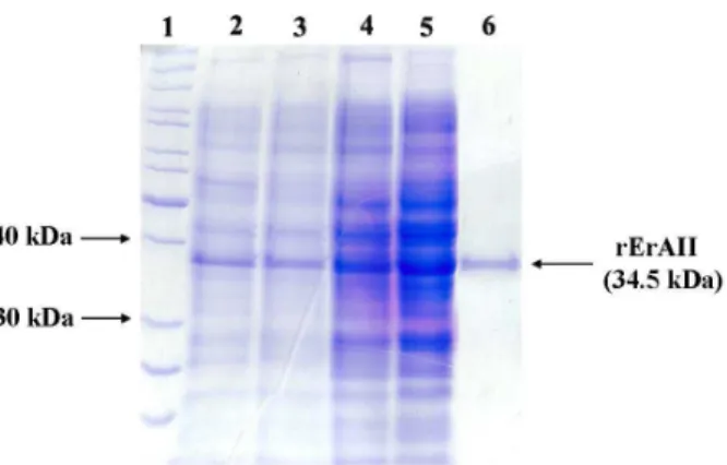
The estimated molecular mass for L-asparaginase II without its signal peptide is 34.5 kDa, which differs from the approximately 39 kDa value observed by SDS-PAGE analysis. The protein may run differently (more slowly) on SDS-PAGE than expected, due to its notably alkaline character (estimated pI value of 8.5) (Walker, 2009).
Optimal Conditions for Soluble Protein Expression in Shaker Cultures
rErAII expression, quantified by densitometry, culture medium preparation convenience, good inducer control and temperature, the final selected conditions for rErAII in shake flask cultures were E. coli C43 (DE3) strain, 30 °C, TB culture medium and 1 mM IPTG. As presented in Figure 1, rErAII expression represented 19.3% of the total protein content in the soluble cell fraction at 6 hours after induction.
Figure 1: rErAII SDS-PAGE (12%) analysis. Lanes: 1, PageRuler unstained protein ladder (Fermentas Life Sciences); 2, 3 and 4, total cell protein content from E. coli cultures grown under different conditions: shake flask cultivation in TB medium at 30 ºC, shake flask cultivation in TB medium at 37 ºC and fed-batch cultivation, respectively; 5, dialyzed crude extract loaded onto a HiPrep 16/10 SP XL (GE Healthcare, United Kingdom) cation exchange column; 6, homo-geneous rErAII pool eluted from the ion-exchange column.
Bioreactor Cultivations
The optimized growth conditions from the shaker cultivations were applied to the bioreactor batch and fed-batch cultures. We employed the Terrific Broth culture medium (TB) in all experiments.
The transition from shaken flasks to bioreactors can reduce the recombinant protein yield (Goyal et al., 2009). Therefore, optimizing the nutritional and metabolic parameters for bioreactor cultivation is important for protein production. A successful cultivation procedure (Panda et al., 1999; Vallejo et al., 2002; Khalilzadeh et al., 2004) with a pre-determined exponential-feeding strategy was applied to maximize the production of rErAII (Korz et al., 1995).
A liquid culture of transformed E. coli C43 (DE3) was started from the MCB under batch cultivation in 1 L TB medium at 30 ºC, with a growth rate of 0.60 h-1. We monitored acetate consumption and pO2 increase
to verify the transition from exponential to stationary cell growth and to define the right moment to initiate
fed-batch culture. In the presence of abundant nutrients and/or low pO2, bacterial cells produce and
excrete acetate into their environment. However, when the bacteria switch to a slower growth rate (when under nutrient deficits or high pO2) they
import and utilize the excreted acetate, which is a process known as the “acetate switch” (Wolfe, 2005).
The exponential addition of concentrated feeding solution started when the biomass of the culture reached 6.0 grams dry cell weight per liter of culture brothat a specific growth rate of μset = 0.1 h-1 (μreal =
0.13 h-1), corresponding to phase 1 of fed-cultivation. Phase 2 started at 18 hours of cultivation when the biomass concentration reached 25.9 gram dry cell weight per liter of culture medium. At this time, the culture was induced with 1 mM IPTG and the μset
was lowered from a value of 0.1 h-1 to 0.08 h-1 for 2 hours before decreasing further (μreal = 0.02 h-1).
IPTG induction was accompanied by a decrease in the bacterial growth rate (Hortsch and Weuster-Botz, 2011). Lowering the μset is a proven method that
maintains the substrate supply between the maintenance and threshold levels (Lee, 1996; Panda
et al., 1999; Johnston et al., 2002; Khalilzadeh et al., 2004). The real specific growth rate in both cultivation phases differed from the set value despite controlling the acetate formation under inhibitory levels. This phenomenon has been reported by other researchers (Panda et al., 1999; Vallejo et al., 2002). At the end of the glucose-controlled fed-batch fermentation, rErAII represented about 10% of the total protein in the soluble protein fraction (Figure 1). Additionally, rErAII had the highest activity in the crude extract at 10 h post-induction, while longer culture times decreased its activity (Figure 2).
Brazilian Journal of Chemical Engineering Vol. 30, No. 02, pp. 245 - 256, April - June, 2013
L-Asparaginases from different organisms have been cloned and successfully expressed in bacterial and yeast expression systems (Khushoo et al., 2005; Kotzia and Labrou, 2005; Ferrara et al., 2006; Kotzia and Labrou, 2007; Wink et al., 2010). Kumar et al. (2009) have grown and maximized glutaminase-free L-asparaginase expression from Pectobacterium carotovorum MTCC 1428 in a bioreactor, achieving 28.87 U mg-1 of protein and a productivity of 1282.5 U L-1 h-1. Our work on recombinant Erwinia carotovora
L-asparaginase II production in Escherichia coli fed-batch cultures achieved a final biomass concentration of 30.7 grams of dry cell weight per liter of culture broth with a specific product concentration of 29.3 mg rErAII per gram of dry cell weight, corresponding to a volumetric yield of 0.9 grams of soluble rErAII per liter of culture medium. Considering the purified rErAII specific activity for L-Asn (Figure 3(a)) of 86.76 U mg-1, we achieved a productivity of 2602.8 U L-1 h-1. To the best of our knowledge, we could not find any report in the literature where recombinant
E. carotovora L-asparaginase II was produced achieving such yields.
Purification and Mass Spectrometry
Our protocol varied from previously published protocols because it involved cellular disruption through a modified French press, which is an effective and economical technique for cell-free extract preparation providing high enzyme recovery. Purified biologically active rErAII protein was obtained by loading the soluble protein fraction of the cell-free extract onto a HiPrep 16/10 SP XL cation exchange column at pH 5.5 and eluting the proteins using 20 mM potassium phosphate buffer on a linear pH gradient profile from 5.5 to 8.5. Owing to its notably alkaline character (estimated pI of 8.5), the rErAII loaded on the cation exchange column was tightly adsorbed to the resin, whereas most of the E. coli proteins, particularly the L-asparaginases with acidic pI values, were washed off of the column (Figure 1), thereby ensuring separation of the various proteins. The established one-step purification protocol yielded ~5 mg of rErAII per gram of dry cell weight, corresponding to a volumetric yield of 168 mg of rErAII per liter of culture medium.
Purified rErAII samples were desalted and digested with trypsin and were subjected to LC-MS/MS analysis. We obtained 1626 spectra that corresponded to 24 different peptides (approximately 88% of the rErAII sequence), which ensured the protein’s identity. The mass spectrometric analysis showed that rErAII was separated from its E. coli
counterpart, which is important for preparing therapeutic doses of E. carotovora L-asparaginases
II because these two enzymes have distinct immunological specificities and provide alternative therapies for patients who develop hypersensitivity to one of the L-asparaginases (Duval, 2002).
L-Asparaginase Activity Measured by Isothermal Titration Calorimetry
L-asparaginases from E. coli and E. chrysanthemi
and the alternative enzyme from E. carotovora have unique but comparable kinetic parameters, as determined by well-established colorimetric assays through discontinuous Nessler (Wink et al., 2010) and AHA methods (Derst et al., 2000), continuous direct (Krasotkina et al., 2004) assays and continuous coupled (Kotzia and Labrou, 2005) assays. This enzymatic comparison has generated discussions about the various enzymes’ substrate affinity (L-asparagine and L-glutamine) (Derst et al., 2000; Krasotkina et al., 2004) and their toxicological consequences in leukemia treatment (Duval, 2002; Vrooman et al., 2010). The anti-tumor properties of the enzyme are related to its asparaginase activity (Oettgen et al., 1967), but the enzyme’s glutaminase activity has been implicated in causing side effects during ALL treatment (Pui et al., 1981; Priest et al., 1982; Sahu et al., 1998; Hawkins et al., 2004). Therefore, both L-asparaginase and L-glutaminase activities should be considered to improve this enzyme’s therapeutic benefit.
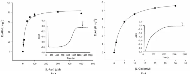
Isothermal titration calorimetry (ITC) is a sensitive technique that directly determines the thermodynamic and kinetic parameters of enzymatic reactions by measuring the heat absorbed or released during a chemical reaction (binding, dilution, or transformation) (Bianconi, 2007). This nondestruc-tive method is completely general, thereby enabling precise analysis of reactions in spectroscopically opaque solutions, such as crude cell extract, using physiological substrates. As described by Bianconi (2003), the calorimetric enthalpy is the sum of the different heat effects that take place during the reaction. The downward baseline displacement after enzymatic injection indicates an exothermic reaction (Figure insets 3 and 4). ITC was employed to compare the thermodynamic and kinetic parameters of two different L-asparaginases: rErAII (purified as described in Materials and Methods) and Escherichia coli L-asparaginase II (EcAII) (purchased from Medac, Germany). The rErAII ΔHapp was 9.6 kcal mol-1 for
L-asparagine hydrolysis and 7.0 kcal mol-1 for L-glutamine hydrolysis (Figure insets 3(a) and 3(b), respectively). The EcAII ΔHapp was 6.1 kcal mol-1
(a) (b)
Figure 3: rErAII kinetic and thermodynamic parameters. Initial velocity experiments at different L-Asn (a) and L-Gln (b) concentrations, as measured by ITC. Heat flow as a function of time (μcal s-1) for the consumption of L-Asn (Inset A) and L-Gln (Inset B), at 25 ºC in potassium phosphate buffer (pH 7.0). The arrow indicates the time of the second injection.
(a) (b)
Figure 4: EcAII kinetic and thermodynamic parameters. Initial velocity experiments at different L-Asn (a) and L-Gln (b) concentrations, as measured by ITC. Heat flow as a function of time (μcal s-1) for the consumption of L-Asn (Inset (a)) and L-Gln (Inset (b)), at 25 ºC in potassium phosphate buffer (pH 7.0). The arrow indicates the time of the second injection.
The kinetic parameters obtained by ITC for the purified rErAII were compared with those of EcAII (Table 1). Other researchers have reported that calorimetric and spectrophotometric data agree well (Todd and Gomez, 2001; Bianconi, 2003). Our enzyme is a pharmaceutically relevant alternative because L-Asparaginases with low Km
values for L-Asn and large values for L-Gln are preferable during the course of anti-cancer therapy (Hawkins et al., 2004). The kcat/Km ratio is an
apparent second-order rate constant that determines the specificity of the enzyme for competing substrates (Fersht, 1999). Accordingly, the ratio of the L-Asn kcat/Km value to the L-Gln kcat/Km value
indicates that both enzymes convert the substrates
equally well: rErAII exhibited a 2.8x10³-fold increase, compared to EcAII’s 2.8x10³-fold increase. Moreover, the Km value for L-Gln hydrolysis
catalyzed by rErAII (10.3 mM) is larger than that found for L-asparaginases II from E. carotovora
Brazilian Journal of Chemical Engineering Vol. 30, No. 02, pp. 245 - 256, April - June, 2013
Table 1: Kinetic parameters for the hydrolysis of L-Asn and L-Gln by rErAII and EcAII (Elspar®) at 25 °C and pH 7.0
L-asparagine L-glutamine Km (μM) kcat (s-1) kcat/Km (M-1.s-1) Km (mM) kcat (s-1) kcat/Km (M-1.s-1)
rErAII 33±6 49.8±3.8 1.50x10⁶±0.29 10.3±0.4 5.6±0.1 0.53x10³±0.02
EcAII (Elspar®) 18±3 53.2±2.0 2.95x10⁶±0.47 3.7±0.2 3.9±0.1 1.04x10³±0.05
In addition to determining the thermodynamic and kinetic parameters, the calorimetric assays were implemented to define the crude extract activity during cultivation of rErAII (Figure 2). Due to the low ε215 (102.5 M
-1
cm-1 (Howard and Carpenter, 1972)) of the -amide bond of L-asparagine or L-glutamine and the fact that the crude extracts are opaque solutions, rErAII activity cannot be measured by direct continuous assay. Figure 2 shows an increase in rErAII specific activity during fed-batch cultivation, reaching maximal values 10 h after IPTG induction. This proposed method for measuring L-asparaginase activity could help the biopharmaceuti-cal industry because the equipment and method are simple and are easily implemented to monitor the activity during cultivation and to control the quality of the final product.
CONCLUSIONS
As already pointed out by other researchers (Lee, 1996; Choi et al., 2006), recombinant protein produc-tion methods have to improve continuously to meet commercial demands. In this work, we produced an active purified recombinant E. carotovora asparaginase II from the soluble cell fraction of fed-batch cultivated IPTG-induced E. coli with cell and enzyme productivity that agreed with and outweigh those in the literature. It is important to point out that, to the best of our knowledge, this is the first time that recombinant E. carotovora asparaginase II has been produced in a laboratory scale bioreactor in
E. coli cultures. Furthermore, the previous work on asparaginase from our research group (Wink et al., 2010) was extended, achieving a higher volumetric yield and specific rErAII concentration. Additionally, the recombinant enzyme from Erwinia carotovora
may be a promising alternative therapy for treating ALL because of its lower glutaminase activity compared to Escherichia coli L-asparaginase II. The straightforward calorimetric techniques for determin-ing the enzyme kinetics are accurate and precise for both purified enzyme and crude extracts compared to well-established spectrophotometric
assays. We present a method for the production of a low affinity L-Gln L-asparaginase II using a recombinant expression system. This represents an important step for future implementation by the biopharmaceutical sector.
NOMENCLATURE
DCW Dry Cell Weight g.L-1
dQ/dt heat flow μcal.s-1
DTT Dithiothreitol EcAII Escherichia coli
L-asparaginase II (Asparaginase Elspar®) IPTG isopropyl-
-D-thiogalactopyranoside ITC isothermal titration
calorimetry
kcat/Km apparent second-order rate
constant
M-1.s-1
kcat apparent unimolecular rate
constant
s-1
kDa: kilodalton
Km concentration at which the
rate of the enzyme reaction is half of Vmax
M
L-Asn L-asparagine M
L-Gln L-glutamine M
MCB master cell bank pO2 dissolved oxygen
concentration
%
rErAII recombinant Erwinia carotovora L-asparaginase II
SDS-PAGE
sodium dodecyl sulfate-polyacrylamide gel electrophoresis TFA trifluoroacetic acid U amount of enzyme that
catalyzed the formation of 1 μmol ammonia per minute at 25 ºC
μmol.min-1.mg-1
V calorimetric cell volume L
Greek Letters
ΔHapp total reaction enthalpy
variation
cal.mol 1
ε215 molar extinction coefficient M 1.cm 1
μ specific growth rate h 1
ACKNOWLEDGMENTS
Financial support for this work was provided by FINEP (Financiadora de Estudos e Projetos) to Quatro G Pesquisa & Desenvolvimento Ltda.
REFERENCES
Bae, N., Pollak, A. and Lubec, G., Proteins from
Erwinia asparaginase Erwinase (R) and E. coli
asparaginase 2 MEDAC (R) for treatment of human leukemia, show a multitude of modifications for which the consequences are completely unclear. Electrophoresis, 32, 14, 1824-1828 (2011). Bianconi, M. L., Calorimetric determination of
thermodynamic parameters of reaction reveals different enthalpic compensations of the yeast hexokinase isozymes. The Journal of Biological Chemistry, 278, 21, 18709-18713 (2003).
Bianconi, M. L., Calorimetry of enzyme-catalyzed reactions. Biophysical Chemistry, 126, 1-3, 59-64 (2007).
Bradford, M. M., A Rapid and sensitive method for the quantitation of microgram quantities of protein utilizing the principle of protein-dye binding. Analytical Biochemistry, 72, 248-254 (1976). Broome, J. D., Evidence that the L-asparaginase of
Guinea Pig serum is responsible for its antilym-phoma effects. The Journal of Experimental Medicine, 118, 99-120 (1963).
Choi, J., Keum, K. and Lee, S., Production of recombinant proteins by high cell density culture of Escherichia coli. Chemical Engineering Science, 61, 3, 876-885 (2006).
Covini, D., Tardito, S., Bussolati, O., Chiarelli, L. R., Pasquetto, M. V., Digilio, R., Valentini, G. and Scotti, C., Expanding targets for a metabolic therapy of cancer: L-asparaginase. Recent Patents on Anti-Cancer Drug Discovery, v. 7, n 1, pp. 4-13, (10) (2012).
Derst, C., Henseling, J. and Röhm, K. H., Engineering the substrate specificity of
Escherichia coli asparaginase II. Selective reduction of glutaminase activity by amino acid replacements at position 248. Protein Science, 9, 10, 2009-2017 (2000).
Duval, M., Comparison of Escherichia coli -asparaginase with Erwinia-asparaginase in the treatment of childhood lymphoid malignancies: Results of a randomized European Organisation for Research and Treatment of Cancer–Children's Leukemia Group phase 3 trial. Blood, 99, 8, 2734-2739 (2002).
European Medicines Agency, Evaluation of medicines for human use: Assessment of the paediatric needs chemotherapy products (Part I). London, (2006). Available in: <http://www.ema.europa.eu/docs/ en_GB/document_library/Other/2009/10/WC500 004053.pdf>. (Acessed in July 5, 2011).
Evans, W. E., Tsiatis, A., Rivera, G., Murphy, S. B., Dahl, G. V., Denison, M., Crom, W. R., Barker, L. F. and Mauer, A. M., Anaphylactoid reactions to Escherichia coli and Erwinia asparaginase in children with leukemia and lymphoma. Cancer, 49, 7, 1378-1383 (1982).
Ferrara, M., Severino, N., Mansure, J., Martins, A., Oliveira, E., Siani, A., Pereira Jr., N., Torres, F. and Bon, E., Asparaginase production by a recombinant Pichia pastoris strain harbouring
Saccharomyces cerevisiae ASP3 gene. Enzyme and Microbial Technology, 39, 7, 1457-1463 (2006). Fersht, A., Structure and mechanism in protein
science: A guide to enzyme catalysis and protein folding. W. H. Freeman, New York, 1st Ed. (1999). Goyal, D., Sahni, G. and Sahoo, D. K., Enhanced
production of recombinant streptokinase in
Escherichia coli using fed-batch culture. Biore-source Technology, 100, 19, 4468-4474 (2009). Hawkins, D. S., Park, J. R., Thomson, B. G.,
Felgenhauer, J. L., Holcenberg, J. S., Panosyan, E. H. and Avramis, V. I., Asparaginase pharma-cokinetics after intensive polyethylene glycol-conjugated L-asparaginase therapy for children with relapsed acute lymphoblastic leukemia. Clinical Cancer Research : An Official Journal of the American Association for Cancer Research, 10, 16, 5335-5341 (2004).
Hortsch, R. and Weuster-Botz, D., Growth and recombinant protein expression with Escherichia coli in different batch cultivation media. Applied Microbiology and Biotechnology, 90, 1, 69-76 (2011).
Howard, J. B. and Carpenter, F. H., L-Asparaginase from Erwinia carotovora. The Journal of Biological Chemistry, 247, 4, 1020-1030 (1972).
Brazilian Journal of Chemical Engineering Vol. 30, No. 02, pp. 245 - 256, April - June, 2013
Khalilzadeh, R., Shojaosadati, S. A., Maghsoudi, N., Mohammadian-Mosaabadi, J., Mohammadi, M. R., Bahrami, A., Maleksabet, N., Nassiri-Khalilli, M. A., Ebrahimi, M. and Naderimanesh, H., Process development for production of recombi-nant human interferon-gamma expressed in
Escherichia coli. Journal of Industrial Microbiology & Biotechnology, 31, 2, 63-69 (2004).
Khushoo, A., Pal, Y. and Mukherjee, K. J., Optimi-zation of extracellular production of recombinant asparaginase in Escherichia coli in shake-flask and bioreactor. Applied Microbiology and Bio-technology, 68, 2, 189-197 (2005).
Klammer, A. A. and Maccoss, M. J., Effects of modified digestion schemes on the identification of proteins from complex mixtures. Journal of Proteome Research, 5, 3, 695-700 (2006).
Korz, D. J., Rinas, U., Hellmuth, K., Sanders, E. A. and Deckwer, W.-D., Simple fed-batch technique for high cell density cultivation of Escherichia coli. Journal of Biotechnology, 39, 59-65 (1995). Kotzia, G. A. and Labrou, N. E., Cloning, expression
and characterisation of Erwinia carotovora L-asparaginase. Journal of Biotechnology, 119, 4, 309-323 (2005).
Kotzia, G. A. and Labrou, N. E., L-Asparaginase from Erwinia chrysanthemi 3937: Cloning, expression and characterization. Journal of Biotechnology, 127, 4, 657-669 (2007).
Krasotkina, J., Borisova, A. A., Gervaziev, Y. V. and Sokolov, N. N., One-step purification and kinetic properties of the recombinant l-asparaginase from
Erwinia carotovora. Biotechnology and Applied Biochemistry, 39, 2, 215-221 (2004).
Kumar, S., Pakshirajan, K. and Venkata Dasu, V., Development of medium for enhanced produc-tion of glutaminase-free L-asparaginase from
Pectobacterium carotovorum MTCC 1428. Applied Microbiology and Biotechnology, 84, 3, 477-486 (2009).
Lee, S. Y., High cell-density culture of Escherichia coli. Trends in Biotechnology, 14, 3, 98-105 (1996). Montgomery, D. C., Design and Analysis of
Experiments. Wiley, New York (1997).
Moola, Z. B., Scawen, M. D., Atkinson, T. and Nicholls, D. J., Erwinia chrysanthemi L-asparagi-nase: epitope mapping and production of antigenically modified enzymes. Biochemical Journal, 302, 921-927 (1994).
Moritz, R. L., Configuration, Column Construction, and Column Packing for a Capillary Liquid Chromatography System. Cold Spring Harbor Protocols, 2007, 2, pdb.prot4578 (2010).
Oettgen, H. F., Old, L. J., Boyse, E. A., Campbell, H. A. and Philips, F. S., Inhibition of leukemias in man by L-asparaginase. Cancer Research, 27, 2619-2631 (1967).
Onishi, Y., Yano, S., Thongsanit, J., Takagi, K., Yoshimune, K. and Wakayama, M., Expression in Escherichia coli of a gene encoding type II l-asparaginase from Bacillus subtilis, and charac-terization of its unique properties. Annals of Microbiology, (2010).
Panda, A. K., Khan, R. H., Appa Rao, K. B. C. and Totey, S. M., Kinetics of inclusion body production in batch and high cell density fed-batch culture of Escherichia coli expressing ovine growth hormone. Journal of Biotechnology, 75, 161-172 (1999).
Priest, J. R., Ramsay, N. K., Steinherz, P. G., Tubergen, D. G., Cairo, M. S., Sitarz, A. L., Bishop, A. J., White, L., Trigg, M. E., Levitt, C. J., Cich, J. A. and Coccia, P. F., A syndrome of thrombosis and hemorrhage complicating L-asparaginase therapy for childhood acute lymphoblastic leukemia. Journal of Pediatrics, 100, 6, 984-989 (1982). Pui, C. H., Burghen, G. A., Bowman, W. P. and Aur,
R. J., Risk factors for hyperglycemia in children with leukemia receiving L-asparaginase and prednisone. The Journal of Pediatrics, 99, 1, 46-50 (1981).
Riesenberg, D. and Guthke, R., High-cell-density cultivation of microorganisms. Applied Microbiol-ogy and BiotechnolMicrobiol-ogy, 51, 422-430 (1999).
Sahu, S., Saika, S., Pai, S. K. and Advani, S. H., L-asparaginase (Leunase) induced pancreatitis in childhood acute lymphoblastic leukemia. Journal of Pediatric Hematology/Oncology, 15, 6, 533-538 (1998).
Schwartz, J. H., Reevesi, J. Y. and Broome, J. D., Two L-asparaginases from E. coli and their action against tumors. Biochemistry, 56, 1516-1519 (1966). Todd, M. J. and Gomez, J., Enzyme kinetics
determined using calorimetry: a general assay for enzyme activity. Analytical Biochemistry, 296, 2, 179-187 (2001).
Vallejo, L. F., Brokelmann, M., Marten, S., Trappe, S., Cabrera-Crespo, J., Hoffmann, A., Gross, G., Weich, H. A. and Rinas, U., Renaturation and purification of bone morphogenetic protein-2 produced as inclusion bodies in high-cell-density cultures of recombinant Escherichia coli. Journal of Biotechnology, 94, 185-194 (2002).
Cohen, H. J., Sallan, S. E. and Silverman, L. B.,
Erwinia asparaginase after allergy to E. coli
asparaginase in children with acute lymphoblastic leukemia. Pediatric Blood & Cancer, 54, 2, 199-205 (2010).
Walker, J. M., The protein protocols handbook. Springer (2009).
Wink, P. L., Bogdawa, H. M., Renard, G., Chies, J.
M., Basso, L. A. and Santos, D. S., Comparison between two Erwinia carotovora L-asparaginase II constructions: Cloning, heterologous expression, purification, and kinetic characterization. Journal of Microbial & Biochemical Technology, 2, 1, 13-19 (2010).