*e-mail: [email protected]
Hydrolytic Degradation of Composite Resins:
Effects on the Microhardness
Josué Martosa*, Prudêncio Willy Rodo Osinagaa, Elisabeth de Oliveirab, Luis Antônio Suita de Castroc
aDepartment of Clinics and Restorative Dentistry, Pelotas School of Dentistry, Federal University of Pelotas, Pelotas - RS, Brazil
bDepartment of Chemistry, Chemistry Institute, University of São Paulo, São Paulo - SP, Brazil
cEMBRAPA-CPACT; Laboratory of Immunology and Microscopy, Pelotas - RS, Brazil
Received: March 6, 2003; July 26, 2003
The purpose of this investigation was to evaluate the microhardness of two laboratory-proc-essed composites (Artglass; belleGlass) and two direct placement composites (Filtek Z250; Alert), after aging in distilled water. Twenty cylinders (8 mm diameter; 2 mm height) per tested material were prepared and stored in 10 ml of distilled water. Five Knoop hardness measurements were made on the surface of the specimens with a Miniload Hardness Tester under a load of 50 g for 30 s at 10 min, 24 h, 30 and 90 days. Statistical analysis was perfomed using two-way ANOVA, followed by a SNK multiple comparison test (p < 0.05). The analysis showed statistically signifi-cant difference among hardness means recorded at the different aging time and the tested materi-als. It may be concluded that all materials presented hydrolytic degradation due to aging in aque-ous environment.
Keywords:composite resin, hydrolytic degradation, microhardness
1. Introduction
Composite resins are currently one of the most widely utilized materials in restorative dentistry and the satisfac-tory clinical performance is largely determined by its
re-sistance to degradation in the oral environment1,2.
Mechani-cal properties of composite resins are vastly influenced not only by their chemical composition, but also by the envi-ronment to wich they are exposed. The corrosion process promoted by the water and the presence of a constant load on the surface of the resin are responsible for the appear-ance and propagation of interfacial debonding, matrix crack-ing, superficial flaws, filler dissolution and filler particle dislodgment3-5.
The hydrolytic degradation of these materials happens mainly because of accumulation of water between the filler-matrix interface that promotes the displacement of inorganic
particles6 or due to the slow development of superficial flaws
related to preexistent corrosive processes7,8. The dissolution
or elution of leachable components of composite resins, mainly inorganic ions or filler particles, may present, at short or long period, a deleterious effect in the polymeric net-work of the material, modifying its structure physically and chemically.
The surface microhardness of dental composites may be significantly affected by both water absorption and the
contact time with the aqueous media9.
A significant reduction of Knoop microhardness in re-storative composite resins was observed after storage in
dis-tilled water during 30 days10, 12 months11 and a long period
of time12. However, the different types of restorative
com-posite resins and polymerization method, may yield differ-ent performances with respect to the hydrolytic degradation process.
resistance to intraoral softening13,14.
The aim of this investigation was to assess the surface hardness of four commercial composite resins after aging in water.
2. Material and Methods
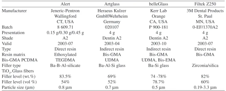
Four commercial composite resins were evaluated in this study: two laboratory-processed composite, Artglass (Heraeus Kulzer GmbH, Wehrheim, DE 63450) and belleGlass HP (Kerr Corporation, Orange, CA 92867) an hybrid composite resin, Filtek Z250 (3M Dental Products, St Paul, MN 55144) and an condensable composite resin, Alert (Jeneric-Pentron, Wallingford, CT 06492). Details about the tested materials with their compositions, specifi-cations and manufacturers are listed on Table 1.
Twenty cylindrical specimens of each material were pre-pared using a split stainless steel mould with 8 mm in diam-eter by 2 mm in height according to the manufacturer’s speci-fications. The restorative materials Filtek Z250 and Alert were light-cured for 60 s on their top surfaces through clear polyester matrix strip using a visible light-curing unit (KM 100-R, DMC Equipaments, São Carlos, SP) with an
inten-sity of 400 mW/cm2 determined with a radiometer (Curing
Lightmeter 105, DMC Equipaments, São Carlos, SP). The laboratory-processed composite resin Artglass were cured with the xenon stroboscopic light curing unit (UniXS, Heraeus Kulzer GmbH, Wehrheim, DE 63450) for 180 s with a polymerization rate between 450-500 nm and strobe frequency of 20 Hz in each 10 ms. The laboratory-proc-essed composite belleGlass HP were cured with a metal halide lamp (Tek Lite, Kerr Corporation, Orange, CA 92867) for 60 s with a wavelenght of 400-500 nm, and an
addi-tional polymerization (HP Curing Unit, Kerr Corporation,
Orange, CA 92867) was made under 60 psi (29 lb/pol2)
ni-trogen pressurized at an elevated temperature of 120 to 140 °C for 20 min.
The top surfaces of each specimen were ground with water-lubricated silicon-carbide (SiC) paper and polishing with 2000 grit paper (Wetordry, 3M Dental Products, St Paul, MN 55144) on an automated polisher (Struers A/S, Copen-hagen, Denmark) to produce a smooth, uniform surface. Polished specimens were then stored in distilled water at 37 °C.
Five measurements of Knoop surface hardness were re-corded from each specimen using a Miniload Hardness Tester (Durimet, Ernst Leitz GmbH, Wetzlar, Germany). A Knoop diamond indenter was applied under a load of 50 g for 30 s and the length of the indentation’s long diagonal measured at 400× magnification after the applied load was removed. The surface hardness measurements were recorded at end of the polymerization period (time zero) and at aging times of 24 h, 30 and 90 days of storage in distilled water.
The Knoop hardness number (KHN) for each indenta-tion was determined and means and standard deviaindenta-tion were calculated at each time interval for each group of specimens. The values were compared by factorial analysis of variance (ANOVA) using the SPSS software (SPSS 8.0, SPSS Inc.,
Chicago, IL 60611). When F-tests were significant,
Post-hoc Student-Newman-Keuls multiple comparison intervals
were further performed to identify statistically homogene-ous subsets (p = 0.05).
Additionally, the surface texture of each two randomly selected specimens and two control samples of the four com-posite resins were qualitatively evaluated by SEM using a
Table 1. Description of materials.
Alert Artglass belleGlass Filtek Z250
Manufacturer Jeneric-Pentron Heraeus Kulzer Kerr Lab 3M Dental Products
Wallingford GmbHWehrheim Orange St. Paul
CT, USA Germany CA, USA MN, USA
Batch 8 609.71 020107 P 900-181 0-EF/1370A2
Presentation 0.15 g/0.30 g/0.45 g 4 g 4 g 4 g
Shade A2 Dentin A2 Dentin A2 A2
Valid 2003-07 2003-04 2003-10 2003-07
Type Direct resin Indirect resin Indirect resin Direct resin
Resin matrix Ethoxylated Bis-GMA Bis-GMA Bis-GMA
Bis-GMA PCDMA TEGDMA UDMA UDMA, Bis-EMA
Filler type Ba-B-Al-silicate Ba-Al-Si glass Ba-Si glass Zirconia/silica
TiO2,Glass fibers
Filler level (wt.%) 83.5% 69% 74 -78% 82%
Filler level (vol %) 54% 52% 78.7% 60%
digital scanning microscope (Zeiss DSM 940A, Carl Zeiss, Oberkochen, Germany)
3. Results
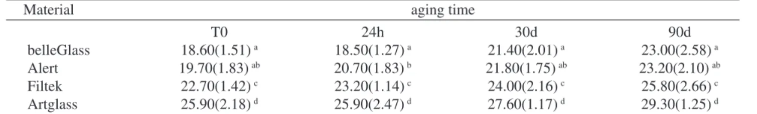
Microhardness means recorded at the different aging times and standard deviation are summarized in Table 2.
An analysis of variance demonstrated that all liquid-stored in an aqueous environment were softened compared
to dry samples (p < 0.05). Aging in water for 24 h did not
significantly change the KHN for any of the tested compos-ites. Continued aging in water beyond 30 d generally caused a significant reduction in the microhardness for each com-posite (p < 0.05). After that, a slow gradual decrease in hard-ness was observed (p < 0.05).
It was observed that the differences in microhardness for Alert and Belleglass between the unage and 30 and 90 d
aging were not significant (p > 0.05). The final hardness
values, measured at 90 d, were significantly lower (p < 0.05) than the corresponding values measured under dry condi-tions. A more pronounced decrease of microhardness of the wet-stored samples was found during the final wet storage. Statistical analysis of the data revealed significant
dif-ference (P < 0.05) among the experimental groups.
Com-paring the materials, regardless of the microhardness, belleGlass HP and Alert showed statistical similarity and yielded the highest means. On the other hand, Artglass and Filtek Z250 presented the lowest microhardness mean.
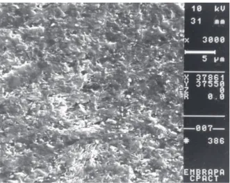
The SEM examinations of dry-stored controls and speci-mens kept in an aqueous environment showed few surface alterations. The most evident was the presence of voids and porosity in some areas, there were not apparent loss of fill-ers or topography alterations after aging time (Fig. 1-8).
4. Discussion
The results of this investigation showed that the all four composites stored in distilled water suffered a reduction in surface hardness. All liquid-stored specimens showed a sig-nificant drop of Knoop hardness compared to the dry-stored controls.
Table 2. Microhardness values (KHN) of tested materials after different storage time periods.
Material aging time
T0 24h 30d 90d
belleGlass 18.60(1.51) a 18.50(1.27) a 21.40(2.01) a 23.00(2.58) a
Alert 19.70(1.83) ab 20.70(1.83) b 21.80(1.75) ab 23.20(2.10) ab
Filtek 22.70(1.42) c 23.20(1.14) c 24.00(2.16) c 25.80(2.66) c
Artglass 25.90(2.18) d 25.90(2.47) d 27.60(1.17) d 29.30(1.25) d
Means followed by the same superscript letters in the column indicate no significant difference (p < 0.05).
The fact that composites subjected to the visible light-curing experienced a reduction in properties wich closely paralleled the reduction experienced by the laboratory-proc-essed specimens suggests that the reduction has a similar etiology in both cases. Studies have attributed the decrease in hardness of dental composites immersed in water to
hy-drolysis of ester groups in the resin matrix15.
The mechanism of hydrolytic degradation is enhanced if the filler particles have metallic ions in their
composi-tion4,16. The explanation of this effect is that some ions in
the filler particles, such as zinc and barium, are electroposi-tive and tend to react with water. With the loss of these ele-ments into water, the charge balance inside the silica net-work is changed and reestablished with the penetration of hydrogen ions of the water in the spaces occupied by the zinc and barium. As a result of the increase of the concen-tration of hydroxy ions, the siloxane (Si-O-Si) bonds of the silica network start to break, and there is formation of an
autocatalytic cycle of surface degradation4,17,18. This
mecha-nism would explain the continuity of the superficial soften-ing with agsoften-ing time.
The short duration of immersion is therefore aimed at investigating surface changes in hardness that will influ-ence mechanical wear. It has been suggested that resistance to initial softening will improve the abrasion resistance of
dental composite restorations19,20.
The present study agrees with the results of others stud-ies10,11,12. Decreased in Knoop microhardness was found in
the commercially available composites stored in water for
30 days10, and Helvatjoglou (1991) assumed that the changes
in superficial hardness were due to the plasticization by water
in the material11. The results of the present study were also
consistent with the results of Ferracane et al.(1998) for the
unaged and aged specimens. Contrary to these findings,
Chadwick et al. (1990) found that composite resin stored in
water did not significantly influence surface microhardness during the 1-year study period.
The heat and pressure treatment, alone or in combination with the additional light exposure, was expected to increase the degree of conversion of the polymer. However, no evi-dence for this was revealed, though this is a technique used in at least one commercial curing system (belleGlass HP). The lack of a significant difference suggests that the degradative effects of water are independent of cure state. This conclusion supports a clinical study which reported no difference in resistance for inlays that were light-cured
only vs. those that were exposed to a heat treatment after
the initial light-curing21 and after aging in water12,22.
In addition, the SEM evaluation of dry-stored controls
and specimens kept in an aqueous environment revealed changes in surface texture. The wet-stored samples were significantly rougher than the dry-stored specimens and showed a fine highly porous structure. This surface rough-ness appeared to be a discernible loss of material and crack formation. So long as inorganic fillers of the types currently used are present the surface of composite resins will be
rough, either because of loss or projection of particles23,24.
Composites containing zinc and barium glasses have been shown to be more susceptible to aqueous attack than
those containing quartz16,25,26. From the large softening
ob-served with Filtek Z250 in water, it appears that zirconia Figure 1. SEM micrography of Alert surface at aging time 0 (SEM
original magnification 10000×).
Figure 4. SEM micrography of Artglass surface after 90 days of aging (SEM original magnification 10000×).
Figure 2. SEM micrography of Alert surface after 90 days of ag-ing (SEM original magnification 10000×).
Figure 5. SEM micrography of Belleglass surface at aging time 0 (SEM original magnification 10000×).
Figure 6. SEM micrography of Belleglass surface after 90 days of aging (SEM original magnification 10000×).
Figure 7. SEM micrography of Filtek Z250 surface at aging time 0 (SEM original magnification 10000×).
Figure 8. SEM micrography of Filtek Z250 surface after 90 days of aging (SEM original magnification 10000×).
silicate fillers are also susceptible to aqueous attack. This may be compounded by the smaller filler surface area asso-ciated with the spherical shape of zirconia/silica fillers that may decrease bonding of fillers to the resin matrix.
The filler particle dimension and chemistry of the four tested composites did not represent substantial differences among the materials. However, there were statistically sig-nificant differences in the hardness results of the various materials. It would appear that the filler is not responsible for the observed differences in properties of the test materi-als in this study. It may be that the volume percent of the filler content, the amount of residual monomer or matrix polymers is manifest in the different results.
A variety of mechanisms has been suggested in this pa-per to explain the effects of the storage media upon the sur-face hardness of all four materials. Further investigation is necessary to more completely characterize the hydrolytic degradation of composites that occurs as a result of acceler-ated aging.
5. Conclusions
Under the simulated conditions of this study, the fol-lowing conclusions have been drawn.
Materials stored in an aqueous environment were sof-tened compared to dry samples.
Within the limits of this study, belleGlass HP was the hardest material, and the composites in order of decreasing hardness were Alert, Artglass and Filtek Z250.
Acknowledgements
The authors gratefully thank to Miss Nara Eliane Moreira Rocha for the assistance with the specimens preparation to SEM analysis at EMBRAPA-CPACT.
References
1. Oilo, G. Adv Dent Res, v.6, p. 50-54, 1992.
2. Roulet, J.F.; Wälti, C. J Prosthet Dent, v. 52, p. 182-189,
1984.
3. Sarkar, N.K. J Biomed Mater Res, v. 53, p. 371-380, 2000.
4. Söderholm, K-J.M.; Zigan, M.; Ragan, M.;
Fischlschweiger, W.; Bergman, M. J Dent Res, v. 63,
p. 1248-1254, 1984.
5. Groeningen, G.V.; Jongebloed, W.; Arends, J. Dent Mater,
v. 2, p. 225-227, 1986.
6. Bowen, R.L.; Reed, L.E. J Dent Res, v. 55, p. 738-747,
1976.
7. Michalske, T.A.; Freiman, S.W.; Nature, v. 295,
p. 511-512, 1982.
8. Michalske, T.A.; Freiman, S.W. J Americ Ceramic Soc,
v. 66, p. 284-288, 1983.
9. Hansen, E.K. Scand J Dent Res, v. 91, p. 406-410, 1983.
10. Watts, D.C.; Mcnaughton, V.; Grant, A.A. J Dent, v. 14,
p. 169-174, 1986.
11. Helvatjoglou-Antoniadi, M.; Papadogianis, Y.;
Koliniotou-Kubia, E.; Kubias, S. J Prosthet Dent, v. 65,
p. 215-220, 1991.
12. Ferracane, J.L.; Berge, H.X.; Condon, J.R. J Biomed
Mater Res, v. 42, p. 465-472, 1998.
13. Watts, D.C. Dent Mater, v. 3, p. 265-269, 1987.
14. Asmussen, E. Scand J Dent Res, v. 90, p. 484-489, 1982.
15. Chadwick, R.G. Dent Mater, v. 6, p. 123-128, 1990.
16. Söderholm, K-J.M. J Dent Res, v. 62, p. 126-130, 1983.
17. Charles, R.J. J Appl Physics, v. 29, p. 1549-1553, 1958.
18. Lovell, M.R.; Dalgliesh, M.; Richardson, R.M.; Barnes,
A.C.; Enderby, J.E.; Evans, B. Phys Chem Chem Phys,
v. 1, p. 2379-2381, 1999.
19. Wu, W.; Mckinney, J.E. J Dent Res,v. 61, p. 1180-1183,
1982.
20. Mante, F.; Saleh, N.; Mante, M. Dent Mater, v. 9, p. 325-331, 1993.
21. Wendt, S.L.; Leinfelder, K.F. J Am Dent Assoc, v. 120,
p. 177-181, 1990.
22. De Gee, A.J. Dent Mater, v. 6, p. 266-270, 1990.
23. Sarrett, D.C.; Ray, S. Dent Mater, v. 10, p. 6-10, 1994.
24. El-Din, I.M.; El-Sayed, A.E. Egypt Dent J, v. 40,
p. 765-774, 1994.
25. ∅ysaed, H.; Ruyter, I.E. J Dent Res, v. 65, p. 1315-1318, 1986.
26. Söderholm, K-J.M.; Mukherjee, R.; Longmate, J. J Dent