*e-mail: [email protected]
Trabalho apresentado no XV CBECIMAT, Natal - RN, Novembro de 2002.
Er Rare-earth Ion Incorporation in Sol-Gel SnO
2Evandro Augusto Moraisa, Luis Vicente de Andrade Scalvib*, Viviany Geraldoa,
Sidney José Lima Ribeiroc, Celso Valentim Santillic
aInstituto de Física de São Carlos - USP
C.P. 369, 13560-970 São Carlos - SP, Brazil bDepartamento de Física - FC - UNESP
C.P. 473, 17033-360 Bauru - SP, Brazil cInstituto de Química Araraquara - UNESP
C.P. 355, 14801-907 Araraquara - SP, Brazil
Received: January 29, 2003; Revised: June 30, 2003
Er-doped SnO2 thin films and xerogels are obtained by sol-gel technique. In order to
under-stand the Er3+ rare-earth ion activity in SnO
2 matrix, a characterization of Er incorporation is done
through emission and excitation spectra of xerogels, besides thin film electrical characterization. Effects to grain dimensions are also analyzed based on X-ray diffraction data showing the particle growth with annealing temperature and inhibition of this growth by Er doping. Electrical charac-terization results suggest that Er3+ has an acceptor-like character in SnO
2, and that codoping with
Yb3+ allows an energy transfer process Yb3+→ Er3+.
Keywords: tin-dioxide, sol-gel, erbium, X-ray diffraction
1. Introduction
In the last decades, rare-earth doped materials have been obtained widespread interest, since they can contribute for technological innovation. Rare-earth incor-poration in semiconductors has many applications in optoelectronic devices. Er3+ ion presents several radiative transition concerning decay from several excited core lev-els to ground state, yielding emission from visible to in-frared. Particularly the 4f transition about 1540 nm coin-cides with minimum absorption of fiber optics1, being of great interest for optical communication. In the other hand, tin dioxide is a wide bandgap semiconductor (about 3.5-4.0 eV) 2, which has been widely applied due to its physi-cal and chemiphysi-cal properties. SnO2 thin films are charac-terized by high electrical conductivity and transparency about 80-90% in the visible and high reflectivity in the infrared2. Undoped SnO
2 is an n-type semiconductor since oxygen vacancies or interstitial Sn4+ are donor sites. In the case of sol-gel films, crystallites are rather small (3-10 nm) 3 and a lot of oxygen is adsorbed at boundary layer, trapping electrons from the conduction band4. Then con-ductivity can be greatly increased by eliminating oxygen
from grain boundary layer, which can be done by anneal-ing at proper temperature and gas composition of the an-nealing chamber5. Er3+ is expected to be an acceptor in SnO2 since when it substitutes Sn4+ in the rutile SnO
2 struc-ture, removes an electron from valence band, leaving a hole, which may recombine with a free electron.
The main goal of our investigation is doping a high trans-parency matrix with an attractive core transition ion, con-tributing for production of devices with high capacity of optical transmission, where there is low optical loss, and doping with optically active elements6. The first investiga-tion of rare earth doping in SnO2 concerns Eu and Tb7-10. Therefore some conclusions drawn in this paper refers to comparison to Eu-doped SnO2.
2. Experimental
Colloidal suspensions have been prepared by sol-gel process7. To an aqueous solution 0.2 molar of SnCl
4.5H2O was added the desired amount of ErCl3.6H2O. Under mag-netic stirring, NH4OH was added until pH reaches 11. Then the suspension is submitted to dialysis with distilled water by 10 days in order to eliminate Cl- and NH
4
+ ions. After
conclusion of this process, the sol presents semitransparent aspect.
The xerogel is obtained by keeping the sol at rest, at room temperature by one week. Yb3+ adsorption is obtained by add-ing SnO2:Er powder to an aqueous solution of YbCl3.6H2O, and waiting 24 h before washing with distilled water.
Thin films were prepared at room temperature upon deposition on a borosilicate substrate through dip-coating technique, with a withdrawing rate of 10 cm/min until 30 dips.
For experiments of emission and excitation, xerogels are annealed at 1000 °C by 6 h. For X-ray diffraction measure-ments, they are treated at different temperatures from 100 to 1000 °C. For emission and excitation spectra it was used a xenon lamp of 450 W, a SPEX F212I Fluorimeter and a Germanium detector. For X-ray diffraction measurements it was used a Rigaku diffractometer coupled with a Cu source of 40 kV and 20 mA of current. Detector rate is 3 degrees per minute with a 0.02-degree step. To electrical characteri-zation of thin films, in electrodes are evaporated on the sam-ple through a shadow mask in a Edwards evaporation sys-tem, and submitted to annealing at 150 °C by 20 min. In order to perform measurements in the range of 25 to 300 K we use an Air Products cryogenic chamber.
3. Results and Discussion
Figure 1 shows excitation spectra for SnO2:4%Er and SnO2:0.1%Er xerogels, keeping emission at 1530 nm, which corresponds to radiative transition from excited 4I
13/2 state to ground 4I
15/2 state. The inset in Fig. 1 is the emission spec-tra of SnO2:4%Er under excitation at 328 nm. Curves in Fig. 1 are separated to better visualization. Besides slit open-ing is different and then, no comparison between intensities is possible. The obtained signal corresponds to best resolu-tion allowed by our system. No significant difference in wavelength of observed bands is seen, which means that the spectra due to Er transitions are present in both cases. The most intense band takes place about 328 nm, corre-sponding to SnO2 bandgap and related to electron-hole re-combination. Er3+ intra-ff transitions are present, concern-ing the smaller peaks in Fig. 1, which states 4F
7/2, 2H
11/2, 4S
3/2 and 4F
9/2. On SnO2 xerogel the intense SnO2 forbidden gap transition suggests that Er3+ substitutes Sn4+ on cassiterite structure11. The inset in Fig. 1 shows peaks at 1512, 1525, 1543, 1562 and 1578 nm, which corresponds to Er3+
transi-Figure 1. Excitation spectra for SnO2+4%Er and SnO2+0.1%Er xerogels, emission fixed at 1530 nm. Inset-Emission spectra for SnO2+4%Er xerogel, excitation fixed at 328 nm.
tions of an ion located on a Sn4+ site in SnO
2 cassiterite structures11. All the features shown in Fig. 1 assure that Er is actually incorporated in the SnO2 matrix. We have also observed an increase in luminescence of xerogels by intro-duction of Yb in codoped samples, which takes place due to an energy transfer process Yb3+→ Er3+. This process is shown to be effective since Er luminescence is obtained by pump-ing with 980 nm (Yb3+ transition)12.
which can be easily observed in the curve of the experi-ment carried out at 210 °C, shown in the inset of Fig. 2.
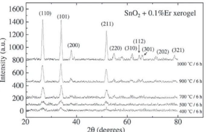
Figure 3 shows X-ray diffraction results to a SnO2:0.1%Er xerogel under several annealing temperatures. It is clearly seen that the higher the annealing temperature, the lower the width and more intense are the diffraction peaks, which means that the crystallinity is increased. Com-paring these results with cassiterite pattern15, there is good agreement as indicated by crystal direction shown in Fig. 3. The average particle size (t) can be determined from dif-fraction peaks, using Scherrer16 method, where t decreases with half width increase. The Scherrer equation is given by16:
t =
where B is the broadening of half width at highest intensity and K is a constant, which is between 0.84 and 0.89, de-pending on geometry. B is evaluated considering contribu-tions of broadening due to grain size and due to instrument broadening16.
From diffraction peaks shown in Fig. 3, crystallite size has been evaluated, which is shown in Table 1. Only (110) and (101) directions are taken into account, since they present the highest intensity in X-ray diffraction pattern of SnO2, being (110) the dominant17. Then these directions are good enough to allow an interpretation of diffraction spec-tra concerning evolution of crystallite dimensions. Particle size increases from 7.3 to 13.2 nm on (110) direction and a larger increase is observed for (101) direction, from 7.2 to 16 nm. This new crystallite dimensions means that there is
Figure 2. Resistivity as function of temperature for some Er-doped thin films. Inset-Current-voltage characteristics above room tem-perature for SnO2:2%Er.
Table 1. Average particle size of SnO2:0.1%Er xerogel.
Temperature Direction (110) Direction (101)
(°C) (nm) (nm)
400 7.3 7.2
500 6.2 6.5
700 8.4 10.7
900 10.0 12.6
1000 13.2 16
Figure 3. X-ray diffraction data for SnO2+0.1%Er xerogel as function of annealing temperature. Lines separated for better visualization.
a significant increase on material crystalinity.
Figure 4 shows the same kind of approach to a SnO2:4%Er xerogel for some different annealing tempera-tures. Crystallite size evaluated from these results is shown in Table 2. Although there is also an increase in the crystallite dimension with annealing temperatures, the particle size is smaller than those obtained by the same treatment on SnO2:0.1%Er. This result suggests that in-corporation of Er impurity controls the grain growth. This dependency of particle size with Er3+ doping concentra-tion is in good agreement with Eu3+-doped SnO
2, since the excess of Eu3+ ions segregates at particle surface and re-tards crystallization compared to undoped xerogels18. An-other important feature for this xerogel is that as the tem-perature is increased, there is a much larger growth on crystallite size on direction (110) than (101), which sug-gests a exchange on preferential direction for growth from (101) to (110), which is in good agreement with Lantto
Table 2. Average particle size of SnO2:4%Er xerogel.
Temperature Direction (110) Direction (101)
(°C) (nm) (nm)
700 6.2 8.7
900 8.7 9.2
1000 10.7 11.2
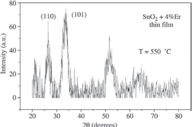
Figure 5. X-ray diffraction data for SnO2:4%Er thin film treated at 550 °C.
sharply, as evaluated from X-ray diffraction data. Incor-poration of Er into SnO2 xerogel inhibits the crystallite growth. Investigation of influence of co-doping with Yb3+ is under progress and the first results, showing the energy transfer process from Yb3+ to Er3+ are being published else-where12.
Acknowledgments
The authors wish to thank Prof. Ligia O. Ruggiero for the help with the technical set-up, and CAPES, FAPESP, CNPq and MCT - PRONEX for the financial resources.
References
1. Coffa, S.; Franzó, G.; Priolo, F.; Polman, A.; Serna, R.
Phys. Rev. B, v. 49, p. 16313, 1994.
2. Ray, S.C.; Karanjai, M.K.; Dasgupta, D. Surface & Coat-ings Technol., v. 102, p. 73, 1998.
3. Souza, A.E.; Monteiro, S.H.; Santilli, C.V.; Pulcinelli, S.H. J. Mater. Sci. Mater. Electron., v. 8, p. 265, 1997. 4. Messias, F.R.; Scalvi, L.V.A.; Siu Li, M.; Santilli, C.V.;
Pulcinelli, S.H. Rad. Eff. & Def. Solids, v. 146, p. 199, 1999. 5. Messias, F.R.; Vega, B.A.V.; Scalvi, L.V.A.; Siu Li, M.; Santilli, C.V.; Pulcinelli, S.H., J. Non-Crystalline Solids, v. 247, p. 171, 1999.
6. Gonçalves, R.R.; Ferrari, M.; Chiasera, A.; Montagna, M.; Morais, E.A.; Scalvi, L.V.A; Santilli, C.V.; Messaddeq, Y.; Ribeiro, S.J.L. J. Metastable and Nanocrystalline Materials, v. 14, p. 107, 2002.
7. Ribeiro, S.J.L.; Pulcinelli, S.H.; Santilli, C.V. Chem. Phys. Lett., v. 190, p. 64, 1992.
8. Ribeiro, S.J.L.; Hiratsuka, R.S.; Massabni, A.M.G.; Davolos, M.R.; Santilli, C.V. J. Non-Crystalline Solids, v. 147&148, p. 162, 1992.
Figure 4. X-ray diffraction data for SnO2+4%Er xerogel as function of annealing temperature. Lines are separated for better visualization.
For comparison, we show in Fig. 5 a plot of X-ray dif-fraction results for a SnO2:4%Er thin film, which has been treated at 550 °C. Scherrer equation applied to this result, yields an average grain size of 3.9 and 3.5 nm in the (110) and (101) directions, respectively. All the results of evalua-tion of grain size for the xerogel yields grains size higher than for thin film. Such a smaller grain size may contribute to increase the grain boundary scattering and, thus, to the observed high resistivity of these films, in conjunction with Er acceptor-like nature.
4. Conclusion
Excitation and emission spectra shows the incorpora-tion of Er3+ in SnO
2 xerogel matrix. Keeping emission fixed at 1530 nm, no band shift on excitation spectra is observed upon increase in doping concentration. Emission spectra under excitation at 328 nm shows several peaks from 1520 to 1580 nm, corresponding to Er3+ core transitions, charac-teristic of a Er3+ located at Sn4+ on SnO
2 structure. Electri-cal characterization of SnO2 thin films suggests the accep-tor like character of Er impurity which leaves the SnO2 matrix practically insulating.
9. Kynev, K.; Gutzov, S.; Peneva, S.K.; Apostolov, A.A.
Cryst. Res. Technol., v. 30, p. 281, 1995.
10. Zupanc Meznar, L.; Pracek, B.; Orel, B.; Bukocek, P.
Thin Solid Films, v. 317, p. 336, 1998.
11. Ribeiro, S.J.L.; Santilli, C.V.; Pulcinelli, S.H.; Fortes, F.L.; Oliveira, L.F.C. J. Sol-Gel Sci. Technol., v. 2, p. 263, 1994.
12. Morais, E.A.; Ribeiro, S.J.L.; Scalvi, L.V.A.; Santilli, C.V.; Ruggiero, L. O.; Pulcinelli, S.H.; Messadeq, Y. J. Alloys and Comp., v. 344, p. 217, 2002.
13. Matsuoka, T.; Tohda, T.; Nitta, T. J. Electrochem. Soc.,
v. 130, p. 417, 1983.
14. Chang, J.P.; Lin, Y.S. Appl. Phys. Lett., v. 79, p. 3666, 2001. 15. Powder Diffraction File, Inorganic v. 21, Publ. By the
JCPDS, Swathmore, 1983.
16. Cullity, B.D. Elements of X-ray Diffraction, Addison-Wesley Pub. Comp., Massachusetts, 1978.
17. Lantto, V.; Rantala, T.T.; Rantala, T.S. J. European Ce-ramic Society, v. 21, p. 1961, 2001.