# $ % $ &
'
$
(
)*
+
(
,- ) . '
$ "
/
President of the jury: Dr. Luís Paulo Rebelo.
Members of the jury: Dr. Mª João Saraiva; Dr. Cláudio Gomes; Dr. Carlos Farinha; Dr. Mamede de Carvalho; Dr. Maria Helena Santos.
By author: NSC%34 cells used as mammalian cell model in this Thesis, visualized by inverted microscopy. Crystal structure of dimeric human Copper%Zinc Superoxide Dismutase (adapted from Valentine et al., 2005). Two of the pathological characteristics of Amyotrophic Lateral Sclerosis mimicked in NSC%34 cells upon mutant SOD1 overexpression: mutant SOD1 aggregation (green) detected by fluorescence microscopy and Golgi apparatus fragmentation (red) detected by immunofluorescence microscopy using an antibody anti%GM130, a cis%Golgi marker.
© 2010
Catarina Heitor Gomes Ph.D. Thesis
ISBN: 978%989%20%1883%6 Laboratório de Glicobiologia
Instituto de Tecnologia Química e Biológica, Universidade Nova de Lisboa Av. Da República, Estação Agronómica Nacional
people to whom I want to express my sincere gratefulness.
To Prof. Júlia Costa, my supervisor, that five years ago received me in her laboratory and gave me the opportunity to study this “puzzling” disease. Thank you for your support and encouragement throughout these years. For always seeing the bright and positive side of things, when I was only seeing the dark ones. Thank you for helping me to strengthen my strong points and overcome the weakest ones.
To Prof. Mamede de Carvalho for showing me how the “bed side” story of ALS is important for the “bench side”. For sharing endless knowledge and support.
To Prof. Peter Altevogt for fruitful discussions and valuable suggestions. Thank you also for welcoming me in your laboratory and for the great help in the field of exosomes.
To Prof. Helena Santos for the willingness to discuss the work and give a different perspective to it.
To Dr. Cláudio Gomes for the help, discussion, advise and for being always available.
To Prof. Michael Swash for fruitful discussions and for sharing profound knowledge about ALS.
To Dr. Helena Vieira e Dr. Marlene Carmo for sharing knowledge and help with the FACS analysis.
To the next door colleagues, from Animal Cell technology lab, specially for being always available to share and help.
To all “ITQB corridor” colleagues with whom I had the opportunity to share concerns and ideas.
for a new perspective of science.
To Fundação para a Ciência e Tecnologia (FCT) and Fundo Social Europeu (FSE) for finantial support (SFRH/BD/18694/2004)
Aos meus amigos da EXPO, Mariana, Gustavo e Gonçalo pelo seu interesse e apoio. À Rita, Susi e Ana pelos momentos que partilhamos desde a universidade. À Marta, cuja amizade o destino aproximou e o tempo perpetuou.
À Sónia que desde o secundário acredita em mim e me faz ver o outro lado das experiências vividas.
Ao Sr. Valdemar e à D. Alice por me (nos) terem sempre apoiado. Ao Luís e à Nanda pelos bons momentos em família.
Aos meus sobrinhos, Duarte, Joana, Rodrigo e Gonçalo, e afilhados, Beatriz e Francisco, por me mostrarem como a vida é bonita e cujo sorriso me deu sempre força e inspiração para continuar.
À minha avó e tio por terem sempre uma palavra de apoio para mim. Sem esquecer os “bolinhos da avó” que, ao longo destes tempos, foram um consolo para a barriga e para a alma.
Ao meu irmão e à minha cunhada pelo apoio e carinho.
Marco, obrigada pela tua paciência. Obrigada pelo teu amor. Obrigada por estares presente. Obrigada por me mostrares que há vida para além da ciência.
Amyotrophic Lateral Sclerosis (ALS) is the most severe and
common adult onset disorder that affects motor neurons in the spinal cord, brainstem and cortex, resulting in progressive weakness and death from
respiratory failure within two to five years of symptoms onset.
The majority of the ALS cases are sporadic, but 5%10% of cases are familial (FALS), nevertheless they are clinically and pathologically very
similar, which suggests they share common pathogenic mechanisms. The discovery of the copper%zinc superoxide dismutase (SOD1) mutated gene
15 years ago, that accounts for 20% of the FALS cases, allowed the creation of cellular and animal models of ALS, and numerous studies have
been performed to investigate the toxicity of the mutant enzyme. These models also allow the design and testing of therapeutic strategies. It is
accepted that SOD1%linked FALS arises through a toxic gain%of%function, and several mechanisms have been proposed. These include protein
misfolding and toxicity from intracellular aggregates, disruption of the neurofilament network and intracellular trafficking along neurofilaments,
and involvement of non%neuronal cells in the vicinity of motor neurons. One major goal of this Thesis has been to establish cellular models of FALS through overexpression of mutant SOD1G93A in model cells of
motor neurons, NSC%34 cells, to mimic pathological characteristics associated with the disease. The importance of cellular models to study
pathological mechanisms associated with the disease was emphasized. In Chapter 2, we have started by constructing and characterizing
stable NSC%34 cell lines overexpressing human mutant SOD1 with the substitution of a glycine by an alanine at position 93 (SOD1G93A). Mutant
GM130, a cis%Golgi marker. A higher percentage of cells overexpressing mutant SOD1 showed Golgi fragmentation when compared with cells
overexpressing wild%type SOD1. This was found not to be due to apoptosis as no caspase%3 activation, loss of mitochondrial transmembrane potential
or nuclear fragmentation were detected by Western blot, fluorescence activated cell sorting (FACS) analysis or fluorescence microscopy,
respectively. Therefore, NSC%34/hSOD1G93A cells constituted a model of FALS with respect to Golgi fragmentation.
In Chapter 3, primary cultures of spinal cord cells from transgenic
SOD1G93A rat embryos E14 were established and characterized. Two types
of cultures were studied, spinal cord sections or dissociated spinal cords. Both cultures were found to contain neurons, including motor neurons, and astrocytes as evaluated using marker antibodies by immunofluorescence
microscopy. The morphology of the trans%Golgi Network and the cis%Golgi apparatus from wild%type and transgenic cells were found to be similar
when detected with antibodies anti%TGN38, anti%GM130 or anti%GRASP65 by immunofluorescence microscopy. These cell cultures were found to
resemble the spinal cord environment and, thus, constituted a good system to study the different cell types involved in the ALS pathology.
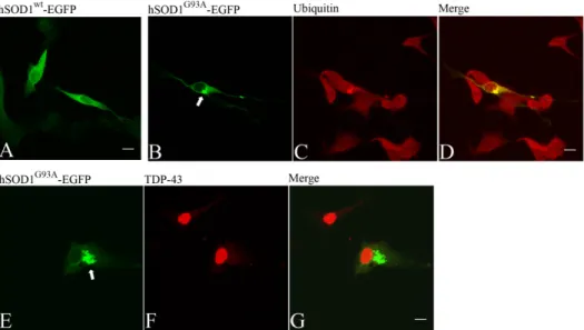
In Chapter 4, NSC%34 cells were transiently transfected with mutant SOD1G93A fused to the enhanced green fluorescent protein, which formed
aggregates as found in tissues from FALS patients. These aggregates were detected with an anti%ubiquitin antibody. Another feature was that endogenous TAR DNA%binding protein of 43 kDa was found predominantly
in the nucleus but not in the aggregates, similarly to that reported for patients with mutant SOD1 associated FALS but contrary to patients with
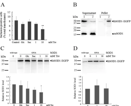
percentage of cells containing mutant SOD1 aggregates and the amount of
detergent insoluble protein were also found to be decreased upon exposure to trehalose at millimolar concentrations.
In Chapter 4, mutant SOD1 overexpression was also found to cause decreased levels of three concomitantly overexpressed glycoproteins % two
secretory, β%trace protein and erythropoietin, and one from the plasma membrane, L1, irrespective of the presence of mutant SOD1 aggregates.
Furthermore, there was no intracellular accumulation of these proteins nor alterations in their N%glycosylation, which indicated that Golgi fragmentation did not cause detectable changes in glycoprotein trafficking nor
glycosylation. As it is known that mutant SOD1 triggers endoplasmic
reticulum stress, which in turn reduces global protein synthesis the decreased levels of concomitantly overexpressed glycoproteins maybe the consequence of endoplasmic reticulum stress.
Previously in the literature, cytosolic SOD1 protein was found extracellularly. In Chapter 5, wild%type and mutant SOD1 were found to be
secreted in association with membrane vesicles from NSC%34 cells. The separation of those vesicles by sucrose density gradient showed that SOD1
co%localized with the exosomal marker CD9. Therefore, SOD1 secretion occurred via exosomes. Furthermore, exosomes produced by NSC%34 cells
were found to interact with the same cells, which may constitute a way to transfer toxicity between cells.
A Esclerose Lateral Amiotrófica (ELA) é a doença neurológica mais
grave e comum do adulto que afecta os neurónios motores na espinal medula, tronco cerebral e cérebro, a qual resulta em fraqueza progressiva.
A morte por insuficiência respiratória ocorre no prazo de dois a cinco anos após o início dos sintomas.
A maioria dos casos de ELA são esporádicos e 5%10% são
hereditários, no entanto, ambos são clínica e patologicamente muito semelhantes, o que sugere que partilham mecanismos fisio%patológicos
comuns. A descoberta de mutações no gene da cobre%zinco superóxido dismutase (SOD1) há 15 anos atrás, as quais correspondem a 20% dos
casos familiares, permitiu o desenvolvimento de modelos celulares e animais de ELA, e têm sido realizados inúmeros estudos para investigar a
toxicidade do enzima mutado. Estes modelos permitem também o desenho e o teste de estratégias terapêuticas. Actualmente, sabe%se que as
mutações na SOD1 relacionadas com a forma familiar de ELA, advêm de um ganho de função tóxica, e têm sido propostos vários mecanismos.
Estes incluem desnaturação de proteína e toxicidade de agregados intracelulares, alterações dos neurofilamentos e do tráfego intracelular ao longo destes, e envolvimento de células não%neuronais nas proximidades
dos neurónios motores.
Um dos principais objectivos desta Tese foi a constituição de
modelos celulares da forma familiar de ELA através da sobre%expressão da SOD1 humana mutante, na qual a glicina, na posição 93, foi substituída
por uma alanina (SOD1G93A), em células modelo de neurónios motores, células NSC%34, para mimetizar as características patológicas associadas
mutada de SOD1G93A. As células que expressavam a forma mutante de SOD1 revelaram o decréscimo das capacidades de proliferação e
diferenciação. Uma das características patológicas dos pacientes com ELA é a fragmentação do complexo de Golgi. Analisou%se nas células
construídas a morfologia do complexo de Golgi por microscopia de imunofluorescência utilizando o anticorpo anti%GM130, que marca o cis% Golgi. Observou%se que uma maior percentagem de células que sobre% expressavam SOD1 mutante apresentava fragmentação do complexo de Golgi, quando comparado com as células que sobre%expressavam a forma
wild type da mesma proteína. Esta fragmentação não era consequência de apoptose, uma vez que não se verificou activação da caspase%3, perda de potencial transmembranar mitocondrial ou fragmentação nuclear por Western blot, fluorescence activated cell sorting (FACS) ou por microscopia de fluorescência, respectivamente. Por conseguinte, as células NSC%34/hSOD1G93A constituem um modelo da forma hereditária de
ELA no que diz respeito à fragmentação do complexo de Golgi.
No capítulo 3, foram estabelecidas e caracterizadas culturas
primárias de células de espinal medula de embriões E14 de ratos transgénicos SOD1G93A. Estudaram%se dois tipos de culturas, secções de
espinal medula ou células da espinal medula dissociadas. Ambas as culturas possuíam neurónios, incluindo neurónios motores, e astrócitos,
identificados com anticorpos marcadores por microscopia de imunofluorescência. A morfologia do trans%Golgi reticular e do cis%Golgi de células provenientes do animal wild type ou transgénico revelou%se semelhante quando detectada com anticorpos anti%TGN38, anti%GM130 ou anti%GRASP65 por microscopia de imunofluorescência. Estas culturas de
envolvidas na patologia de ELA.
No capítulo 4, células NSC%34 foram transientemente transfectadas com o vector codificante da SOD1G93A mutante acoplada à proteína
fluorescente verde que formou agregados semelhantes aos encontrados em tecidos de pacientes com forma familiar de ELA. Estes agregados
foram detectados com um anticorpo anti%ubiquitina. A proteína endógena de 43 kDa, TAR DNA binding protein TDP%43, foi encontrada predominantemente no núcleo, e não nos agregados, de forma semelhante ao descrito nos pacientes com SOD1 mutante associada à forma hereditária de ELA mas, contrário ao descrito para pacientes com ELA
esporádica. Pequenas moléculas, como a trealose, têm sido usadas para
diminuir a agregação de proteína em modelos de doenças neurodegenerativas, devido às suas propriedades como chaperone químico. Aqui, a percentagem de células que possuíam agregados de
SOD1 mutante e a quantidade de proteína insolúvel em detergente foram reduzidas na presença de trealose em concentrações na ordem de
milimolar.
No capítulo 4, observou%se ainda que a sobre%expressão de SOD1
mutante provocava a diminuição dos níveis de três glicoproteínas concomitantemente sobre%expressas % duas de secreção, proteína β trace e eritropoietina, e uma de membrana plasmática, L1, independentemente da presença de agregados de SOD1 mutante. Além disso, não houve
acumulação intracelular destas proteínas nem alterações na sua N% glicosilação, o que indicou que a fragmentação do complexo de Golgi não provocou alterações detectáveis no tráfego de glicoproteínas nem na sua
glicosilação. Uma vez que se sabe que SOD1 mutante provoca stress do retículo endoplasmático que, por sua vez, reduz a síntese proteica global,
mutante eram secretadas em associação com vesículas membranares em células NSC%34. O fraccionamento dessas vesículas por gradiente de
densidade de sacarose mostrou que a SOD1 co%localizava com o marcador exosomal, CD9. Por conseguinte, a secreção de SOD1 ocorreu
via exossomas. Além disso, os exossomas produzidos pelas células NSC% 34 interagiram com as mesmas células, pelo que podem constituir uma
forma de transferência de toxicidade entre células.
Em conclusão, o trabalho descrito nesta Tese fornece evidência de que os modelos celulares, através da sobre%expressão de SOD1 mutante,
são ferramentas importantes no estudo dos mecanismos patogénicos
associados à forma familiar de ELA, tais como, a fragmentação do complexo de Golgi, agregação e secreção de SOD1 mutante. Estes modelos podem também ser usados como ferramentas para testar
Outline of the Thesis List of Figures
List of Tables Abbreviations
!– General introduction 1.1. Amyotrophic Lateral Sclerosis
1.1.1. Epidemiological and clinical aspects
1.1.2. Neuropathological features of ALS 1.2. Genetics of familial and sporadic ALS
1.2.1. FALS 1.2.1.1. SOD1
1.2.1.2. Other mutations 1.2.2. SALS
1.3. Understanding cellular pathogenic mechanisms in ALS 1.3.1. Protein instability and SOD1 aggregation
1.3.2. SOD1 structure, folding and stability
1.3.3. Mechanisms of mutant SOD1 degradation
1.3.4. Morphology of Golgi apparatus and intracellular protein
trafficking
1.3.5. Apoptosis in ALS
1.3.6. Non%neuronal cells affect motor neurons 1.3.7. Other mechanisms
1.4. ALS treatment
1.5. Aims of this Thesis work
apparatus disruption occurs independently from apoptosis 2.1. Abstract
2.2. Introduction
2.3. Materials and methods 2.3.1. NSC%34 cell culture 2.3.2. Protein analysis
2.3.3. Antibodies
2.3.4. Immunofluorescence microscopy
2.3.5. FACS analysis of cell viability and mitochondrial
transmembrane potential 2.4. Results
2.4.1. NSC%34/hSOD1G93A cells show decreased proliferation and differentiation
2.4.2. NSC%34/hSOD1G93A cells have increased levels of Golgi apparatus disruption
2.4.3. Golgi apparatus disruption was not due to apoptosis or Golgi apparatus protein degradation
2.5. Discussion
2.6. Acknowledgments
#– Primary cultures of spinal cord cells from rat embryo: wild type vs. transgenic SOD1G93A
3.1. Abstract 3.2. Introduction
3.3. Materials and methods 3.3.1. Animal Model
3.3.2. Primary cell culture from rat embryos SC
3.3.4. SOD1 analysis of SC from wild%type and transgenic adult
rat model of ALS SOD1G93A 3.3.5. Western blot analysis
3.4. Results
3.4.1. SC derived cell cultures
3.4.2. Monitoring of MN Golgi morphology in the SC derived cell cultures
3.4.3. Analysis of SOD1 from SC of wild%type and transgenic rat model of ALS (SOD1G93A)
3.5. Discussion
3.6. Acknowledgments
$ – Mutant SOD1 does not cause intracellular accumulation of secretory glycoproteins in a cell model of Amyotrophic Lateral Sclerosis
4.1. Abstract 4.2. Introduction
4.3. Materials and methods 4.3.1. NSC%34 cell culture
4.3.2. Detergent insolubility and Western blot analysis 4.3.3. Protein deglycosylation
4.3.4. Fluorescence microscopy 4.3.5. Statistical analysis
4.4. Results and discussion
accumulation
4.4.3. Trehalose diminishes mutant SOD1 aggregates and
detergent insolubility 4.5. Conclusions 4.6. Acknowledgments
%– Evidence for secretion of Cu,Zn superoxide dismutase via exosomes from a cell model of amyotrophic lateral sclerosis
5.1. Abstract 5.2. Introduction
5.3. Materials and methods 5.3.1. Protein analysis
5.3.2. Isolation of membrane vesicles 5.3.3. Sucrose density gradient fractionation
5.3.4. Carboxyfluorescein diacetate, succinimidyl ester labeling of exosomes
5.3.5. Immunofluorescence microscopy of CFSE labeled exosomes
5.3.6. Western blot analysis 5.4. Results
5.5. Discussion
5.6. Acknowledgments
79
84
87 88
91
92 93
93 93
94
94
94
95 95
6.1. General discussion and perspectives
6.1.1. Cellular models overexpressing mutant SOD1 to study ALS
6.1.2. Morphology of Golgi apparatus and intracellular protein trafficking in cellular models of ALS
6.1.3. Reducing mutant SOD1 aggregation in cellular models of ALS
6.1.4. Crosstalk between neuronal and non%neuronal cells via exosomes in ALS
6.2. General conclusions
References
105
110
114
117 120
The work described in this Thesis is concerned with the study of mammalian cell models of Amyotrophic Lateral Sclerosis (ALS) by
overexpression of mutant SOD1G93A. ALS is a devastating neurodegenerative disease that results from the death of motor neurons in the brainstem, cortex and spinal cord, leading to progressive muscular
atrophy and paralysis, and for which no effective therapy has been found yet. Since the discovery of mutations in the gene coding for Cu,Zn
superoxide dismutase (SOD1) 15 years ago, that account for 2% of all ALS cases, several mechanisms to explain the death of motor neurons have
been proposed but more studies are needed to fully understand this disease.
This dissertation starts with a general introduction to ALS, mainly epidemiological, clinical and neuropathological aspects of the disease are
presented. Particular relevance is given to the genetics of familial ALS, especially to SOD1 mutations. This is followed by an overview on the cellular pathogenic mechanisms in ALS, with emphasis to protein instability
and SOD1 aggregation, SOD1 structure, folding and stability, mechanisms of mutant SOD1 degradation, morphology of Golgi apparatus and
intracellular trafficking, apoptosis and the role of non%neuronal cells in ALS. Finally, a summary of clinical trials for ALS treatment is presented.
Chapter 2 describes the establishment of a cell model of the ALS disease by overexpression of mutant SOD1G93A in NSC%34 cells where
Golgi apparatus disruption occurs independently from apoptosis.
In Chapter 3, two types of primary cultures of spinal cord, sections
the effect of trehalose, a chemical chaperone, in preventing mutant SOD1 aggregation.
In Chapter 5, NSC%34 cells overexpressing mutant SOD1G93A are used to study the secretion of cytosolic SOD1 via exosomes. These
membrane vesicles could explain cell%to%cell transfer of mutant toxicity as they can interact with NSC%34 cells.
Figure 1 Page 5 Pathological features described for ALS.
Figure 2 Page 14 Schematic representation of proposed mechanisms that could converge in ALS and cause MN degeneration. Figure 3 Page 17 Crystal structure of metal bound dimeric human SOD1. Figure 4 Page 22 Schematic representation of the secretory and endocytic
pathways.
Figure 5 Page 42 Construction and characterization of stable NSC%34 cell lines expressing recombinant hSOD1wt and hSOD1G93A. Figure 6 Page 44 Immunofluorescence microscopy monitoring of Golgi
apparatus disruption in NSC%34/hSOD1G93A cells.
Figure 7 Page 46 Biparametric DiOC6(3)%PI flow cytometry analysis of NSC%34/hSOD1wt and NSC%34/hSOD1G93A cells.
Figure 8 Page 47 Western blot analysis of caspase%3 cleavage products. Figure 9 Page 61 Monitoring the SC cultures by inverted microscopy. Figure 10 Page 62 Identification of different cells in the cultures of SC
sections by immunofluorescence microscopy.
Figure 11 Page 63 Monitoring of SC dissociated cultures by immunofluorescence microscopy.
Figure 12 Page 64 Monitoring of cis%Golgi in the SC dissociated cultures by immunofluorescence microscopy.
Figure 13 Page 65 Immunofluorescence microscopy of SOD1 in the SC dissociated cell cultures.
Figure 14 Page 66 Western blot analysis of SOD1 in the SC from rat expressing wild%type rat or G93A human SOD1.
Figure 15 Page 78 Immunofluorescence microscopy of mutant SOD1G93A% EGFP aggregates in NSC%34 cells.
insolubility in NSC%34 cells.
Figure 18 Page 96 Western blot analysis of SOD1 protein from the supernatant of NSC%34/hSOD1wt and NSC% 34/hSOD1G93A cells.
Figure 19 Page 97 Western blot analysis of SOD1 protein after sucrose gradient fractionation.
Figure 20 Page 98 NSC%34 cells exposed to labeled exosomes analyzed by confocal microscopy.
*
Table 1 Page 8 Genetics of FALS.
Abbreviation Full form AD ALS ANG AR BDNF BSA CFSE CHO CNS CNTF COX82 CSF DAPI
DiOC6(3) DOC EAAT2 ECL EDTA EE EGFP Endo H EPO ER FACS FALS FBS FCS
Autosomal Dominant
Amyotrophic Lateral Sclerosis ANGiogenin
Autosomal Recessive
Brain%Derived Neurotrophic Factor Bovine Serum Albumin
CarboxyFluoresceindiacetate Succinimidyl Ester Chinese Hamster Ovary
Central Nervous System Ciliary NeuroTrophic Factor CycloOXygenase 2 CerebroSpinal Fluid
4',6%DiAmidino%2%PhenylIndole 3,3’%DihexylOxaCarbocyne iodide sodium DeOxyCholate
astroglial Excitatory Amino Acid Transporter 2 Enhanced ChemiLuminescent
EthyleneDiamineTetra%Acetic acid Early Endosome
Enhanced Green%Fluorescent Protein Endoglycosidase H
ErythroPOietin
Endoplasmic Reticulum
Fluorescence Activated Cell Sorting Familial Amyotrophic Lateral Sclerosis Foetal Bovine Serum
FDA FIG4 FITC FTD FUS GDNF GFAP GFP Glc GM130 GRASP55 GRASP65 GTP HEK HMM HRP hSOD1 Hsp IGF81 IgG Inher LC LE Lyso MAPT MCP81 MN
Food and Drug Administration Factor%Induced Gene 4 protein Fluorescein IsoThioCyanate FrontoTemporal Dementia FUsed in Sarcoma
Glial cell line%Derived Neurotrophic Factor Glial Fibrillary Acidic Protein
Green%Fluorescent Protein Glucose
cis Golgi Matrix protein of 130 kDa
GolgiReAssembly Stacking Potein of 55 kDa GolgiReAssembly Stacking Potein of 65 kDa Guanoside%5’%TriPhosphate
Human Embryonic Kidney High Molecular Mass HorseRadish Peroxidase
human copper%zinc SuperOxide Dismutase 1 Heat%shock protein
Insulin%like Growth Factor%1 Immunoglobulin G
Inheritance Light Chain Late Endosome Lysosome
mSOD1 MVB NB NF NF8L NGFR NO PAGE PBS PCR PDL PI PNGase F p115 PVDF rSOD1 RT SALS SC SD SDS SETX SNARE SOD1 STS Suc
mouse copper%zinc SuperOxide Dismutase 1 MultiVesicular Body
NeuroBasal NeuroFilament
NeuroFilament%Light chain Nerve Growth Factor Receptor Nitric Oxide
PolyAcrylamide Gel Electrophoresis Phosphate Buffered Saline
Polymerase Chain Reaction Poly%D%Lysine
Propidium Iodide
Peptide: %GlycosidaseF protein of 115 kDa PolyVinyledene DiFluoride
Ratcopper%zinc SuperOxide Dismutase 1 Room Temperature
Sporadic Amyotrophic Lateral Sclerosis Spinal Cord
Sprague Dawley
Sodium Dodecyl Sulphate SEnaTaXin
Soluble %ethylmaleimide%sensitive factor Attachment protein REceptor
Copper%Zinc SuperOxide Dismutase 1 STauroSporine
TBS
TDP843
TgN
TGN
TLS
TNFα
Tre
Tris
TRITC
TUDCA
TX8100
VAPB
VEGF
VSVG
WB
wt
β8TP
Tris Buffer Saline
Tar DNA%binding Protein%43 TransgeNic
rans%Golgi Network Translated in LipoSarcoma Tumor Necrosis Factor α Trehalose
Tris(hydroxymethyl)aminomethane Tetramethyl Rhodamine Iso%ThioCyanate TauroUrsoDeoxyCholic Acid
Triton X%100
Vesicle%Associated membrane Protein B Vascular Endothelium Growth Factor Vesicular Stomatitis Virus G protein Western Blot
wild%type
Abbreviations Amino acid name Ala Arg Asn Asp Cys Gln Glu Gly His Ile Leu Lys Met Phe Pro Ser Thr Trp Tyr Val A R N D C Q E G H I L K M F P S T W Y V Alanine Arginine Asparagine
Aspartate (Aspartic Acid) Cysteine
Glutamine
The most common adult onset disorder of motor neuron (MN) is
Amyotrophic Lateral Sclerosis (ALS). This neurodegenerative pathology
was first described by the French neurobiologist and physician Jean+Martin
Charcot in 1874 (Charcot, 1874), being initially known as Charcot’s
sclerosis (Pasinelli and Brown, 2006).
ALS is a fatal neurodegenerative disease that results from selective
dysfunction and death of upper and lower MN in the spinal cord (SC),
brainstem and cortex, which leads to generalized weakness and muscular
atrophy, and death occurs due to respiratory failure within 2+5 years (Bruijn
et al., 2004). The aetiology is likely to be multifactorial, as it involves
complex interactions between genes and environmental factors that initiate
the disease and lead to MN cell death (Majoor+Krakauer et al., 2003).
Despite more than a century of research, there is currently no cure and the
available therapy, the Riluzole, prolongs survival by only a few months
(Majoor+Krakauer et al., 2003).
Approximately 90+95% of ALS cases are sporadic (SALS), whereas
5+10% of cases have a family history, familial ALS (FALS). The majority of
familial cases are clinically and pathologically very similar to sporadic
cases, leading to the hypothesis that they share common pathogenic
mechanisms (Goodall and Morrison, 2006).
ALS is a relatively rare neurodegenerative disease with an annual
incidence rate (number of new cases per year) ranging from 0.4 to 2.4 per
100,000 individuals in developed countries (Mayeux, 2003), with a
individuals. ALS is an age+dependent disorder in which incidence and
mortality rates are higher with increasing age. The average age at onset of
ALS is 63 years but it varies from ages 37 to 88 (Mayeux, 2003).
There are four well+studied geographic areas with 50+100 times
higher prevalence of ALS (Wijesekera and Leigh, 2009): in the Chamorro
population of the Western pacific islands of Guam and Rota, the Japanese
Kii peninsula, the Western coast of Irian Jaya in Indonesia and the isolated
tribe at Anguru on Groote Eylandt in the Gulf of Carpentaria in North
Australia (Majoor+Krakauer et al., 2003). In these endemic areas in the
South Pacific, ALS accounts for about one in ten deaths. For the Chamorro
population of Guam a connection between ALS with dementia and
Parkinsonism has been established in some cases, which is recognized as
a clinical subtype of ALS, the Guamanian ALS/parkinsonism dementia
complex.
The present lack of a biomarker makes it impossible to diagnose
ALS in a pre+clinical stage before irreversible cell death occurs (Eisen,
2009). The El Escorial Criteria, developed in the late 1980s, form the basis
of the clinical diagnosis in the absence of examination
(Mayeux, 2003).
The initial features of this disease can include muscle twitching,
cramping or stiffness; muscle weakness affecting an arm or a leg; slurred
and nasal speech; or difficulty chewing or swallowing (Mayeux, 2003).
The pathological hallmarks of ALS are the degeneration and loss of
MN evident in cortex, brainstem and SC with the presence of intraneuronal
inclusions in degenerating neurons (Figure 1A+C) and glia (Strong et al.,
(A) Intensely argyrophilic, punctuate aggregates corresponding to the Bunina bodies are apparent (arrow) (magnification 40x). (B) Ubiquitin+immunoreactive threads and aggregates are observed as early markers of aberrant protein aggregation in otherwise healthy appearing MN. (C) Axonal spheroids are intensely stained using a monoclonal antibody recognizing highly phosphorylated neurofilament+heavy chain (black arrow), whereas perikarial neurofilament aggregates are more amorphous and heterogeneous (red arrow) (magnification 40x). (D) Hyaline inclusion from the transverse section of the SC from FALS stained with anti+SOD1 (magnification 225x) (E) Abnormal cytoplasmic staining of TDP+43 in lower MN of a SALS patient. (F) Glial inclusion displaying abnormal cytoplasmic immunoreactivity for TDP+43 in
The number of lower MN can be reduced by up to 50% at autopsy.
The remaining neurons are atrophic and contain intraneuronal aggregates
of the following types: bunina bodies, ubiquitinated inclusions and hyaline
conglomerate inclusions (Goodall and Morrison, 2006; Wijesekera and
Leigh, 2009). Bunina bodies are small round, single or branched
eosinophilic (they bind eosin that is acidic), and they have not been widely
described in other diseases (Figure 1A). They stain positive for cystatin and
transferrin and are present in 70+100% of cases. Ubiquitinated inclusions
can be divided according to morphology in “skein+like” (filamentous) or
“Lewy body+like” (round and compact) inclusions (Figure 1B). They are
detected in up to 95% of ALS cases. Hyaline conglomerate inclusions have
the appearance of glass after staining with eosin or haematoxylin, they are
argyrophilic, they contain phosphorylated and non+phosphorylated
neurofilaments (Figure 1C), in some FALS cases they stain for SOD1
(Figure 1D) and they are less specific for the disease (Wood et al., 2003;
Wijesekera and Leigh, 2009).
Recently, TDP+43 has been identified as the major protein in
ubiquitinated inclusions of neurons (Figure 1E) and glial cells (Figure 1F).
This year a mutant form of the DNA/RNA+binding protein FUS/TLS
has been found to be associated with MN degeneration and ALS. The
mutant protein forms large globular and elongated aggregates in the
cytoplasm in SC MN and dystrophic neurites (Figure 1G) (Kwiatkowski et
al., 2009; Vance et al., 2009).
Other pathological hallmarks in ALS are activation of microglia
(microgliosis, Figure 1H) and proliferation of astrocytes (astrogliosis, Figure
1I) (Strong et al., 2005; Lobsiger and Cleveland, 2007).
Another consistent neuropathological feature of upper and lower MN
in ALS is the atrophy and fragmentation of neuronal Golgi apparatus
(Figure 1J) observed for the three clinical subtypes of ALS: SALS, FALS
ALS/parkinsonism dementia complex (Mourelatos et al., 1994). The
fragmentation consists of dispersion of the normal network of large and
irregular elements of the Golgi apparatus into numerous shortened
disconnected cisternae (Gonatas et al., 2006). Importantly, lesions of the
Golgi apparatus were detected in spinal MN of presymptomatic transgenic
SOD1G93A mice, suggesting that the Golgi apparatus is targeted early in disease progression. Golgi apparatus fragmentation may lead to functional
impairment of intracellular protein trafficking essential for secretion and
axonal transport (Mourelatos et al., 1996). Transfection of CHO cells with
mutant SOD1 induced Golgi apparatus dispersion and dysfunction of the
secretory pathway (Stieber et al., 2004).
ALS has been considered a sporadic disorder, but in 10% of
patients the disease is familial (FALS), transmitted as either an autosomal
dominant or recessive disease. In 1991, the first ALS gene ( ) for an
autosomal+dominant form of FALS, was mapped to chromosome 21q. Two
years later the cytosolic copper+zinc superoxide dismutase (SOD1) gene
was shown to be the gene that accounts for 20% of the FALS cases,
and only 1+2% of all patients with ALS (Gros+Louis et al., 2006), but 2+7%
of apparently sporadic patients have also SOD1 mutations. To date,
molecular genetic analysis allowed the identification of several genetic loci
and genes (Table 1) as well as genetic polymorphisms, as risk factors
involved in ALS (Majoor+Krakauer et al., 2003; Valdmanis et al., 2009).
Environmental risk factors, including behavioral and occupational
exposures, such as smoking, heavy exercise or trauma, exposure to
pesticides, lead or solvents, chemicals used in agricultural work, diet high in
glutamate content, common environmental exposure in a particular region,
investigated in ALS patients, but no significant factor has been consistently
related to ALS (Gros+Louis et al., 2006; Valdmanis and Rouleau, 2008).
The SOD1 gene spans 11 kilobases of genomic DNA, comprises
five exons and four introns and encodes a highly conserved 153+amino acid
long protein of 16 kDa (Vucic and Kiernan, 2009).
Loci identified for the inherited form of ALS
are presented.
! " #
ALS1 21q22 AD/AR Adult/
Juvenile
(Siddique et al.,1991; Rosen 1993)
ALS2 2q33 AR Juvenile (Hentati et al., 1994; Hadano et
al., 2001; Yang et al., 2001)
ALS3 18q21 Unknown AD Adult (Hand et al., 2002)
ALS4 9q34 AD Juvenile (Chance et al., 1998; Chen et
al., 2004) ALS5 ALS6 ALS7 ALS8 ALS9 ALS10 ALS11 ALS+FTD
ALS with Parkinsonism and dementia 15q15 16q12 20ptel 20q13.3 14q11.2 1p36 6q21 9q21+q22 17q21.1 Unknown ! " Unknown # $% & ' %$ (&) Unknown * $ AR AD AD AD AD AD AD AD AD Juvenile Adult Adult Adult Adult Adult Adult Adult Adult
(Hentati et al., 1998) (Kwiatkowski et al., 2009; Vance et al., 2009) (Sapp et al., 2003) (Nishimura et al., 2004a; Nishimura et al., 2004b) (Greenway et al., 2004; Greenway et al., 2006) (Yokoseki et al.,2008; Sreedharan et al., 2008) (Chow et al., 2009) (Hosler et al., 2000) (Hutton et al., 1998)
aAbbreviations are: AD, autosomal dominant; ANG, angiogenin; ALS, amyotrophic lateral sclerosis; AR,
autosomal recessive; FIG4, factor+induced gene 4 protein; FTD, frontotemporal dementia; FUS, fused in
sarcoma; Inher., inheritance; MAPT, microtubule+associated protein Tau; SETX, Senataxin; SOD1,
superoxide dismutase 1; TARDBP, TAR DNA binding protein; TLS, translated in liposarcoma; VAPB,
To date, one hundred and forty one mutations spanning the entire
SOD1 polypeptide chain have been identified (http://alsod.iop.kcl.ac.uk/
Als/reports/report Summary.aspx), including one hundred and fifteen amino
acid substitutions, three insertions and four deletions (Simpson and Al+
Chalabi, 2006; Vucic and Kiernan, 2009). The mutations that are either
nonsense or deletion mutations, either introduce novel nucleotides or
remove existing ones, resulting in alteration of the polypeptide length. All
SOD1 mutations described to date are associated with autosomal+dominant
late onset ALS (typical ALS), except for two mutations, the substitution of
an aspartate by an alanine at position 90 or by a glutamine at position 96
(D90A or D96N, respectively), which can both cause dominant and
recessive ALS (Bruijn et al., 2004; Valdmanis and Rouleau, 2008). The
most common SOD1 mutations are D90A, followed by A4V and I113T. The
A4V gives raise to the most aggressive form of ALS with reduced survival
time after onset (Cudkowicz et al., 1997; Vucic and Kiernan, 2009).
Clinical characteristics, penetrance, age of onset, disease
progression and survival, vary greatly between and among different
mutations (Majoor+Krakauer et al., 2003; Gros+Louis et al., 2006)
suggesting that environmental and genetic factors are modifying the
phenotype (Simpson and Al+Chalabi, 2006). For example, some SOD1
mutations are associated with survivals longer than 17 years (G37R, G41R,
E100K, H46R), others with survivals around 10 years (G93C), or with
survivals of 5+6 years (E100G, G85R), some are associated with shorter
survivals such as 1+3 years (L37V, H43R, G93A), or 1 year (A4V) and
some have highly variable survival times (I113T, D90A) (Valentine et al.,
2005).
It is still not understood at present the mechanisms underlying the
toxicity of each SOD1 mutant to MN, causing ALS, but it seems to result
that SOD1 knockout mice did not develop MN disease supporting this
notion.
The first description of a rare juvenile+onset recessive form of FALS
( ) appeared in 1994, and was linked to chromosome 2 (Hentati et al.,
1994). Later, the gene was identified primarily in families of Arabic
origin (Tunisian and Kuwaiti families), displaying a juvenile onset (age 3 to
23 years old) with a relatively long survival (15 to 20 years). The
gene comprises 34 exons that encode for Alsin, a 1,657+amino acid protein
with three motifs homologous to guanine+nucleotide exchange factors that
include several cell+signaling motifs. To date, there are ten reported
mutations in the gene, eight of which are deleterious mutations, one
is a nonsense mutation and one is a splice site mutation. They all lead to
premature termination of the transcript and a truncated Alsin protein
suggesting a loss of the guanine+nucleotide exchange factor function that
results in a deficit in the intracellular trafficking (Hadano et al., 2001; Vucic
and Kiernan, 2009).
A rare autosomal dominant form of ALS with juvenile onset (mean
age at onset 17 years) and slow progression has been localized to a locus
on chromosome 9q34 ( )). Different missense mutations in the
+ gene ( ) have been identified (Chen et al., 2004). The
encoded protein is 2,667+amino acid long and contains a C+terminal
domain found in the superfamily 1 of DNA/RNA helicases. It is conceivable
that the abnormal SETX protein impairs the capacity of neurons to maintain
DNA and produce error+free mature mRNA, thereby resulting in
neurodegeneration.
One additional loci for adult onset autosomal+dominant form of
FALS ( ,) was assigned to chromosome 16q12.1+16q21 (Abalkhail et
sequence variant in exon 15 of the fused in sarcoma/translated in
liposarcoma ( ! also known as ) gene (Kwiatkowski et al., 2009;
Vance et al., 2009). So far 16 mutations in the 15 exons of the ! gene
have been described in patients with FALS, but none in patients with SALS
(Valdmanis et al., 2009). It is estimated that FUS/TLS mutations are
detected in about 4% of FALS cases. FUS/TLS is a 526+amino acid long
protein ubiquitously expressed and normally located in the nucleus, that is
involved in DNA repair and the regulation of transcription, RNA splicing,
and export to the cytoplasm. The FUS/TLS mutations described led to
cytoplasmic retention and apparent aggregation of FUS/TLS in ALS
patients (Figure 1G) (Lagier+Tourenne and Cleveland, 2009; Van Damme
and Robberecht, 2009).
A locus mapped at chromosome 20q13.33, causative of an
autosomal+dominant late+onset atypical ALS ( -), was identified in a
large Caucasian+Brazilian family. Further work led to the identification of a
missense P56S mutation in the vesicle associated membrane
protein/synaptobrevin associated protein B gene (# $%) (Nishimura et al.,
2004b). # $% has six exons that encode for a ubiquitously expressed 33
kDa homodimer protein named VAMP+B or VAP33. VAMP+B is a
membrane protein found at the endoplasmic reticulum (ER). The P56S
causes VAMP+B aggregation and Golgi apparatus fragmentation (Teuling
et al., 2007). These findings suggest that ALS may be caused by
dysfunction in the intracellular membrane trafficking and secretion or
malfunction of VAPB in mediating unfolded protein response (Suzuki et al.,
2009; Vucic and Kiernan, 2009).
In a group of typical+onset patients with ALS from Ireland and
Scotland, candidate+based gene sequencing resulted in the detection of
seven amino acid changes in the angiogenin . &/gene at the 0locus (Greenway et al., 2006). The angiogenin is a 123+amino acid long protein
angiogenesis (like vascular endothelium growth factor, discussed in section
1.3.7.) and it is a potent inducer of neovascularisation 1 1 , needed for cell proliferation. Several of the missense changes identified reside within
highly conserved residues in the catalytic domains of the angiogenin and
were shown to reduce the wild+type activity of the protein (Valdmanis et al.,
2009).
2 is a recently described typical form of ALS linked to the
' %$ gene on chromosome 1p36.22, in which mutations in both
sporadic and familial ALS cases, as well as in frontotemporal dementia
(FTD) were found. This gene codes for the 43+kDa TAR DNA+binding
protein (TDP+43), which binds RNA and DNA, thereby regulating the
transcription and splicing of nuclear material (Sreedharan et al., 2008;
Wijesekera and Leigh, 2009). Other reported functions of TDP+43 include
cell cycle regulation and apoptosis, mRNA transport and regulation of local
translation at synapses (Geser et al., 2009). To date 30 mutations have
been identified and the overall ' %$ mutation frequency in FALS is 2 to
5% (Van Damme and Robberecht, 2009; Wijesekera and Leigh, 2009).
With the exception of the Y374X truncation mutation, all other ' %$
mutations are missense mutations mostly affecting highly conserved and
protein+protein interacting amino residues in the C+terminal region
(Neumann, 2009). TDP+43 is a 414+amino acid protein ubiquitously
expressed, present in neuronal and glial nuclei (more than 90%). Mutant
TDP+43 has been found in ubiquitin+positive cytoplasmic aggregates
present in neurons and glial cells (Wang et al., 2008a). Biochemical
analysis revealed a characteristic pathologic profile due to N+terminal
truncation, abnormal hyperphosphorylation and ubiquitination (Neumann,
2009). Hence, it is plausible that neural toxicity is mediated by disruption of
Several studies have identified gene polymorphisms as risk factors
for the more common SALS, which may influence the onset and
progression of ALS (Beleza+Meireles and Al+Chalabi, 2009). Some of the
protein products of the genetic polymorphisms reported include:
neurofilament heavy/light chain subunit (intermediate filaments expressed
in adult neurons) (Xu et al., 1993; Figlewicz et al., 1994); glutamate
excitatory amino acid transporter 2, EAAT2 (Rothstein et al., 1995) and
glutamate receptor, AMPA (Carriedo et al., 1996); ciliary neurotrophic factor
(Orrell et al., 1995); and, vascular endothelium growth factor (angiogenesis
factor) (Lambrechts et al., 2003).
$ %
As familial and sporadic ALS are clinically and pathologically similar,
understanding the pathophysiological processes in FALS may provide a
greater understanding of the mechanisms underlying neurodegeneration in
SALS (Vucic and Kiernan, 2009). Indeed, the identification of pathogenic
alleles of SOD1 has led to the generation of cellular and transgenic rodent
models for the study of ALS. The exact pathophysiological mechanisms
underlying neurodegeneration remain elusive, but they seem to be
multifactorial (Figure 2), with evidence emerging of a complex interaction
between genetic factors and molecular pathways.
The combination of genetic, pathological and biochemical studies of
both human ALS tissue obtained and from biopsies, and
animal models, led to the identification of intrinsic pathogenic
characteristics of ALS MN, including: formation of protein aggregates,
proteasome dysfunction, ER stress pathways, increased sensitivity to cell
death signals, glial cell pathology, neuroinflammation, altered mitochondrial
oxidative stress+mediated damage, glutamate excitotoxicity, cytoskeletal
abnormalities and impaired axonal transport, growth factor deficiency and
aberrant RNA metabolism (Rothstein, 2009).
& '( . Adapted from Pasinelli and
Brown (2006).
3 $ 4 5
Several lines of evidence point out that the toxicity of SOD1 mutants
causing ALS arise from the acquired gain of a toxic function, resulting from
the propensity of SOD1 mutants to form large aggregates. In transgenic
rodents with mutant SOD1+mediated ALS and in some human ALS cases,
aggregates that are immunoreactive for SOD1 are detected in MN and
within the astrocytes surrounding them (Vucic and Kiernan, 2009). In these
onset and increase in abundance with disease (Johnston et al., 2000;
Wang et al., 2002).
Aggregates found in SALS and FALS patients, as well as mouse
models, may also contain ubiquitin (Ince et al., 1998; Watanabe et al.,
2001), a protein adduct which typically targets proteins for disposal via the
proteasome. Misaccumulation of ubiquitinated, misfolded proteins might
adversely affect the proteasome machinery and impair normal protein
degradation.
( 1 studies have shown that mutant proteins oligomerize over
time to form small pore+like structures that are similar to some forms of β– amyloid proteins (Matsumoto et al., 2006; Nordlund and Oliveberg, 2006).
In fact, fibrillar SOD1 species generated 1 from wild+type and mutant
SOD1 bind the amyloid markers Congo Red and Thioflavin+T, suggesting
the presence of amyloid structure. Micrometer+scale SOD1 aggregates in
transgenic mice have also stained positive with amyloid indicators such as
Thioflavin S, suggesting the presence of amyloid structures (Shaw and
Valentine, 2007; Chattopadhyay and Valentine, 2009).
As in other neurodegenerative disorders, it remains unclear whether
protein aggregates directly cause cellular toxicity and have a key role in
pathogenesis, or aggregates are innocent by+products of the
neurodegeneration process, or their formation may actually be a beneficial
process by being part of a defence mechanism to reduce intracellular
concentrations of toxic proteins (Cleveland and Rothstein, 2001;
Wijesekera and Leigh, 2009). It has been proposed that they could
deregulate organelle function including Golgi apparatus, ER and
mitochondria, or cause axonal transport defects possibly linked to aberrant
accumulations of intermediate filaments, overwhelming the capacity of the
protein folding chaperones and/or of ubiquitin proteasome system to
degrade important cellular regulatory factors (Vucic and Kiernan, 2009).
only of mutant SOD1 but also of other proteins, and sequestering proteins
that are essential for cellular processes, such as heat+shock proteins (Hsp)
(Julien and Kriz, 2006; Pasinelli and Brown, 2006). Indeed, mutant SOD1
directly interacts with Hsp70, Hsp40, Hsp27 and αβ+crystallin (Shinder et al., 2001; Wang et al., 2003).
It is currently understood that mutant SOD1 aggregation is a
multistep pathway of sequential protein monomerisation, demetallation and
oligomerisation, but the precise toxic biochemical species of SOD1,
whether monomeric, multimeric, soluble, insoluble or disulfide reduced,
remain elusive (Khare et al., 2004; Turner and Talbot, 2008a).
3 6 4 5
The SOD1 protein is a homodimer of two subunits of 16 kDa, in
which each monomer consists primarily of an eight+stranded antiparallel
Greek+key β+barrel, with two large loops, the so+called “electrostatic” loop
and the “metal binding” loop (Figure 3). The electrostatic loop has a number
of charged residues. The metal binding loop contains many of the residues
necessary for binding of the metals (Valentine et al., 2005; Rakhit and
Chakrabartty, 2006). Each subunit can form an intramolecular disulfide
bond between cysteines 57 and 146, which is unusual for an intracellular
protein, as it is present in the reducing environment of the cytoplasm. It can
coordinate one catalytic Cu2+ ion and one Zn2+ ion in its active site. The catalytic copper is bound by four histidines. The zinc ion is thought to play a
stabilizing structural role and act as a positive charge sink. The Cu2+ and Zn2+ ions are coordinated in close proximity and share a unique bridging histidine side chain (Rakhit and Chakrabartty, 2006; Shaw and Valentine,
2007). The dimer is held together primarily by numerous main+chain to
main+chain hydrogen bonds, water+mediated hydrogen bonds, and
reduces the solvent accessible surface area, greatly increasing its stability
(Rakhit and Chakrabartty, 2006).
$: ") . Copper and
zinc ions are shown as blue and orange spheres, respectively. The zinc loop is depicted in orange and the electrostatic loop in teal. The intrasubunit disulfide bond is shown in red. Adapted from Valentine et al. (2005).
The main function of the SOD1 enzyme involves free radical
scavenging whereby, through cyclical reduction and oxidation (dismutation)
of the copper ion, catalyses the conversion of the superoxide anion (O2+), a toxic by+product of mitochondrial oxidative phosphorylation, to molecular
oxygen (O2) and hydrogen peroxide (H2O2), which in turn is reduced to water by glutathione peroxidase and catalase, thus preventing oxidative
damage.
This ubiquitously expressed metalloenzyme is highly expressed
constituting 0.5+1% of soluble protein in the brain and SC. It is mainly
located within the cytosol, with a smaller fraction at the intermembrane
space of mitochondria (Furukawa and O'Halloran, 2006).
SOD1 is a highly stable protein in its fully metallated (holo),
disulfide+oxidized form and remains folded at temperatures near the boiling
dimeric quaternary structure in 1% sodium dodecyl sulphate (SDS) and
enzymatic activity after 1 h in 4% SDS, 8 M urea or 6 M guanidine+HCl.
Additionally, it has also been shown to be resistant to proteolytic digestion
(Valentine et al., 2005; Chattopadhyay and Valentine, 2009).
There are minimal changes in the crystal structure of fully metallated
mutant SOD1, nevertheless there are popular hypotheses to explain effects
of mutations in SOD1, which include decreased thermostability of holo or
metal+deficient (apo) SOD1, increased hydrophobicity and aggregation
propensity, loss of metals, aberrant chemistry and increased susceptibility
to posttranslational modifications, including augmented vulnerability to
disulfide reduction (Hart, 2006; Chattopadhyayand Valentine, 2009). Mutant
SOD1 has been shown to have decreased half+life 1 1 , but to different
degrees depending on the mutation. The faster degradation of the mutants
seems to involve the ubiquitin+proteasome system (Valentine et al., 2005).
SOD1 enzymatic activity varies greatly depending on which
mutation is present and does not correlate with disease severity, with some
mutant forms maintaining full activity, some being inactive (H46R) and
others near normal (G93A) (Bruijn et al., 2004). Nevertheless, it was shown
that loss of SOD1 stability, faster and higher aggregation propensities
relate to faster death of ALS patients, revealing that protein instability and
aggregation propensity are synergistic risk factors (Wang et al., 2008b;
Prudencio et al., 2009a).
Understanding how charge, hydrophobicity, local pH, conformational
destabilization and disordered regions of the polypeptide each contribute to
SOD1 aggregation can provide multiple approaches to stop SOD1
aggregation and to treat or prevent these familial forms of ALS (Shaw and
3 3 *
In every eukaryotic cell, there are two main systems for the
degradation of cytoplasmic proteins: the ubiquitin–proteasome system and
autophagy. The ubiquitin+proteasome system is the main intracellular proteolytic system that accounts for most of the selective intracellular protein degradation and mainly degrades short+lived damaged and
misfolded proteins (Cheroni et al., 2009). Autophagy is less selective and is involved in degradation of long+lived proteins in addition to organelles, such
as mitochondria (Pasquali et al., 2009).
Alterations in the functionality of the ubiquitin+proteasome system
have been proposed to be responsible for the accumulation of potentially harmful ubiquitinated proteins in ALS (Cheroni et al., 2009). Expression of mutant SOD1 leads to proteasomal inhibition that may result in
accumulation of mutant SOD1 and motor neuronal death (Urushitani et al.,
2002; Cheroni et al., 2005). A decrease in constitutive proteasome subunits during disease progression in the SC of transgenic mice expressing the FALS SOD1G93Awas found (Cheroni et al., 2009). However, it is likely that additional degradation pathways such as autophagy participate in SOD1
turnover (Kabuta et al., 2006).
Autophagy is an ubiquitous dynamic process that involves the
formation of double membrane structures, which fuse with lysosomes
where their content is degraded (Klionsky and Emr, 2000). Autophagic
degradation is implicated in the removal of aggregated proteins associated
with pathological conditions such as Alzheimer’s, Parkinson’s, Huntington’s
and prion diseases (Pasquali et al., 2009). ( 1 , autophagy inhibition
induced mutant SOD1+mediated cell death, and increased SOD1 levels in
detergent+soluble and insoluble fractions (Kabuta et al., 2006). Additionally,
stress is also a potent trigger of autophagy that can counterbalance this
stress (Hoyer+Hansen and Jaattela, 2007).
Endoplasmic reticulum stress is triggered by the accumulation of
misfolded proteins within the ER lumen (Nishitoh et al., 2008), it can
activate two adaptive pathways: the conserved ER to nucleus signaling
pathway called the unfolded protein response, and ER+associated protein
degradation. The latter eliminates misfolded or unassembled proteins from
the ER; its targets are selected by a quality control system within the ER
lumen and are ultimately destroyed by the cytoplasmic ubiquitin+
proteasome system. Studies showed that mutant SOD1 aggregated in
association with the ER (Tobisawa et al., 2003), induced ER stress but
specifically impaired ER+associated protein degradation (Nishitoh et al.,
2008). Additionally, an ER stress inhibitor delayed formation of insoluble
aggregates of the mutant SOD1 and suppressed cell death (Oh et al.,
2008). Up+regulation of the full spectrum of unfolded protein response
markers in lumbar cord tissue from human SALS patients was also
demonstrated. Furthermore, increased levels of protein disulphide
isomerase were found in the cerebrospinal fluid (CSF) of these patients
(Atkin et al., 2008). Unfolded protein response, which is known to reduce
global protein synthesis and to induce the synthesis of chaperones
(Harding et al., 2002), is part of a pathophysiological process, contributing
to neuronal death in both familial and sporadic disease (Atkin et al., 2008).
3 ) * 5 & 7
The Golgi apparatus is a cytoplasmic organelle involved in the
transport, processing, and targeting of proteins synthesized in the rough ER
and destined for the secretory pathway (Figure 4) (Nakagomi et al., 2008).
The Golgi apparatus also serves as site for the post+translational
modification of proteins and lipids, mostly glycosylation. In most higher
interconnected cisternae and vesicles that carry molecular “cargo” from one
cisterna to the next by the coordinated fission of vesicles from the lateral
edge of one cisterna and fusion to the next cisterna. It is organized around
the microtubule+organizing center in the perinuclear region (Gonatas et al.,
2006).
The structural and functional integrity of the Golgi apparatus is
maintained by different systems of proteins and structures that include
microtubules and microtubule+associated proteins, the actin+associated
cytoskeleton, the Golgi matrix proteins and proteins that ensure the
targeting and fusion of transport vesicles to the correct compartment, such
as GTP+binding proteins and SNAREs (Gonatas et al., 2006).
Originally, the Golgi apparatus was thought to be a static organelle,
but it is actually a highly dynamic structure. The steady+state structure of
the Golgi stacks is partly a consequence of the balance of anterograde and
retrograde transport through the Golgi (Hicks and Machamer, 2005).
As the Golgi apparatus is involved in numerous important functions
quality control of proteins in the Golgi apparatus and ER must be stringent
to ensure appropriate cellular function. It is reasoned that fragmentation of
the Golgi might have detrimental effects and lead to dysfunction of the
cytoplasmic machinery in neurons as well as in non+neuronal cells
(Nakagomi et al., 2008). Indeed, the fragmentation of the neuronal Golgi
apparatus has been reported in ALS (Fujita and Okamoto, 2005), as well as
in Alzheimer’s disease (Stieber et al., 1996), Parkinson’s disease (Gosavi
et al., 2002), spinocerebellar ataxia type 2 (Huynh et al., 2003), in
Creutzfeldt+Jacob disease (Sakurai et al., 2000), among others (Fan et al.,
2008). Furthermore, α+synuclein expression blocked ER+to+Golgi vesicular
trafficking (Cooper et al., 2006), similarly, misfolded growth hormone
caused Golgi apparatus fragmentation and disrupted the traffic of two
3 8
Evidence has suggested that the ultimate mechanism of neuronal
damage in human ALS occurs through programmed cell death, resembling
apoptosis (Sathasivam and Shaw, 2005a; Pasinelli and Brown, 2006).
Apoptosis is an energy+dependent process characterised by cell shrinkage,
compaction and contraction of cytoplasm organelles, membrane blebbing,
nuclear chromatin condensation and fragmentation (Goodall and Morrison,
2006). Apoptosis can be triggered by three mechanisms: activation of cell
surface receptors of the tumour necrosis family, such as Fas; cytochrome
release from the mitochondria; and stress to the ER. Apoptosis processes
are tightly regulated by a series of proteins including caspases, the Bcl+2
family of pro+ and anti+apoptotic proteins and inhibitors of apoptosis
proteins (Wijesekera and Leigh, 2009). Caspases are cysteine proteases
with aspartate specificity that can be divided into upstream effector (such
as caspase+1, +8 and +9) and downstream+executioner caspases (such as
caspase+3 and +7), the former activating the latter in the apoptotic pathway.
Upon activation, the caspases cleave other intracellular targets including
other caspases resulting in an amplified cell death cascade.
The first evidence of an apoptotic response in ALS was the
increased activation of effector caspases+1 and +9 in the CSF (Ilzecka et
al., 2001) and in the SC tissue (Li et al., 2000; Inoue et al., 2003) of ALS
patients. Further, SC expression of caspase+1 gene was significantly
increased in ALS cases (Anagnostou et al., 2008). Caspase activation is
also very prominent within astrocytes of mutant SOD1 mice (Li et al., 2000;
Pasinelli et al., 2000) raising the possibility that caspase+1 activation may
contribute to an inflammatory pathway causing astrocytosis. It has been
demonstrated that caspase+3 activation in glial cells proteolytically
inactivates the astroglial glutamate excitatory amino acid transporter 2,