INTRODUCTION
Isolation of DNA repair mutants from normal cul-ture cells has proven to be very useful in identifying ge-nes related to cell genome stability maintenance (Collins, 1993). These mutants have been useful in further under-standing the several mechanisms that cells use to cope with DNA damage, mainly through removal by classic nucle-otide excision repair. Simple damage removal is also re-lated to other metabolic pathways, including DNA repli-cation, RNA transcription and cell cycle (Seroz et al., 1995).
A photoreactivation prone cell line (PtK2) from the marsupial Potorous tridactylus (Chiang and Rupert, 1979), is useful for isolating mutants defective in processing ul-traviolet (UV)-induced DNA damage, with a positive se-lection system similar to that described by Rosenstein and Ohlsson-Wilhelm (1979) in ICR 2A frog cells. This sys-tem uses cell ability to eliminate cyclobutane pyrimidine dimers (CPD) enzymatically through exposure to light (300-500 nm) by photoreactivation, to rescue mutant cells unable to process DNA damages by incorporating the pho-tosensitizer nucleotide analogue 5-bromo-2’-deoxyu-ridine (BUdr). Briefly, cells are irradiated with UV, fol-lowed by incubation with BUdr and exposure to black light (310-400 nm), leading to both CPD photoreactivation and mortality of those cells that had incorporated BUdr. Cells surviving this selection procedure, most probably because of failure in replication of damaged templates or DNA repair replication, and consequent failure in BUdr incor-poration, can be isolated. Based on this selection system, we were able to isolate a clone with a slight increase in
UV sensitivity. A detailed characterization of this clone concerning nucleotide excision and post-replication re-pair was made and is described below.
MATERIAL AND METHODS
Cell culture
The PtK2 cell line is derived from the kidney of a male rat kangaroo (Potorous tridactylus), kindly supplied by Dr. C.S. Rupert (University of Texas, USA). The cells were routinely grown at 37oC in a 5% CO
2 atmosphere in
Dulbecco’s modified Eagle’s medium supplemented with 10% calf serum and antibiotics.
UV-irradiation and photoreactivation
The cells were washed twice with prewarmed PBS and irradiated with a low-pressure germicidal lamp (254 nm, dose rate 0.5 J m-2 s-1). Photoreactivation was done in
PBS with cell plates spread in a single layer over fluores-cent (2-15 W) or black light lamps (310-400 nm), sepa-rated from the dishes by a 4-mm glass. Cells were exposed to this light for 2 h at 37oC. After irradiation and
photore-activation, PBS was replaced with normal growth medium.
Selection procedure
Exponentially growing cells were irradiated with UV (15 J/m2) and after incubation in complete medium for 1 h
at 37oC to allow the replicating regions to reach the lesions,
3.3 x 10⁻⁵ M BUdr and 2.5 x 10⁻⁶ M 5-fluoro-2’deoxyuridine
Characterization of a mutant rat kangaroo cell line with alterations
in the cell cycle and DNA repair
E.N. Miyaji1, R.T. Johnson3, C.S. Downes3, E. Eveno4, M. Mezzina4, A.Sarasin4 and C.F.M.Menck2
Abstract
Using a positive selection system for isolating DNA replication and repair related mutants, we isolated a clone from a rat kangaroo cell line (PtK2) that has increased sensitivity to UV light. Characterization of this clone indicated normal post-replication repair after UV irradiation, and normal removal rates of cyclobutane pyrimidine dimers and pyrimidine(6-4)pyrimidone photoproducts by excision repair. However, this cell line has decreased ability to make early incisions on damaged DNA, possibly indicating a defect in preferential repair of actively transcribed genes, and a slower cell proliferation rate, including a longer S-phase. This phenotype reinforces the present notion that control of key mechanisms in cell metabolism, such as cell cycle control, repair, transcription and cell death, can be linked.
1Departamento de Biologia, Instituto de Biociências and 2Departamento de Microbiologia, Instituto de Ciências Biomédicas, USP, São Paulo, SP,
Brasil. Send correspondence to C.F.M.M. Av. Prof. Lineu Prestes, 1374, 05508-900 São Paulo, SP, Brasil. E-mail: [email protected]
3CRC Mammalian Cell DNA Repair Research Group, Department of Zoology, University of Cambridge, UK.
(FUdr) were added to the medium. Cells were cultivated for more 24 h and then exposed to black light for 2 h. This protocol was repeated again after 14 days.
Cell survival
The cells were plated on small Petri dishes (24 cm2)
at low density (103 cells per plate). After 14 days, they
were fixed with 10% formaldehyde and stained with 1% crystal violet. Colonies of more than 20 cells were scored. Survival values correspond to the ratio between the num-ber of treated cell colonies and the numnum-ber of non-irradi-ated cell colonies.
BNDC post-replication repair assay
Exponentially growing cells were UV-irradiated and incubated in fresh medium for 1 h before labeling with
3H-thymidine (20 µCi/ml) for 15 min. Cells were then
re-moved for immediate analysis or cultivated in medium containing 10-5 M dNTPs before analysis. DNA extraction
and BNDC (benzoylated naphtylated diethyl cellulose chro-matography) were performed as described by Pillidge et al. (1986).
Assay for incisions by hydroxyapatite (HAP) chromatography
The incision assay was based on the method described by Squires et al. (1982). Briefly, cells were incubated for 72 h with 3H-thymidine (0.2 µCi/ml) in growth medium to
prelabel the DNA, replated in fresh medium and further incubated for 24 h. The DNA synthesis inhibitor hydrox-yurea (HU, 10-2 M) and (1-β-D-arabinofuranosyl)cytosine
(araC, 10-4 M) were added to the medium, either before
and after UV-irradiation or in pulses after irradiation. Re-pair ended by washing the cells with ice-cold PBS and lys-ing them for 25 min on ice in an alkaline DNA-unwindlys-ing buffer (0.15 M NaOH, 0.3 M NaCl and 0.01 M EDTA in 5% sucrose). After neutralization with 0.15 M KH2PO4,
DNA size was reduced by sonication for 8 s at 24 kHz, and at a 8-µm amplitude. Hydroxyapatite column chromatog-raphy and DNA break calibration were carried out as de-scribed by Squires et al. (1982).
Measurements of UV-endonuclease-sensitive sites (ESS)
The number of ESSs was determined essentially as described by Menck and Meneghini (1982). Basically, pu-rified DNA was incubated with a Micrococcus luteus ex-tract with UV-endonuclease activity. Untreated and treated DNA molecular weights were measured by alkaline sucrose gradient sedimentation. From these two values, ESS fre-quency, taken as CPD frequency per unit length of DNA, was calculated.
Determination of pyrimidine (6-4) pyrimidone photo-products ((6-4)PPs) by immuno-slot-blots
For immuno-slot-blot analysis, 0.5 µg purified DNA was loaded on nitrocellulose membrane (BA 83-S, Schleicher & Schuell). Membranes were saturated with PBS containing 5% lyophilized no-fat milk (nfm) for 1 h at 37oC (or several hours at 4oC), and then incubated with
64-2 antibodies (diluted 1:500 in 0.5% nfm PBS) for 1 h at 37oC (Mezzina et al., 1994). After extensive washing
with this buffer, blots were incubated with a 1:2000 dilu-tion of a second anti-mouse horseradish peroxidase-con-jugated antibody (Calbag, San Francisco, USA) for 30 min at room temperature. Blots were then extensively washed with 0.5% nfm PBS and PBS before processing with ECL (Amersham) solutions and exposure of X-ray films. For precise (6-4)PPs quantification, films of different expo-sure times were scanned in order to calculate absorbance values in a linear range.
Synchronization
After reaching confluence, cells were cultivated for another 10 days to make sure all had attained a pre-repli-cative G0 state, then plated at a lower density in normal
growth medium with aphidicolin, a DNA synthesis inhibi-tor. After 24 h, with the cells synchronized at early S-phase, the aphidicolin-containing medium was replaced by nor-mal growth medium, followed by immediate initiation of DNA replication. Pulse labeling was performed by adding
3H-thymidine (2 µCi/ml, Amersham) to the culture
me-dium for 30 min. The incorporated radioactivity was mea-sured (Menck and Meneghini, 1982).
RESULTS
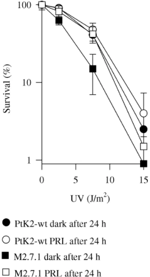
UV was also expected, since the positive selection used can favor cells with increased ability to deal with DNA dam-age. One of the sensitive clones, M2.7.1, was chosen for further analysis. M2.7.1 cells were slightly more sensi-tive to UV and exposure to photoreactivating light 24 h after irradiation caused a cell survival increase (Figure 1), which was not observed in wild-type cells. Differences in survival between wild-type and M2.7.1 cells irradiated with 7.5 and 15 J/m2 were significant (P < 0.05, Student t-test),
while differences between survival in cells maintained in the dark or exposed to photoreactivating light were only significant in M2.7.1 cells (P < 0.01, Student t-test). We previously determined that although wild-type cells are still able to eliminate CPDs 24 h after irradiation, there is no increase in survival (Miyaji and Menck, 1995) and that this probably happens due to irreversible events in the apoptosis pathway that have already taken place by this time (Miyaji and Menck, 1996).
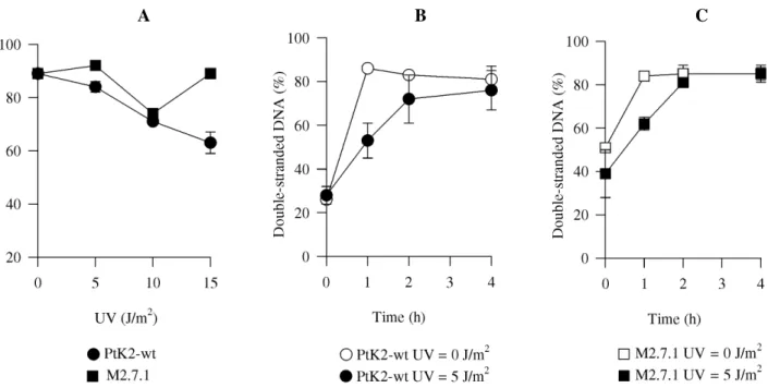
Nascent chain elongation analysis was performed by BNDC chromatography, a technique that allows determina-tion of the percentage of newly synthesized double-stranded DNA, which corresponds to the rate of DNA chain elonga-tion growth after a given treatment (Pillidge et al., 1986). No difference was found between growth of daughter strands from M2.7.1 and wild-type cells irradiated with different UV doses (Figure 2A). The same analysis was performed after longer periods following UV irradiation and, again, no difference was detected between M2.7.1 and wild-type cells (Figure 2B and C). This indicates that there is no defect in mutant ability to elongate newly synthesized DNA in the presence of a damaged template, i.e., translesion synthesis is normal in M2.7.1.
We next assayed M2.7.1 excision repair capacity by quantifying breaks induced by the incision step of the DNA excision repair pathway. This was performed by HAP chro-matography (Squires et al., 1982). Analysis after irradia-tion with different UV doses indicated a significant de-crease in the number of single-strand breaks generated in M2.7.1 when compared to wild-type cells (P < 0.01, Stu-dent t-test) (Figure 3A). Analysis at increased periods af-ter irradiation further confirmed this decrease in M2.7.1 incision activity rate, at least until 6 h after irradiation (Figure 3B). Although these results indicate a DNA exci-sion repair defect, quantification of CPDs (ESS) and (6-4)PPs (Table I), did not indicate any gross difference be-tween M2.7.1 and wild-type cells in the removal of these lesions 24 h after UV exposure. Clearly, very few (if any) CPDs are eliminated in the dark by wild-type and M2.7.1 cells, while (6-4)PPs were not detected after this period of time. Moreover, these cells seem to be equally effi-cient in the removal of CPDs when illuminated with pho-toreactivating light. Interestingly, the data indicate that the cells are also able to remove (6-4)PPs by exposure to photoreactivating light.
Alterations in pool precursors for the DNA synthe-sis could, in principle, also be responsible for the altered M2.7.1 cell phenotype. However, this seems unlikely, since no defect in sealing DNA breaks after UV-irradia-tion was noticed either in mutant or wild-type cells and no differences in M2.7.1 cell phenotype were observed when deoxynucleotides were supplemented in the cell media (data not shown).
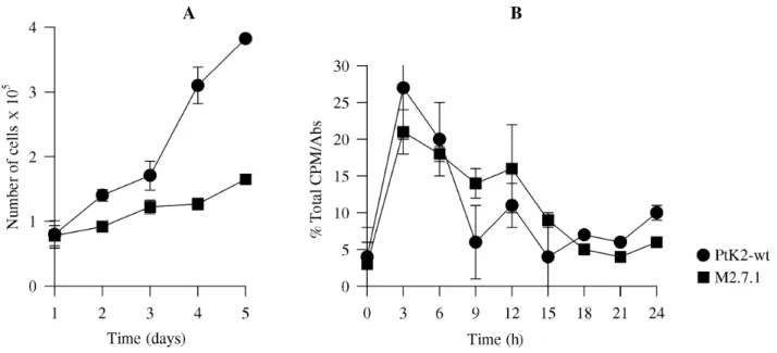
We investigated M2.7.1 and wild-type cell prolifera-tion. A slower growth rate was found for M2.7.1, when com-pared to wild type cells (Figure 4A). Moreover, after re-lease of both from early S-phase arrest and subsequent DNA synthesis analysis, there was a longer S-phase of approxi-mately 15 h in M2.7.1 compared to 9 h in parental cells (Figure 4B).
DISCUSSION
A positive selection procedure was used to obtain cell mutants defective in DNA damage processing after UV ir-Figure 1 - UV survival of the M2.7.1 mutant. PtK2 parental cell line (circles)
Figure 2 - Determination of nascent chain elongation. PtK2 (circles) and M2.7.1 (squares) cells were irradiated with indicated UV doses, maintained in their original medium for 1 h, then incubated with 3H-thymidine. Cells were then incubated for 3 h or for the time indicated in fresh medium with dNTPs and prepared for BNDC-chromatography. A: UV dose response; B: PtK2 time course - 0 J/m2 (open circles) and 5 J/m2 (closed circles); C: M2.7.1 time course - 0 J/m2 (open squares) and 5 J/m2 (closed squares). wt, Wild-type cells.
A B C
Figure 3 - DNA repair measurements by incision assay. A: UV dose response. PtK2 (circles) and M2.7.1 (squares) cells labeled with 3H-thymidine were incubated for 40 min with HU and araC, irradiated with indicated UV doses, incubated with inhibitors for another 40 min and then prepared for HAP chromatography. B: PtK2 (circles) and M2.7.1 (squares) cells labeled with 3H-thymidine were irradiated with UV (5 J/m2), incubated in fresh medium for indicated time before incubation for 40 min with HU and araC and then prepared for HAP chromatography. wt, Wild-type cells.
radiation. We characterized one clone with increased UV sensitivity. These cells have normal DNA chain elongation in the presence of UV-induced lesions. Thus, it is unlikely that post-replication repair mechanisms in these cells are affected. On the other hand, we found that they have a sig-nificant defect in overall DNA excision repair measured
shortly after UV exposure, as incisions accumulated at a lower frequency than in the parental cells. Moreover, these mutants had decreased cell-growth rate and corresponding increased S-phase length.
PtK2 cells did not eliminate CPDs efficiently (less than 10% of CPDs are removed in 24 h) (Miyaji and Menck,
Table I - DNA repair of CPDs (ESS) and (6-4)PPs in PtK2 (wt) and M2.7.1 cells.
Treatmenta % ESSb % (6-4)PPsb
wt PtK2 M2.7.1 wt PtK2 M2.7.1 UV = 15 J/m2 t = 0 h, dark 100 100 100 100 UV = 15 J/m2 t = 0 h, PRL 35 ± 31 55 ± 9 56 ± 15 35 ± 24 UV = 15 J/m2 t = 24 h, dark 99 ± 14 78 ±3 NDc ND UV = 15 J/m2 t = 24 h, PRL 40 ± 22 39 ±7 ND ND The values represent the average from at least three independent experi-ments. aCells were irradiated and incubated for indicated time before expo-sure to photoreactivating light (PRL) or maintenance in the dark for 2 h in PBS. bCPDs were determined by the number of endonuclease sensitive sites (ESS) and pyrimidine (6-4) pyrimidone photoproducts ((6-4)PPs) by immuno detection. cND = Not detected.
Figure 4 - Cell proliferation in PtK2 (circles) and M2.7.1 (squares) cells. A: Growth rate analysis - cells were plated (105 cells/plate) and cell number determined after indicated time. B: Synchronized cells as described in Material and Methods were pulse-labeled for 30 min and harvested at indicated times, for measure-ments of nucleotide incorporation in DNA during S-phase.
1995 and the present work). However, (6-4)PPs photoprod-ucts were reversed efficiently with no lesions detected af-ter 24 h in the dark. The M2.7.1 clone did not show any significant difference in (6-4)PPs removal when compared to parental cells 24 h after irradiation. But initial (6-4)PPs removal rates could be lower than normal during the first few hours after UV in M2.7.1. Alternatively, PtK2 cells may efficiently do preferential repair of CPDs or (6-4)PPs pho-toproducts in actively transcribed genes, as do rodent cells (Bohr et al., 1986). The number of incisions accumulated in these cells is small compared to human cells (about 20% incisions determined for normal human cells) (Squires et al., 1982), possibly reflecting actively transcribed gene repair. If this hypothesis is correct, mutant cells could have
a diminished incision capacity for preferential repair of active genes. Increased UV sensitivity in these cells would be explained by this defect since preferential repair likely has an important function in cell resistance to UV (Bohr et al., 1986).
The slower M2.7.1 growth rate indicates problems in cell cycle control in these cells. Much evidence points to a direct relationship between cell cycle control and DNA dam-age responses. It is well known that eukaryotic cells arrest their cell cycle at G1, S or G2 phases after DNA damage, a response known as checkpoint control (Hunter, 1993). These delays would allow the cell to cope with lesions be-fore proceeding to a subsequent cell cycle phase. If the cells cannot tolerate the lesions, they may initiate a cascade of events leading to cell death through apoptosis (Steller, 1995). We have shown that most PtK2 cells die through apoptosis (Miyaji and Menck, 1996, 1998). Growth rate defect, including the increased S-phase duration observed in M2.7.1 cells, may interfere with cell ability to choose between interrupting the cell cycle or initiating apoptosis in response to DNA damage. Such a defect may also be di-rectly related to the selection procedure used, since less BUdr would be incorporated after irradiation of such cells, a possibility consistent with the observed increase in sur-vival when cells are photoreactivated 24 h after UV irradia-tion, a feature not found in the parental cells (Miyaji and Menck, 1995 and the present work). Significant differences in apoptosis induction (analyzed by induction of inter-nucleosomal DNA breaks) in M2.7.1 were not detected (data not shown).
The PtK2 cells (both parental and mutant cell lines) could eliminate (6-4)PPs faster when illuminated by
toreactivating light, confirming a report by Mitchell et al. (1990), who found that marsupial cells (Monodelphis domestica) were able to photorepair (6-4)PPs, possibly resulting from enhanced repair of these lesions after CPD elimination. However, recent data point to the existence of specific photolyases to (6-4)PPs in Drosophila (Todo et al., 1993), plants (Nakajima et al., 1998) and even in Xeno-pus cells (Hitomi et al., 1997). Thus, the results shown in this study may be due to (6-4)PP photolyases also present in marsupial cells.
Detection of alterations in both repair capability and the cell cycle agrees with the present notion that several key mechanisms in cell metabolism, such as transcription, repair, cell cycle control and apoptosis, can be linked. The transcription factor TFIIH has been shown to consist of at least nine subunits, including XPB and XPD, involved in nucleotide excision repair (Schaeffer et al., 1993, 1994), and a cdk-activating kinase complex (CAK) involved in cell cycle regulation (Adamczewski et al., 1996). Furthermore, CAK seems to be integrated within TFIIH via association with XPB and XPD (Rossignol et al., 1997). XPB binding to P53, which is involved in induction of cell cycle arrest and apoptosis after DNA damage, has also been shown (Wang et al., 1996). Although current knowledge of these mecha-nisms in rat kangaroo cells is scarce, they may be similar to what is observed in human and rodent cells. Thus, alterations detected in M2.7.1 in nucleotide excision repair and cell proliferation could mean there is linkage in the control of key mechanisms in cell metabolism.
ACKNOWLEDGMENTS
Research supported by Fundação de Amparo à Pesquisa do Estado de São Paulo (FAPESP). E.N.M. was the recipient of a PhD fellowship from CNPq (Brasília, Brazil). Publication supported by FAPESP.
RESUMO
Uma linhagem mutante de células de rato-canguru (PtK2) com aumento de sensibilidade à luz ultravioleta (UV) foi isolada a partir de um sistema de seleção positiva. A caracterização desta linhagem indicou que tanto a síntese translesão como o reparo-excisão das lesões mais freqüentemente induzidas por UV, dímeros de pirimidina e (6-4) pirimidina-pirimidonas, encontram-se em níveis normais. No entanto, esta linhagem apresenta uma redução no nível de incisões logo após a indução das lesões, o que poderia representar um defeito no reparo preferencial de genes ativos. Alte-rações na proliferação celular, com uma fase S mais longa, também foram detectadas. Este fenótipo reforça a idéia de que mecanismos de controle do metabolismo celular, como ciclo celular, reparo, trans-crição e morte celular, estão interligados.
REFERENCES
Adamczewski, J.P., Rossignol, M., Tassan, J.-P., Nigg, E.A., Moncollin, V.
and Egly, J.-M. (1996). MAT1, cdk7 and cyclin H form a kinase com-plex which is UV light-sensitive upon association with TFIIH. EMBO
J.15: 1877-1884.
Bohr, V.A., Okumoto, D.S. and Hanawalt, P.C. (1986). Survival of UV-irradi-ated mammalian cells correlates with efficient DNA repair in an essential gene. Proc. Natl. Acad. Sci. USA83: 3830-3833.
Chiang, T. and Rupert, C.S. (1979). Action spectrum for photoreactivation of ultraviolet-irradiated marsupial cells in tissue culture. Photochem. Photobiol. 30: 525-528.
Collins, A.R.S. (1993). Mutant rodent cell lines sensitive to UV, ionizing radiation and cross linking agents: a comprehensive survey of genetic and biochemical characterization. Mutat. Res. DNA Repair 293: 99-118.
Hitomi, K., Kim, S.T., Iwai, S., Harima, N., Otoshi, E., Ikenaga, M. and Todo, T. (1997). Binding and catalytic properties of Xenopus (6-4) photolyase.
J. Biol. Chem.272: 32591-32598.
Hunter, T. (1993). Breaking the cycle. Cell75: 839-841.
Menck, C.F.M. and Meneghini, R. (1982). Resistance of 3T3 mouse cells to UV light in relation to excision and transfer of dimers to daughter strands.
Photochem. Photobiol.35: 507-513.
Mezzina, M., Eveno, E., Chevallier-Lagente, O., Benoit, A., Carreau, M., Vermeulen,W., Hoeijmakers, J.H., Stefanini, M., Lehmann, A.R., We-ber, C.A. and Sarasin, A. (1994). Correction by the ERCC2 gene of UV sensitivity and repair deficiency phenotype in a subset of tricho-thiodystrophy cells. Carcinogenesis15: 1493-1498.
Mitchell, D.L., Applegate, L.A., Nairn, R.S. and Ley, R.D. (1990). Photoreac-tivation of cyclobutane dimers and (6-4) photoproducts in the epider-mis of the marsupial, Monodelphis domestica. Photochem. Photobiol. 51: 653-658.
Miyaji, E.N. and Menck, C.F.M. (1995). Ultraviolet-induced cell death is independent of DNA replication in rat kangaroo cells. Photochem. Photobiol.61: 454-458.
Miyaji, E.N. and Menck, C.F.M. (1996). Photoreversion of ultraviolet in-duced apoptosis in rat kangaroo cells. Apoptosis1: 153-160.
Miyaji, E.N. and Menck, C.F.M. (1998). Human Bcl-2 expression delays ul-traviolet-induced apoptosis in marsupial cells. Photochem. Photobiol. 68: 719-724.
Nakajima, S., Sugiyama, M., Iwai, S., Hitomi, K., Otoshi, E., Kim, S.T., Jiang, C.Z., Todo, T., Britt, A.B. and Yamamoto, K. (1998). Cloning and characterization of a gene (UVR3) required for photorepair of 6-4 photo-products in Arabidopsis thaliana. Nucleic Acids Res.26: 638-644.
Pillidge, L., Downes, C.S. and Johnson, R.T. (1986). Defective post-replica-tion recovery and UV sensitivity in a simian virus 40-transformed In-dian muntjac cell line. Int. J. Radiat. Biol.50: 119-136.
Rosenstein, B. and Ohlsson-Wilhelm, B.M. (1979). Isolation of UV-sensitive clones from a haploid frog cell line. Somatic Cell. Genet.5: 117-128.
Rossignol, M., Kolb-Cheynel, I. and Egly, J.-M. (1997). Substrate specificity of the cdk-activating kinase (CAK) is altered upon association with TFIIH. EMBO J.16: 1628-1637.
Schaeffer, L., Roy, R., Humbert, S., Moncollin, V., Vermeulen, W., Hoeijmakers, J.H.J., Chambon, P. and Egly, J.-M. (1993). DNA repair helicase: a component of BTF2 (TFIIH) basic transcription factor. Sci-ence260: 58-63.
Schaeffer, L., Moncollin, V., Roy, R., Staub, A., Mezzina, M., Sarasin, A., Weeda, G., Hoeijmakers, J.H.J. and Egly, J.-M. (1994). The ERCC2/ DNA repair protein is associated with the class II BTF2/TFIIH tran-scription factor. EMBO J.13: 2388-2392.
Seroz T., Hwang, J.R., Moncollin, V. and Egly, J.M. (1995). TFIIH: a link between transcription, DNA repair and cell cycle regulation. Curr. Opin. Genet. Dev.5: 217-221.
Squires, S., Johnson, R.T. and Collins, A.R.S. (1982). Initial rates of DNA incision in UV-irradiated human cells. Mutat. Res.95: 389-404.
Steller, H. (1995). Mechanisms and genes of cellular suicide. Science267: 1445-1449.
Todo, T., Takemori, H., Ryo, H., Ihara, M., Matsunaga, T., Nikaido, O., Sato, K. and Nomura, T.A. (1993). A new photoreactivating enzyme that spe-cifically repairs ultraviolet light-induced (6-4) photoproducts. Nature 361: 371-374.
Wang, X.W., Vermeulen, W., Coursen, J.D., Gibson, M., Lupold, S.E., Forrester, K., Xu, G., Elmore, L., Yeh, H., Hoeijmakers, J.H.J. and
Harris, C.C. (1996). The XPB and XPD DNA helicases are components of the p53-mediated apoptosis pathway. Genes Dev.10: 1219-1232.