Volume 2012, Article ID 454759,10pages doi:10.1155/2012/454759
Research Article
Effect of Tetramethylammonium Hydroxide on Nucleation,
Surface Modification and Growth of Magnetic Nanoparticles
ˆ
Angela L. Andrade,
1, 2Jos´e D. Fabris,
3Jos´e D. Ardisson,
4Manuel A. Valente,
5and Jos´e M. F. Ferreira
11Department of Ceramics and Glass Engineering, CICECO, University of Aveiro, 3810193 Aveiro, Portugal 2Department of Chemistry, ICEB, Federal University of Ouro Preto, 35400-000 Ouro Preto, MG, Brazil 3Federal University of Jequitinhonha and Mucuri Valleys (UFVJM), 39100-000 Diamantina, MG, Brazil
4Applied Physics Laboratory, Nuclear Technology Development Center, (CDTN)/CNEN, 31270-901 Belo Horizonte, MG, Brazil 5Department of Physics, University of Aveiro, I3N, 3810193 Aveiro, Portugal
Correspondence should be addressed to Jos´e M. F. Ferreira,[email protected] Received 26 March 2012; Accepted 11 June 2012
Academic Editor: Leonard Deepak Francis
Copyright © 2012 ˆAngela L. Andrade et al. This is an open access article distributed under the Creative Commons Attribution License, which permits unrestricted use, distribution, and reproduction in any medium, provided the original work is properly cited.
Nanoparticles of magnetite (Fe3O4) were obtained by reacting ferric chloride with sodium sulphite, through the reduction-precipitation method. The effects of adding tetramethylammonium hydroxide (TMAOH) during or after the precipitation of the iron oxide were studied in an attempt to obtain well-dispersed magnetite nanoparticles. Accordingly, the following experimental conditions were tested: (i) precipitation in absence of TMAOH (sample Mt), (ii) the same as (i) after peptizing with TMAOH (Mt1), (iii) TMAOH added to the reaction mixture during the precipitation of magnetite (Mt2). Analyses with transmission electron microscopy (TEM), X-ray diffraction, M¨ossbauer spectroscopy, attenuated total reflectance Fourier transform infrared spectroscopy (ATR-FTIR), zeta potential, and magnetization measurements up to 2.5 T revealed that magnetite was normally formed also in the medium containing TMAOH. The degree of particles agglomeration was monitored with laser diffraction and technique and inspection of TEM images. The relative contributions of N´eel and Brownian relaxations on the magnetic heat dissipation were studied by investigating the ability of suspensions of these magnetite nanoparticles to release heat in aqueous and in hydrogel media. Based on ATR-FTIR and zeta potential data, it is suggested that the surfaces of the synthesized magnetite particles treated with TMAOH become coated with (CH3)4N+cations.
1. Introduction
Studying fundamental properties of synthetic analogous
magnetite (Fe3O4) nanoparticles keeps drawing much
sci-entific attention and technological interest mainly due to
their actual and many potential uses on data storage [1,2],
biolabeling, and on the separation of biomolecules [3,4].
Most of those applications require chemically stable and uniformly sized nanoparticles that will be well dispersed in liquid media. Usually, a protection layer on the particle surface is necessary to ensure their chemical stability and improve their dispersion ability, in order to increase the
surface area to volume ratios. Fe3O4 nanoparticles are
inherently unstable and tend to spontaneously aggregate to minimize their high surface energies.
Stable concentrated suspensions of magnetic nanopar-ticles in either organic or inorganic solvents are known
as magnetic fluids or ferrofluids [5]. Magnetic fluids were
firstly synthesized in 1964, by Papell [6]. They behave as a
functional fluid and have increasingly found technological applications in a variety of fields, such as electronic packing,
mechanical engineering, aerospace, and bioengineering [7].
Over the recent years, new ferrofluids and their derivatives have been developed for medical diagnosis and therapeutic practices in oncology, specifically, in these cases, for materials
with magnetic ferrofluid hyperthermia (MFH) [8,9], and for
many other biomedical technologies [10–13].
placing the magnetocaloric material closely to or in direct
contact with the affected internal organ of human body.
Those organs tend to have fewer blood vessels and are less oxygenated than health ones. Consequently, they are more
sensible and die when the local temperature goes above 43◦C.
The heat dissipation from magnetic particles is caused by the delay of the relaxation of the magnetic moment through either the rotation within the particle (N´eel) or the rotation of the particle itself (Brownian), when they are exposed to an AC-magnetic field with magnetic field reversal times shorter than the magnetic relaxation times of
the particles [14]. In 2009, Suto et al. [14] showed that the
relative contribution between N´eel and Brownian relaxations
varies depending upon the particle size. Differently from
the Brownian relaxation, the heat dissipated through N´eel relaxation is not influenced by viscosity of the medium. For example, if the viscosity of the medium is high or if the rotational degree of freedom of the particle restricted, the heat dissipated may either diminish or even completely seize. It is always necessary to determine the relative simultaneous contribution of heat from N´eel and Brownian relaxations, to estimate the possible minimum and maximum heat that
could be generated within vivoexperiments.
Berger et al. [15] have successfully prepared a
fer-rofluid of Fe3O4 by reacting iron (II) and iron (III) ions
in an aqueous ammonia solution with the addition of tetramethylammonium hydroxide (TMAOH), in order to chemically stabilize the magnetic nanoparticles in a colloidal
suspension. Cheng et al. [16] have synthesized ferrofluids
of magnetite by coprecipitating iron (II) and iron (III) ions in an aqueous solution alkalinized with TMAOH.
According to Jolivet et al. [17], TMAOH acts as a surfactant
to nanoparticles by absorbing the cationic species at the surface OH groups thus creating an electrostatic repulsion layer surrounding the particles keeping them apart. On the
other hand, a number of earlier studies by Yang et al. [18–
23] on hydrothermal synthesis of TiO2[18–22] andα-Al2O3
nanoparticles [23] confirmed that TMAOH plays a very
active peptizing role, enhancing the state of dispersion and the crystallinity of the synthesized materials.
In the present work, Fe3O4 nanoparticles were
syn-thetizedviaprecipitation of partially reduced ferric chloride
with sodium sulfide, in an alkaline solution. The effects
of adding TMAOH either during the precipitation or after the synthesis of magnetite on the characteristics of the nanoparticles and on the colloidal stability of the suspensions will be discussed in this report.
2. Experimental Procedure
2.1. Reagents. All chemicals used, such as iron (III)
chloride hexahydrate, FeCl3·6H2O (Riedel-de Haen,
France); sodium sulfite, Na2SO3 (Sigma–Aldrich, Japan);
ammonium hydroxide, NH4OH (Fluka, Germany); 25%
aqueous tetramethylammonium hydroxide solution,
C4H13NO·5H2O (TMAOH) (Aldrich, Germany, Japan) and hydrochloric acid, HCl (Sigma-Aldrich) were of analytical grade and used as received.
2.2. Synthesis of Iron Oxide Nanoparticles. Samples of Fe3O4
nanoparticles were obtainedviathe reduction-precipitation
method, following the procedure described in details
else-where [24]. In one of them, the iron ions solution was
alkalinized with ammonium hydroxide and the particles were peptized with TMAOH. In the other, the iron ions solution was directly alkalinized with the peptizing agent (TMAOH), as described below.
2.2.1. Alkalinizing with Ammonia. 30 mL of a FeCl3.6H2O
stock solution containing 0.5 mol L−1 (dissolved in
0.5 mol L−1 HCl), 20 mL of a Na2SO3 stock solution
with 1 mol L−1, and 50.8 mL of a NH4OH solution diluted
to a total volume of 800 mL were used. Just after mixing Fe3+
and SO3−2, the color of the solution changed from yellow to
red and, after few minutes, the yellow color reappeared. A diluted ammonia solution was then quickly poured into the mixture under vigorous stirring; a black precipitate was then formed. Stirring continued for an additional 30 min. The pH of the solution was monitored to be maintained at 10.0
±0.1. The suspension containing the precipitated powder
was centrifuged at 2,000 rpm for 3 min; the supernatant was discarded. This procedure was repeated for five times by redispersing the resultant cakes in distilled water, each time. The as-obtained precipitate sample was labeled Mt. The Mt nanoparticles were peptized with TMAOH. Typically, 1 mL of 25 mass% TMAOH solution was added to each centrifuge tube containing an amount of wet cake corresponding to about 1 g of dry powder and re-dispersed by stirring with a thin glass rod until obtaining homogeneous suspensions. These suspensions were then dried to obtain the powders
for the different measurements. This sample peptized with
TMAOH was labeled Mt1.
2.2.2. Alkalinizing with TMAOH. In this case, magnetite nanoparticles were synthesized in the same way as described above, but alkalinizing was made by quickly pouring 25
mass% TMAOH solution until a pH of about 10.0±0.1 was
obtained. A precipitate was formed. The mixture was stirred for further 30 min. The obtained suspension was centrifuged at 2,000 rpm for 3 min, and the supernatant was discarded. This procedure was repeated five times by redispersing the resultant cakes in distilled water. The centrifuged cakes were dried to obtain the powders to be used for further characterization. The obtained solid sample, precipitated in medium containing TMAOH, was labeled Mt2.
2.3. Characterization Techniques. For transmission electron microscopy analysis (TEM), a drop of the suspension of the magnetite sample was placed onto a copper mesh coated with an amorphous carbon film and then dried in an evacuated desiccator. TEM micrographs were recorded on a transmission electron microscope (Hitachi, 9000 NA, Japan) and the particle size was measured with the software Image J. The crystalline phases were determined by X-ray powder
diffraction (XRD) analysis (Rigaku Geigerflex D/Max,
C Series; CuKα radiation; 2θ angle range 20◦–70◦; step
identified by comparing the experimental X-ray patterns with standard files compiled by the International Centre
for Diffraction Data. The Rietveld structural refinement
was performed with FULLPROF 2010 program. M¨ossbauer spectra were collected in constant acceleration transmission
mode with a∼50 mCi57Co/Rh gamma-ray source. Spectra
at 298 K and 100 K were obtained with a spectrometer equipped with a transducer (CMTE model MA250) controlled by a linear function driving unit (CMTE model MR351). Values of M¨ossbauer isomer shifts are quoted
relatively toα-Fe. The experimental reflections were fitted
to Lorentzian functions by least-square fitting with software NORMOS-90 (this NORMOS package was developed by R. A. Brand, Laboratorium f¨ur Angewandte Physik, Universit¨at Duisburg, D-47048, Duisburg-Germany). The ATR–FTIR data were collected with a FT-IR model Mattson Galaxy
S-7000, 32 scans, and resolution of 4 cm−1.
The determination of zeta potential (ξ) was performed
on a COULTER DELSA 440SX, by using a stock suspension of the ground material in deionized water, prepared and homogenized by ultrasonication for 15 min. Drops of this stock suspension were then added to an aqueous solution
of KCl 10−3mol L−1 for the zeta potential measurements.
The pH of the measuring solution was varied and adjusted to several values, in the pH range of 3.5 to 10.5, by using
10−3mol L−1aqueous solutions of KOH and HNO3.
The agglomerate grain sizes in suspension were determined by dynamic light scattering (DSL) using a Zetasizer Nano ZS instrument (Malvern Instruments
Ltd., UK) that allows specific measurements of the z
-average diameter (defined as the intensity-weighted -average hydrodynamic diameter of the particles being measured). An aliquot of a stock suspension of the ground material in deionized water was prepared and homogenized by ultrasonication for 5 min. The used water was purified by
passing through a 0.22µm Millipore filter. The z-average
diameter was calculated as the average of six measurements. The DC magnetic measurements were performed on powder samples (100–150 mg) using a vibrating sample mag-netometer (VSM) with a cryogen-free magnet (cryogenic-cryofree), at the University of Aveiro, Portugal. Typical
hys-teresis curves were obtained at 300±0.1 K with a magnetic
field varying between−2.5 and 2.5 T. The magnetic
param-eters such as saturation magnetization (Ms), coercivity (Hc), and retentivity (Mr) were obtained from the VSM results.
Heat dissipation experiments were carried out by trans-fering the suspensions with dispersed magnetite in both water and hydrogel into a test tube. This tube was placed at the center of a three-loop coil of the VSM equipment, con-sisting of a power supply (Nova Star 5 kW RF Power Supply, Ameritherm, Inc, Scottsville, NY, USA) and a heating station (Induction atmospheres). The sample concentration was
approximately 2 mg mL−1in water and 2 mg g−1in polyvinyl
alcohol hydrogel. In the hydrogel dispersed samples, rotation of particles was restricted and the magnetic moment relaxed only through N´eel relaxation. The temperature of the magnetic suspension was measured with an optical fiber thermometer. Results were taken as the mean of triplicate measurements.
The polyvinyl alcohol hydrogel was based on the method
earlier proposed by Hyon et al. [25], with minor
modifi-cations. A homogeneous polyvinyl alcohol (PVA) solution with a PVA concentration of 15 wt% was obtained by heating the mixture of PVA and a mixed water/dimethyl sulfoxide
(DMSO) solvent at 140◦C for 2 hrs. The mixing ratio of water
to DMSO was kept to 20/80 by weight. Then the casted PVA
was placed in a freezer at−20◦C for 24 h.
3. Results and Discussion
Centrifuging the just formed precipitate, still in aqueous suspension, in order to finally obtain the Mt2 sample, at 2,000 rpm for 3 min was found to be enough condition to separate the solid particles from the liquid phase, whereas for the corresponding Mt1 sample, centrifugation even at
12,000 rpm for 6 min was insufficient to separate the two
phases. This suggests that Mt2 particles are coarser and/or strongly agglomerated than Mt1.
The obtained Mt, Mt1, and Mt2 samples were black in color and exhibited magnetic behavior, as it could be observed from their response to the magnetic field of a small hand magnet. The Mt1 suspension kept their colloidal characteristics for up to 5 months with negligible sedimentation in this ferrofluid. Contrarily, the Mt2 sample was completely sedimented when the ferrofluid flask was in rest for 1 h, evidencing again the coarser and/or aggregated nature of its particles.
Thez-average diameter of the Mt, Mt1, and Mt2 samples
in water was as follows: 243.7 ± 18.59, 85.86 ± 2.736,
and 1,906±495.8 nm, respectively. The large individual or
aggregated grains, as observed for sample Mt2, explains the reason by which it sedimented even faster than the Mt1 sample. The Mt1 ferrofluid is also much better dispersed than Mt and Mt2.
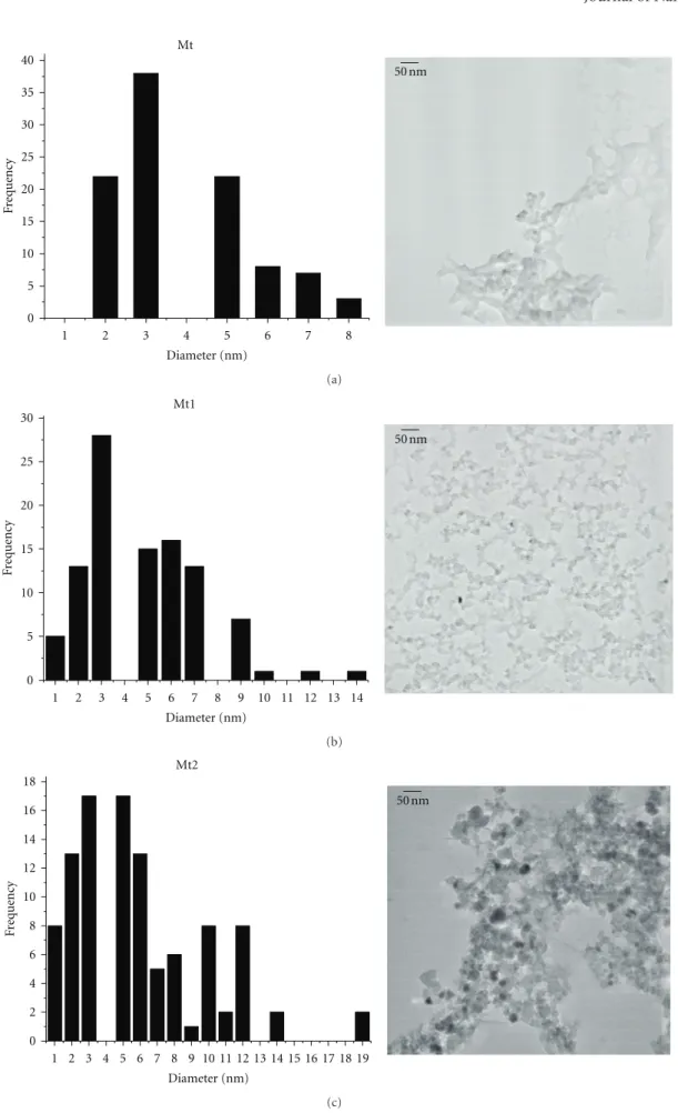
Figure 1 shows TEM images of Mt, Mt1, and Mt2
nanoparticles. The mean particle size diameters for the
samples Mt, Mt1, and Mt2 were found to be about 5.7 ±
2.9 nm, 5.8 ±2.7 nm, and 9.7 ±6.4 nm, respectively. The
mean size of the particles/aggregates is larger in the case of Mt2 sample. The higher tendency of particles in sample Mt2 to be aggregated can be explained by the bigger particle
sizes but also by their higher crystallinity (Figure 2), well
favored by the addition of the peptizing agent (TMAOH). Larger particles of well-crystallized magnetite in which the bulk properties supersede surface properties, and they are expected to magnetically attract more strongly each other.
Hosono et al. [26] suggested that the bigger particles could
be a consequence of higher concentration of charged ions in the solution since agglomeration between particles occurs by shrinkage of electric double layer. Moreover, the role of the ionic strength on the particle size also is largely dependent on the nature of the electrolyte. The smallest cations being the best screening ions, their influence on the surface charge is highest. The surface of iron oxides is negatively charged
at pH >PZC (point of zero charge). These characteristics
explain the strong influence of NH4+ on reducing particle
0 5 10 15 20 25 30 35 40
Diameter (nm)
F
requency
1 2 3 4 5 6 7 8
Mt
50 nm
(a)
0 5 10 15 20 25 30
Diameter (nm)
F
requency
1 2 3 4 5 6 7 8 9 10 11 12 13 14
Mt1
50 nm
(b)
50 nm Mt2
Diameter (nm)
F
requency
1 2 3 4 5 6 7 8 9 10 11 12 13 14 15 16 17 18 19 0
2 4 6 8 10 12 14 16 18
(c)
20 25 30 35 40 45 50 55 60 65 70 (220)
(311)
(400) (511) (440)
Mt2 Mt1 Mt
600 c
ounts
2θ(degrees Cu Kα)
ICCD card no. 019-0629
Figure2: X-ray diffraction patterns of samples: Mt, Mt1, Mt2, and
magnetite (ICDD file no. 019-0629).
preventing particles aggregation. This interpretation is in line
with that of Bacri et al. [27,28], who proposed a decreasing
stability of the iron oxide nanoparticles as a consequence of a decrease of the electrical double layer thickness around the particles, driven by the increased ionic strength of the media upon adding TMAOH.
The crystalline structure of the synthesized iron oxide
species analyzed by powder XRD (Figure2) shows that Fe3O4
was identified in these Mt, Mt1, and Mt2 samples. The most
intense reflections for this phase were registered at 2θ =
30.095◦, 35.423◦, 43.053◦, 57.22◦and 62.76◦, corresponding
to the planes of (220), (311), (400), (511), and (440),
respectively, of the iron oxide spinel. The peaks in the diff
rac-tograms match well with the PDF data for magnetite from
the powder diffraction file (PDF) from International Centre
for Diffraction Data (ICDD) card number 019-0629. The
average particle sizes for Mt, Mt1, and Mt2 magnetites, from breadths of reflection 311, estimated with Scherrer equation
were 11, 10, and 13 nm, respectively [29]. From routine
powder XRD data only, it may be difficult to differentiate
the coexisting maghemite (γFe2O3) and magnetite (Fe3O4)
in a same sample since structure is cubic with relatively
close unit cell dimension. The diffraction patterns for both
phases are quite similar. Therefore, one might presume that magnetite and maghemite are components of the obtained ferromagnetic phases, as oxidation processes could not be totally avoided during chemical synthesis and solid powder
washings and handling in air. Results in Figure 2confirm
that the sample Mt that has not been in contact with the peptizing agent exhibits the lowest degree of crystallinity as it can be drawn from the noisy background and from the
broader diffraction peaks. Contrarily, the Mt2 sample that
has been synthesized from the very beginning in presence
of TMAOH presents the sharpest XRD diffraction peaks,
which is in good consistence with its coarser morphology
(Figure 1). When the peptizing agent was added to the
precipitated Mt sample, it promoted a better dispersion of the partially amorphous nanoparticles and enhanced the degree of spatial arrangement of the matter, thus increasing
the crystallinity. This effect was reportedly proposed for
other systems [18–23]. These results suggest that, in the case
M
ag
n
etic
OH
OH
OH +
+
+
CH3
CH3
CH3
CH3
CH3
CH3
CH3
CH3
CH3
CH3
CH3
CH3
N
N
N
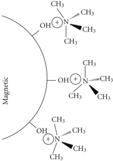
Figure3: Peptization exerted by tetramethyl ammonium cations
adsorbed onto the surface of magnetic nanoparticles thus, creating repulsive forces and enabling the particle growing species to be added to the surface in an organized manner.
of Mt2, the [(CH3)4N]+ cations are prone to readily adsorb
specifically onto the surface of the early nuclei being formed,
as schematized in Figure 3, hindering some of them to
achieve the critical size for growing. Under this perspective,
a smaller number of effective nuclei would be formed,
explaining the greater size of the resulting nanoparticles in the case of Mt2 sample.
M¨ossbauer spectra obtained at variable temperatures
(Figure4) reveal that these nanosized particles do contain
ferric iron oxides in small particle sizes, more clearly for samples Mt and Mt2.
These magnetic nanoparticles in a liquid medium expe-rience reorientations of the magnetization vector, in single
anisotropic magnetic domains (N´eel relaxation time, τN)
but also due to viscous rotation of the particle (Brownian
relaxation time,τB) [30,31].
The global magnetic relaxation times (τ) of colloids are
the sum of the N´eel and Brownian relaxation rates, which is
given by [31,32]
1
τ =
1
τB
+ 1
τN. (1)
These relaxations explain the obtained M¨ossbauer
spec-tra with a relatively intense censpec-tral Fe3+doublet along with a
magnetic sextet, as it was more clearly observed for samples Mt and Mt2. However, lowering the sample temperature
tends to progressively block the relaxations effect and
promote the magnetic ordering. The coexistence of these two major spectral contributions, that is, superparamagnetic
and magnetically ordered patterns, in different proportions,
means that particle sizes distributions are different in each
Spectral area=70% Spectral area=30%
Spectral area=100%
Spectral area=64%
1
0.99
0.98
1
0.98 1 0.99
0.98
0.97
R
elati
ve t
ransmission
−12−9−6−3 0 3 6 9 1210 20 30 40 50
HHF(Tesla)
Velocity (mm/s) Mt
Mt1
Mt2
PH
HF
Spectral area=36%
(a)
1
0.98
0.96 1
0.98
1
0.99
0.98
R
elati
ve t
ransmission
−12−9−6−3 0 3 6 9 12 10 20 30 40 50 60
HHF(Tesla)
Velocity (mm/s) Mt
Mt1
Mt2
PH
HF
Spectral area=30%
Spectral area=100%
Spectral area=64% Spectral area=36% Spectral area=70%
(b)
Figure4: M¨ossbauer spectra of Mt, Mt1, and Mt2 at (a) RT, and ( b) 80 K.
1400 1200 1000 800 600 400
FeO
TMAOH
Mt
Mt1
Mt2 CN
Int
ensit
y (a.u.)
Wavenumber (cm−1) CH3
CH3
Figure5: ATR-FTIR spectra of TMAOH, Mt, Mt1, and Mt2.
than for the other two samples. Values of hyperfine mag-netic fields at maximum probability for all these magmag-netic samples steadily increase on going from 297 K to 80 K, following values well around those expected for maghemite
(γFe2O3).
The apparent contradiction with evidences from hyper-thermic measurements that point to sample Mt2 as the sample containing bigger particles than Mt and Mt1 is explained by assuming that the Brownian relaxation rate in sample Mt2, with more strongly aggregated nanoparticles, is much slower so to produce a more significant hyperthermic
effect. However, M¨ossbauer spectra are necessarily collected
with the solid sample. In this condition, the global relaxation time is only influenced by the N´eel relaxation, for which magnitude, particularly for samples Mt and Mt2, would be comparable or below the time-scale window for M¨ossbauer
spectroscopy, or∼10−7s.
A M¨ossbauer spectrum results from the hyperfine cou-pling arising from extranuclear electric and magnetic fields that influence the energy levels of the probe nucleus. The fast fluctuation of the magnetization vector between easy
directions in the monodomain nanosystem, relatively to
the lifetime decay of the 57Fe 14.4 keV M¨ossbauer level,
would lead to an electric quadrupole coupling, resulting in a spectral superparamagnetic doublet. Particles aggregation that is promoted by coating their surface with surfac-tant or polymerizing organic materials may be enough to change the hyperthermic behavior of the material but not necessarily the hyperfine structure of the solid material. Aggregates of fine particles may present typical hyperthermic behavior as of bigger particles, essentially due to the increasing Brownian reorientation energy in the colloidal medium, but their hyperfine structure still accounts for local interactions at the atomic nuclear level in the superparamagnetic state of the solid mate-rial.
Figure5compares the ATR–FTIR spectra of the TMAOH
and of the Mt, Mt1, and Mt2 samples. The ATR–FTIR
spectrum of the TMAOH shows a strong band at 1490 cm−1,
assigned to the asymmetric methyl deformation mode,
δasym(CH3). A smaller one assigned to the symmetrical
methyl deformation mode δsym(CH3) appears near to
1430 cm−1. A single band appeared at 950 cm−1is attributed
to theυasym(C–N) mode which is generally observed in the
domain 900–1000 cm−1 [33]. The Mt sample shows a band
at 550 cm−1. Since magnetite has an inverse spinel-type
structure, it shows bands indicating the vibrations MT–O–
MO(ν1≈600–550 cm−1), where MTand MOcorrespond to
the metal occupying tetrahedral and octahedral positions,
respectively [34–36]. The ATR–FTIR spectra of the Mt1 and
Mt2 samples also show the band at 550 cm−1 characteristic
of magnetite and the characteristic vibration of Fe-O in
maghemite (γ-Fe2O3) at 690 cm−1[37–39], as well as the
transmittance bands typical of TMAOH, proving that the peptizing agent has been strongly adsorbed at the surface of the nanoparticles.
Zeta potential data are a powerful tool being extensively
used to evaluate the effects of the presence of
surface-active agents in different systems. In the present work, this
0 1 2 3 4 5 6 7 8 9 10 11 12 0
10 20
30 Mt2
Mt1 Mt
Z
eta pot
ential (mV
)
pH
−10
−20
−30
Figure6: Variation of zeta potential of samples: Mt, Mt1, and Mt2.
0
1 2 3
0 20 40
60 Mt1Mt2 Mt
M
o
ment (em
u/g)
B(Tesla)
−3 −2 −1
−20
−40
−60
Figure7: Magnetization curves of samples: Mt, Mt1, and Mt2.
added TMAOH on the solid/liquid interface properties of
magnetite nanoparticles. Results presented in Figure6show
that the pH values corresponding to the isoelectric points (pHIEP) of the Mt and Mt1 samples were found to be about 6.4 and 6.8, respectively. This is in good agreement with the reported data in the scientific literature for magnetite
[40,41]. In the Mt2 sample, the pHIEP was about 7.3. The
increase in pHIEPof the Mt1 and Mt2 samples, with respect
to that determined for Mt sample, indicates that [(CH3)4N]+
cations were specifically adsorbed onto the surface of the
synthesized iron oxide species [42]. This specific adsorption
is partially driven by the negative charge exhibited by
magnetite above its pHIEP (6.4) and has been apparently
more extensive when the peptizing agent was added during
the precipitation. Thus, the adsorbed [(CH3)4N]+ ions tend
to decrease the negative charge of the iron oxide powder at alkaline pH range.
Magnetization curves obtained for Mt, Mt1, and Mt2
samples at 300 K are presented in Figure 7. The curves
indicate a superparamagnetic behavior, for all these samples, as evidenced by zero coercivity and remanence on the
magnetization loop. The saturation magnetization (MS) was
obtained from moment versus magnetic field (B) curves.
For the Mt sample, the saturation magnetization value
is 70.7 emu g−1, being higher than values reported in
the literature (46.3 emu g−1) for samples prepared by the
Table1: The∆Tmaxvalues of the magnetite samples dispersed in
water and hydrogel under 220 Oe AC magnetic field.
Sample ∆Tmax
Water Hydrogel
Mt 23 12
Mt1 11 6
Mt2 19 8
same method [43], whereas for peptized samples Mt1 and
Mt2 with TMAOH the corresponding values are 62.8 and
63.2 emu g−1. The magnetic saturation values of magnetite
nanoparticles are experimentally determined to be in the
range of 30–50 emu g−1, which is lower than the bulk value,
90 emu g−1[44]. Similar values have been obtained for Mt
sample. It was expected that the saturation magnetization was higher for samples Mt1 and Mt2, as they have a higher degree of crystallinity and, in the case of Mt2, a larger particle
size. The differences between the saturation magnetization
between the samples with and without TMAOH can be ascribed to the previous presumption that magnetite and maghemite are both present in these peptized powders with TMAOH.
The magneto caloric behavior, influencing the heating ability of magnetite samples Mt, Mt-1, and Mt-2, is shown
in Figure 8. For each experiment, the temperature rising
was measured for every 1 min up to an accumulated time of 20 min and then every 5 min up to 1 h. Superparamagnetic particles generate heat by conversion of magnetic energy into thermal energy caused by the delay in magnetic relax-ation. According to the N´eel model, the magnetic moment originally locked along the crystal easy axis rotates away from that axis, tending to align with the external field. The N´eel mechanism is analogous to the hysteresis loss in multidomain magnetic particles whereby there is an internal friction due to the movement of the magnetic moment in an external field that results in heat generation. In the Brownian mode, the whole particle rotates in the direction of the field with the moment locked along the crystal axis. The heat generated through N´eel or Brownian relaxation in an applied AC magnetic field depends on the size of the nanoparticles. Smaller nanoparticles generate heat preferentially by the N´eel modell.
The amount of heat generated by pure water and hydrogel in 220 Oe AC magnetic fields was measured to obtain any contribution of water and hydrogel in the increase of temperature by the dispersed nanoparticles. The whole contribution of both pure water and gel was discounted from the final temperature value for each ferrofluid.
The results of heat dissipation experiments of the samples Mt, Mt1, and Mt2 dispersed in water and hydrogel are shown
in Figure8. The ∆Tmax values for magnetite nanoparticles
dispersed in water and hydrogel were determined from the gradients of temperature-time curves obtained by exposing the samples to an AC magnetic field strength of 220 Oe at
0 400 800 1200 1600 2000 2400 2800 3200 3600 0
5 10 15 20 25 30
Mt1 Mt2 Mt
Incr
ease of
t
emper
atur
e (K)
Time (s)
(a)
0 400 800 1200 1600 2000 2400 2800 3200 3600
0 5 10 15 20 25 30
Mt
Mt2 Mt1
Incr
ease of
t
emper
atur
e (K)
Time (s)
(b)
Figure8: Temperature-time curves of the samples Mt, Mt1, and Mt2 dispersed in (a) water and (b) PVA hydrogel.
The ∆Tmax values for dispersed samples Mt, Mt1, and
Mt2 in hydrogel were roughly 52, 54, and 42% less than that of the sole sample dispersed in water. The reduction in heat dissipation was attributed to the inhibition of particle rotation. In other words, particles should have an average Brownian magnetic relaxation time (TB) less than room temperature (RT) and narrow particle size distribution. This suggested that the fraction of the particles that generated heat through Brownian rotation was comparatively large. The sample with an average diameter below 13 nm had large fraction of particles dissipating heat through N´eel relaxation losses. On the other hand, the sample with average diameter larger than 13 nm had large fraction of particles dissipating
heat through Brownian relaxation [14]. Furthermore, these
results suggested that it is important to analyze the relative contributions of N´eel and Brownian relaxations losses to
formulate the appropriate strategy for effective in vivo
treatments. And also, it could be concluded that the samples with blocking temperature below RT are more suitable for
a reliable in vivo MFH treatment. Thus, to derive at the
appropriate sample forin vivoexperiments, similar study on
size-classified particles with TB less than RT and narrow size distribution should be pursued.
Hyperthermia is a cancer therapy that consists in heating selectively tumour in focusing organ zones. Those zones have fewer blood vessels and are less oxygenated than health ones. Consequently, they are more sensible and can die if the
local temperature increases above 43◦C. Thus, a temperature
variation of∆Tmax = 11◦C would be sufficient to produce
therapeutical effects.
4. Conclusions
Tetramethylammonium hydroxide (TMAOH) was revealed
to be an effective peptizing agent for magnetite
manoparti-cles in ferrofluid systems based on. The [(CH3)4N]+cations
specifically adsorbed onto the surface of the synthesized iron oxide species give rise to repulsive stabilization forces that enable the particle growing species to be added to the
surface in a more organized manner. Accordingly, when TMAOH is added during precipitation as in the case of Mt2 samples it promotes the formation of larger particles having a higher degree of crystallinity, including magnetite and maghemite. The addition of TMAOH after the magnetite has been synthesized promotes the colloidal stability of the nanoparticles and enhances the transformation of the amorphous material into more ordered crystalline phases.
Acknowledgments
This work was supported by CNPq (Brazil including Grants nos. 202212/2007-6 and 302479/2010-4), FAPEMIG (includ-ing Grantd no. PPM 00419-10 and no. APQ-00651-11) (Brazil) and by CICECO, (University of Aveiro, Portugal). CAPES (Brazil) grants the Visiting Professor PVNS fellow-ship to JDF at UFVJM.
References
[1] S. Sun, C. B. Murray, D. Weller, L. Folks, and A. Moser, “Monodisperse FePt nanoparticles and ferromagnetic FePt nanocrystal superlattices,” Science, vol. 287, no. 5460, pp. 1989–1992, 2000.
[2] V. Skumryev, S. Stoyanov, Y. Zhang, G. Hadjipanayis, D. Givord, and J. Nogu´es, “Beating the superparamagnetic limit with exchange bias,”Nature, vol. 423, no. 6942, pp. 850–853, 2003.
[3] M. Zhao, L. Josephson, Y. Tang, and R. Weissleder, “Magnetic sensors for protease assays,”Angewandte Chemie, vol. 42, no. 12, pp. 1375–1378, 2003.
[4] P. S. Doyle, J. Bibette, A. Bancaud, and J. L. Viovy, “Self-assembled magnetic matrices for DNA separation chips,”
Science, vol. 295, no. 5563, p. 2237, 2002.
[5] R. E. Rosensweig, “Magnetic fluids,” inFerrohydrodynamics, Cambridge University Press, Cambridge, UK, 1985.
[6] S. S. Papell, US Patent No: 3 215 572, 1965.
[8] A. Jordan, R. Scholz, P. Wust et al., “Endocytosis of dextran and silan-coated magnetite nanoparticles and the effect of intracellular hyperthermia on human mammary carcinoma cellsin vitro,”Journal of Magnetism and Magnetic Materials, vol. 194, no. 1, pp. 185–196, 1999.
[9] D. C. F. Chan, D. B. Kirpotin, and J. P. A. Bunn, “Synthesis and evaluation of colloidal magnetic iron oxides for the site-specific radiofrequency-induced hyperthermia of cancer,”
Journal of Magnetism and Magnetic Materials, vol. 122, no. 1– 3, pp. 374–378, 1993.
[10] C. Grob, K. Buscher, E. Romanus, C. A. Helm, and W. Weitschies, “Characterization of a ferrofluid by atomic force microscopy and photon correlation spectroscopy after mag-netic fractionation,” European Cells and Materials, vol. 3, supplement 2, pp. 163–166, 2002.
[11] L. M. Lacava, Z. G. M. Lacava, M. F. Da Silva et al., “Magnetic resonance of a dextran-coated magnetic fluid intravenously administered in mice,”Biophysical Journal, vol. 80, no. 5, pp. 2483–2486, 2001.
[12] Z. G. M. Lacava, R. B. Azevedo, L. M. Lacava et al., “Toxic effects of ionic magnetic fluids in mice,”Journal of Magnetism and Magnetic Materials, vol. 194, no. 1, pp. 90–95, 1999. [13] M. Rˇacuciu, D. E. Creangˇa, V. Bˇadescu, and N. Sulitanu,
“Microstructural investigation of some biocompatible fer-rofluids,”Journal of Magnetism and Magnetic Materials, vol. 316, no. 2, pp. e772–e775, 2007.
[14] M. Suto, Y. Hirota, H. Mamiya et al., “Heat dissipation mechanism of magnetite nanoparticles in magnetic fluid hyperthermia,”Journal of Magnetism and Magnetic Materials, vol. 321, no. 10, pp. 1493–1496, 2009.
[15] P. Berger, N. B. Adelman, K. J. Beckman, D. J. Campbell, A. B. Ellis, and G. C. Lisensky, “Preparation and properties of an aqueous ferrofluid,”Journal of Chemical Education, vol. 76, no. 7, pp. 943–948, 1999.
[16] F. Y. Cheng, C. H. Su, Y. S. Yang et al., “Characterization of aqueous dispersions of Fe3O4 nanoparticles and their biomedical applications,”Biomaterials, vol. 26, no. 7, pp. 729– 738, 2005.
[17] J. P. Jolivet, R. Massart, and J. M. Fruchart, “Synthesis and physicochemical study of non-surfactant magnetic colloids in an aqueous-medium,” Nouveau Journal De Chimie-New Journal of Chemistry, vol. 7, no. 5, pp. 325–331, 1983. [18] J. Yang, S. Mei, and J. M. F. Ferreira, “Hydrothermal synthesis
of nanosized titania powders: influence of peptization and peptizing agents on the crystalline phases and phase transi-tions,”Journal of the American Ceramic Society, vol. 83, no. 6, pp. 1361–1368, 2000.
[19] J. Yang, S. Mei, and J. M. F. Ferreira, “Hydrothermal synthesis of nanosized titania powders: influence of tetraalkyl ammo-nium hydroxides on particle characteristics,”Journal of the American Ceramic Society, vol. 84, no. 8, pp. 1696–1702, 2001. [20] J. Yang, S. Mei, and J. M. F. Ferreira, “Hydrothermal synthesis of TiO2 nanopowers from tetraalkylammonium hydroxide peptized sols,”Materials Science and Engineering C, vol. 15, no. 1-2, pp. 183–185, 2001.
[21] J. Yang, S. Mei, and J. M. F. Ferreira, “In situ preparation of weakly flocculated aqueous anatase suspensions by a hydrothermal technique,” Journal of Colloid and Interface Science, vol. 260, no. 1, pp. 82–88, 2003.
[22] J. Yang, S. Mei, and J. M. F. Ferreira, “Hydrothermal processing of nanocrystalline anatase films from tetraethylammonium hydroxide peptized titania sols,” Journal of the European Ceramic Society, vol. 24, no. 2, pp. 335–339, 2004.
[23] J. Yang, S. Mei, and J. M. F. Ferreira, “Hydrothermal synthesis of submicrometerα-alumina from seeded tetraethylammo-nium hydroxide-peptized aluminum hydroxide,” Journal of the American Ceramic Society, vol. 86, no. 12, pp. 2055–2058, 2003.
[24] ˆA. L. Andrade, D. M. Souza, M. C. Pereira, J. D. Fabris, and R. Z. Domingues, “pH effect on the synthesis of mag-netite nanoparticles by the chemical reduction-precipitation method,”Quimica Nova, vol. 33, no. 3, pp. 524–527, 2010. [25] S. H. Hyon, W. I. Cha, and Y. Ikada, “Preparation of
transparent poly(vinyl alcohol) hydrogel,” Polymer Bulletin, vol. 22, no. 2, pp. 119–122, 1989.
[26] T. Hosono, H. Takahashi, A. Fujita, R. J. Joseyphus, K. Tohji, and B. Jeyadevan, “Synthesis of magnetite nanoparticles for AC magnetic heating,” Journal of Magnetism and Magnetic Materials, vol. 321, no. 19, pp. 3019–3023, 2009.
[27] J. C. Bacri, R. Perzynski, V. Cabuil, and R. Massart, “Phase diagram of an ionic magnetic colloid: experimental study of the effect of ionic strength,”Journal of Colloid And Interface Science, vol. 132, no. 1, pp. 43–53, 1989.
[28] E. Hasmonay, A. Bee, J. C. Bacri, and R. Perzynski, “pH effect on an ionic ferrofluid: evidence of a thixotropic magnetic phase,”Journal of Physical Chemistry B, vol. 103, no. 31, pp. 6421–6428, 1999.
[29] A. L. Patterson, “The scherrer formula for X-ray particle size determination,”Physical Review, vol. 56, no. 10, pp. 978–982, 1939.
[30] S. Laurent, S. Dutz, U. O. H¨afeli, and M. Mahmoudi, “Magnetic fluid hyperthermia: focus on superparamagnetic iron oxide nanoparticles,”Advances in Colloid and Interface Science, vol. 166, no. 1-2, pp. 8–23, 2011.
[31] S. Laurent, D. Forge, M. Port et al., “Magnetic iron oxide nanoparticles: synthesis, stabilization, vectorization, physic-ochemical characterizations and biological applications,”
Chemical Reviews, vol. 108, no. 6, pp. 2064–2110, 2008. [32] R. E. Rosensweig, Ferrohydrodynamics, Cambrige Univerity
Press, New York, NY, USA, 1985.
[33] A. Ouasri, A. Rhandour, M. C. Dhamelincourt, P. Dhamelin-court, and A. Mazzah, “Vibrational study of (CH3)4NSbCl6 and [(CH3)4N]2SiF6,”Spectrochimica Acta, vol. 58, no. 12, pp. 2779–2788, 2002.
[34] E. Barrado, F. Prieto, J. Medina, and F. A. L ´opez, “Characteri-sation of solid residues obtained on removal of Cr from waste water,”Journal of Alloys and Compounds, vol. 335, no. 1-2, pp. 203–209, 2002.
[35] J. L. Mart´ın De Vidales, A. L ´opez-Delgado, E. Vila, and F. A. L ´opez, “Effect of the starting solution on the physico-chemical properties of zinc ferrite synthesized at low temperature,”
Journal of Alloys and Compounds, vol. 287, no. 1-2, pp. 276– 283, 1999.
[36] M. Ma, Y. Zhang, W. Yu, H. Y. Shen, H. Q. Zhang, and N. Gu, “Preparation and characterization of magnetite nanoparticles coated by amino silane,”Colloids and Surfaces A, vol. 212, no. 2-3, pp. 219–226, 2003.
[37] S. N. Inamdar and S. K. Haram, “Synthesis and characteri-zation of uncappedγ-Fe2O3nanoparticles prepared by flame pyrolysis of ferrocene in ethanol,”Journal of Nanoscience and Nanotechnology, vol. 6, no. 7, pp. 2155–2158, 2006.
[38] M. P. Morales, S. Veintemillas-Verdaguer, M. I. Montero et al., “Surface and internal spin canting inγ-Fe2O3nanoparticles,” Chemistry of Materials, vol. 11, no. 11, pp. 3058–3064, 1999. [39] Z. Jing, “Preparation and magnetic properties of fibrous
gamma iron oxide nanoparticles via a nonaqueous medium,”
[40] E. Baumgartner, M. A. Blesa, and A. J. G. Maroto, “Kinetics of the dissolution of magnetite in thioglycolic acid solutions,”
Journal of the Chemical Society, Dalton Transactions, no. 9, pp. 1649–1654, 1982.
[41] S. C. Pang, S. F. Chin, and M. A. Anderson, “Redox equilibria of iron oxides in aqueous-based magnetite dispersions: effect of pH and redox potential,”Journal of Colloid and Interface Science, vol. 311, no. 1, pp. 94–101, 2007.
[42] M. P. Albano and L. B. Garrido, “Dispersion of concentrated aqueous Si3N4-Y2O3-Al2O3slips with tetramethylammonium hydroxide,”Ceramics International, vol. 25, no. 1, pp. 13–18, 1999.
[43] T. Sato, T. Iijima, M. Seki, and N. Inagaki, “Magnetic properties of ultrafine ferrite particles,”Journal of Magnetism and Magnetic Materials, vol. 65, no. 2-3, pp. 252–256, 1987. [44] A. H. Lu, E. L. Salabas, and F. Sch¨uth, “Magnetic
nanoparti-cles: synthesis, protection, functionalization, and application,”
Submit your manuscripts at
http://www.hindawi.com
Scientifica
Hindawi Publishing Corporation
http://www.hindawi.com Volume 2014 Hindawi Publishing Corporation
http://www.hindawi.com Volume 2014
Hindawi Publishing Corporation
http://www.hindawi.com Volume 2014
Hindawi Publishing Corporation
http://www.hindawi.com Volume 2014
Ceramics
Journal ofHindawi Publishing Corporation
http://www.hindawi.com Volume 2014
Nanoparticles
Journal ofHindawi Publishing Corporation
http://www.hindawi.com Volume 2014
Hindawi Publishing Corporation
http://www.hindawi.com Volume 2014 International Journal of
Biomaterials
Hindawi Publishing Corporation
http://www.hindawi.com Volume 2014
Nanoscience
Journal ofTextiles
Hindawi Publishing Corporation
http://www.hindawi.com Volume 2014
Journal of
Hindawi Publishing Corporation
http://www.hindawi.com Volume 2014
Crystallography
Journal ofHindawi Publishing Corporation
http://www.hindawi.com Volume 2014
The Scientific
World Journal
Hindawi Publishing Corporationhttp://www.hindawi.com Volume 2014
Hindawi Publishing Corporation
http://www.hindawi.com Volume 2014
Coatings
Journal ofAdvances in
Materials Science and Engineering Hindawi Publishing Corporation
http://www.hindawi.com Volume 2014
Hindawi Publishing Corporation
http://www.hindawi.com Volume 2014
Hindawi Publishing Corporation
http://www.hindawi.com Volume 2014
Metallurgy
Journal ofHindawi Publishing Corporation
http://www.hindawi.com Volume 2014
BioMed
Research International
Materials
Journal ofHindawi Publishing Corporation
http://www.hindawi.com Volume 2014
N
a
no
ma
te
ria
ls
Hindawi Publishing Corporation
http://www.hindawi.com Volume 2014
Journal of