Isolation, morphological and molecular characterization of phytate-hydrolysing fungi
by 18S rDNA sequence analysis
Iti Gontia-Mishra, Dhanshree Deshmukh, Niraj Tripathi, Khushboo Bardiya-Bhurat,
Keerti Tantwai, Sharad Tiwari
Biotechnology Centre, Jawaharlal Nehru Agricultural University, Jabalpur, Madhya Pradesh, India.
Submitted: February 26, 2012; Approved: July 2, 2012.
Abstract
Phytate is the primary storage form of phosphate in plants. Monogastric animals like poultry, pigs and fishes have very low or no phytase activities in their digestive tracts therefore, are incapable to ef-ficiently utilize phytate phosphorus from the feed. Phytase from microbial sources are supplemented to feedstuff of these to increase the uptake of phytate phosphorus. In the present work efforts were made to isolate and characterize proficient phytase producing fungi from soil. Phytase producing fungi were isolated using phytate specific medium. Fungal isolates were selected according to their higher phytase activities. These isolates were further characterized and identified by morphological and microscopic analysis and confirmed by amplification of 18SrRNAgene, using specific primers. This gene was subsequently sequenced and phylogenetic affiliations were assigned. Fungal isolates were identified as various species ofAspergillus. Phytases from these fungi could be utilized as a feed additive in poultry and swine industries.
Key words:phytase, phytate, fungi, 18SrRNAgene,Aspergillus.
Introduction
In many plants, phytic acid (myo-inositol 1,2,3,4,5,6 hexakisphosphate) is one of the main storage forms of phosphate, accounting for 60-80% of total phosphorous (P) in grains, legumes and oilseeds (Sharma and Gole, 1978; Wanget al., 2007). Cereal grains and oilseed meals are ma-jor ingredients of animal feed. The amount of phosphorus in cereal grains and oilseed meals would meet the require-ment of optimal growth of animals if all the phosphorus from phytate were available to animals. Phytate phospho-rus is not available to monogastric animals (such as poultry, piggery and fish) due to lack of adequeate levels of phytase enzyme in their gastrointestinal tracts (Brinch-Pedersenet al., 2002; Singh and Satayanarayana, 2010). Phosphate supplementation is required for optimal growth of animals. Phytase catalyzes the dephosphorylation of phytate to inositol and orthophosphate (Wodzinski and Ullah, 1996; Hamadaet al., 2005). Moreover, in the regions with intense animal production, a large amount of undigested and ex-creted phosphate (phytate) contributes significantly to the
environmental pollution. Furthermore, phytic acid is also considered as an anti-nutritional factor, since it causes min-eral deficiency due to efficient chelation of metal ions (Fe3+, Zn2+, Ca2+, Mg2+, K+) and forming complexes with proteins, thus affecting their digestion (Urbano et al., 2000). Microbial phytase produced by fermentation as a feed additive is widely used to manage the nutritional and environmental problems caused by phytate.
Commercial production of phytase as feed additives is mostly focused on fungi and yeasts, as they are the most prolific extracellular producers of this enzyme (Farhatet al., 2008). Most of the naturally occurring phytases having high thermostability and a broad pH range were identified from fungi (Simon and Igbasan, 2002). Due to immense in-dustrial and environmental implication of phytases there is an ongoing interest in isolation of new fungal strain produc-ing phytase and optimization of this enzyme. In the present work efforts were made for isolation, morphological and molecular characterization of proficient phytase producing fungal strains.
Send correspondence to S. Tiwari. Biotechnology Centre, Jawaharlal Nehru Agricultural University, 482004 Jabalpur, Madhya Pradesh, India. E-mail: [email protected].
Materials and Methods
Sample collection
Samples were collected in sterile polythene bags from agricultural land, vermicompost and food based sources such as poultry and pig wastes, and fishery pond located at Jawaharlal Nehru Agricultural University Campus, Jabal-pur, Madhya Pradesh, India (23°17’ N; 72°98’ E).
Isolation of phytase producing fungi
Soil samples (0.2 g) were suspended in 10 mL of 0.9% saline solution and kept on incubator shaker with 150 rpm for 2 h. Soil suspension (1 mL) was inoculated into 100 mL of phytate specific medium (PSM) containing 1.5% glucose, 0.5% (NH4)2SO4, 0.05% KCl, 0.01% MgSO4.7H2O, 0.01% NaCl, 0.01% CaCl2.2H2O, 0.001% FeSO4.7H20, 0.001% MnSO4.H20, pH 6.5 with 0.5% so-dium phytate (Hosseinkhani et al., 2009). Medium was sterilized by autoclaving (15 psi, 121 °C, 20 min), with the exception of sodium phytate, which was sterilized by mem-brane filtration (Millipore, 0.45mm) and added aseptically to cooled autoclaved media. The samples were kept in incu-bator shaker with shaking at 150 rpm at 30 °C for 10 days. Fungal cultures were re-inoculated in fresh PSM medium to ensure enrichment of phytase producing fungi and incu-bated at 30 °C, 165 rpm for 7 days.
Fungal cultures obtained through the enrichment pro-cess were inoculated in PSM agar medium containing cal-cium phytate (0.5%) as sole source of phosphorus. Plates were kept for incubation at 30 °C for 5 days. After incuba-tion zone of clearing around the fungal growth on PSM agar plates were observed. The zone of clearing around the fun-gal growth is indicator of phytase production. The samples which showed clear zone were considered as positive sam-ples. Counterstaining for confirmation of phytase activity was performed for the positive isolates according to the method of Baeet al.(1999).
Phytase enzyme activity assay
Isolates that produced clear zones on screening me-dium were tested for phytase production in PSM broth with sodium phytate. A spore suspension of 1 x 10⁷/ mL was in-oculated in 100 mL of PSM medium in a 500 mL Erlen-meyer flask and incubated at 30 °C with 200 rpm shaking for 7 days. Cultures (2 mL) were centrifuged and the super-natant was used for phytase activity assays. The enzyme ac-tivity was estimated according to the method described by Engelenet al.(1994). The incubation mixture (2 mL) con-tained 1 mL of the culture filtrate, 1mL of substrate solution [10 mM sodium phytate as substrate and 0.2 M sodium ace-tate buffer (pH 5.5)] and incubated at 37 °C for 1 h. The re-action was terminated by the addition of 1mL of the colour developing reagent [250 mL of ammonium hepta-molyb-date solution (10% of ammonium molybhepta-molyb-date in 0.25% am-monia solution), 250 mL of ammonium vanadate solution
(2.35 g of ammonium vanadate in 1 L of 2% (v/v) nitric acid solution), and 165 mL of 65% nitric acid, finally di-luted to 1L with water]. The absorbance was measured at 415 nm. Distilled water was used as a blank. One unit of phytase activity was defined as 1mmol of phosphate pro-duced per min per mL of culture filtrate under the assay condition (pH 5.5, temperature 37 °C and substrate concen-tration, sodium phytate [C6H6O24P6Na12] at 0.0051 mol/L). Standard curve was prepared using potassium dihydrogen phosphate (KH2PO4) in the range 0-1000mmol.
pH and temperature optima of crude phytase enzyme
To determine the pH optimum curve, the enzyme was incubated with sodium phytate prepared in 0.2 M Sodium acetate buffer, pH 3.0, 3.6, 4.2, 4.8, 5.5; 0.2 M citrate buffer, pH 6.0 and 6.6 and 0.2 M Tris, pH 7.0, 7.5 and 8.5 for 1 h at 37 °C and released phosphate ions were assayed. The temperature optimum was determined by incubating the enzyme with substrate, prepared in 0.2 M sodium ace-tate pH 5.5 at different temperatures for 1 h and the phytase activity was assayed.
Morphological characterization
The morphological identification of isolates (DD1-DD3; IG1-IG5) was conducted using four different types of mediaviz.Czapek dox agar (CDA), Czapek yeast agar (CYA), Czapek yeast 20% sucrose agar (CY2S) and Malt yeast agar (MYA). Creatine sucrose agar (CREA) was used to study acid or base production by fungi (Samsonet al., 2007). All media were incubated at 28 °C for 7 days. Colonies on each medium were compared for their diame-ters, overall colors, colors of conidia, reverse colors, tex-ture, zonation and sporulation. All the isolates were also subjected to microscopic analysis for their characterization and identification.
Identification of fungi using 18S rRNA gene analysis
Fungal mycelium or spores were cultured on potato dextrose agar medium (Himedia, India). The plates were incubated at 30 °C for 2 to 3 days. The fungal mycelium was used for DNA isolation. DNA was extracted using method described by Huntet al.(2004).
PCR amplification of 18S rRNA gene
PCR amplification of fungal small-subunit rDNA (18SrRNAgene) was carried out using the primer set EF4 and EF3 (Smitet al., 1999). The EF4 and EF3 primers am-plified a 1.5-kb section of the 18SrRNAgene. Primer
se-quences were as follows: EF4
(5’-GGAAGGG[G/A]TGTATTTATTAG-3’) and EF3
(5’-TCCTCTAAATGACCAAGTTTG-3’). PCR amplifi-cation was performed in a 25mL reaction containing 2.5 U ofTaq DNA polymerase (Sigma), a 10 X dilution of the manufacturer’s buffer (Sigma), 200mM concentrations of
each deoxynucleoside triphosphate (dNTPs), and 20 pM of primers EF4 and EF3 and 50 ng of genomic DNA. The re-action conditions were as follows: initial denaturation at 94 °C for 4 min, 40 amplification cycles of denaturation at 94 °C for 1 min, annealing at 48 °C for 1 min and primer ex-tension at 72 °C for 3 min; followed by a final exex-tension at 72 °C for 10 min. PCR amplifications were carried out us-ing a Thermo-Hybaid PCR thermal cycler (Thermo Fisher Scientific USA). Aliquots of the PCR products (5mL) were analyzed in 1% (w/v) agarose gels (Sigma, USA) by hori-zontal gel electrophoresis. DNAs were visualized by UV excitation after staining with ethidium bromide (0.5 mg/L). The PCR product was purified using Bangalore Genie PCR purification kit following the manufacturer’s instruction. The 18S rRNA nucleotide sequence was determined by PCR-direct sequencing done by Chromous Biotech Pvt. Ltd., Bangalore, India.
Phylogenetic analysis of the 18S rRNA gene se-quences was performed with CLC DNA workbench ver-sion 6. The phylogenetic trees were inferred using the neighbour-joining method (Saitau and Nei, 1987) and boot-strap analyses were performed. The evolutionary distances were computed using the Maximum Composite Likelihood method (Tamuraet al., 2004).
Results and Discussion
Isolation of phytase producing fungi from soil
Twenty-five soil samples were collected from differ-ent locations for the isolation of phytase producing fungi. Out of these only fourteen samples showed fungal growth in phytate specific screening medium containing 1% so-dium phytate. The fungal cultures obtained from these soil samples were re-inoculated in PSM broth for enrichment of these phytate utilizing cultures. For further confirmation of their phytase producing trait, fungal cultures were inocu-lated on PSM agar medium containing 0.5% calcium phy-tate. Eight fungal isolates produced zone of clearing on PSM agar medium. Counterstaining was also performed to visualize the zone of clearing. These fungal isolates were designated as IG 1, IG 2, IG 3, IG 4, IG 5, DD 1, DD 2 and
DD 3. Counterstaining approach was carried out to overcome the selection of false positive isolates for phytase production on PSM medium (Fredrikson et al., 2002; Chadhaet al., 2004). The formation of clear zone is attrib-uted to the production of various acids (acetic acid, maleic acid, etc.), which lowers the pH of the medium and hence increase the solubility of calcium phytate (Baeet al., 1999).
Phytase activity and its optimization
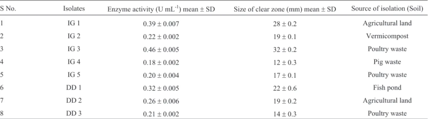
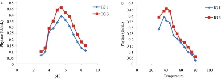
Phytase activity was determined by measuring the amount of liberated inorganic phosphate and its reaction with colour reagent. It was carried out for fungal isolates ir-respective of clear zone formation on PSM medium. It was found that fungal isolates which did not form clear zone on PSM, showed negligible phytase activities in liquid me-dium. Isolate IG 3 and IG 1 showed highest phytase activities which were 0.46 U mL-1and 0.39 U mL-1, respec-tively. Enzyme activities as well as size of zone of clear-ance for different isolates are shown in Table 1. Optimization of phytase activity for isolate IG 1 and IG 3 were carried out to determine their optimum pH and tem-perature. Phytase activity for isolates IG 1 and IG 3 was op-timum at pH 5.5 as depicted from Figure 1a and opop-timum temperature was 37 °C and 40 °C respectively as shown in Figure 1b. Phytase often has a low-pH optimum range (pH 4.5-6.0) with a rapid drop in activity at pH value above 6.0 (Marlidaet al., 2010). It is needed to be determined that phytase activity for the fungal isolates is extracellular or cell-associated.
Morphological and microscopic characterization of fungi
Fungal isolates were grown in different growth me-dium (CDA, CYA, MYA and CY2S) and colonies on each medium were recorded for their diameters, overall colors, colors of conidia, reverse colors, texture, zonation and sporulation as shown in Table 2. Isolates IG 5, DD2 showed similar results to IG 1 and similarly, IG 3 and DD1 showed comparable results on different culture media so only a sin-gle representatives from them were taken into consider-ation and shown in Table 2. On the basis of morphological
Table 1- Phytase activities for different fungal isolates.
S No. Isolates Enzyme activity (U mL-1) mean
±SD Size of clear zone (mm) mean±SD Source of isolation (Soil)
1 IG 1 0.39±0.007 28±0.2 Agricultural land
2 IG 2 0.22±0.002 19±0.1 Vermicompost
3 IG 3 0.46±0.005 32±0.2 Poultry waste
4 IG 4 0.18±0.002 12±0.3 Pig waste
5 IG 5 0.20±0.004 17±0.1 Poultry waste
6 DD 1 0.32±0.005 22±0.6 Fish pond
7 DD 2 0.26±0.006 19±0.2 Agricultural land
characters and growth pattern on different media, isolates IG 1, IG 5, DD 2 were suggested to beAspergillus niger
(Sharma and Pandey, 2010). Likewise, isolates IG 2 and IG 4 were reported to beA. fumigatusandA. terreus, respec-tively whereas isolates DD1 and IG 3 showed similarity to
A. awamori(McClenny, 2005; Zainet al., 2009; Perroneet al., 2011). Growth abilities of isolates were tested on CREA medium. CREA is the semi-selective media useful for classification of various fungal cultures (Samsonet al., 2007). On CREA, characteristics of colonial growth, pro-duction of acid (turning of the medium from purple to yel-low) can be used as diagnostic features. Isolate IG 1, IG 3, IG 4, IG 5 and DD 1, DD 2 showed moderate growth and good acid having production resulting in large yellowish halo around the colonies on CREA medium. Similar results forA. nigerwere reported by Samsonet al.(2007), showing this as a characteristic feature for distinguishing biseriate species (A. niger,A. awamoriandA. terreus) from uniseri-ate species. IG 2 showed poor growth and limited acid pro-duction on CREA medium as characteristic feature of uniseriate species.
Microscopic characterization for definitive identifi-cation of the isolates was carried out. In case of isolate IG 2 the microscopic analysis showed that the mycelium was wide, septate and hyaline with acute angle branching. Coni-dial head was uniseriate and columnar. Isolates IG 1, IG 5 and DD 2, also showed wide, septate and hyaline mycelium with acute angle branching but their conidial head was biseriate and radiate and conidia were attached in chains. Similar results were reported forA. fumigatusandA. niger, respectively by McClenney (2005), thus isolates IG1, IG 5, DD2 could be predicted as A. niger and IG 2 as A. fumigatus. In microscopic analysis of IG 4, mycelium was found to be wide, septate and conidiophores were long, co-lumnar and hyaline. Conidial head were globose to slightly elliptical and biseriate. Additionally, hyaline accessory conidia were also visible on the hyphae.A. terreusis the only member of the genusAspergillusthat produces such
structures (Balajee, 2009). The Isolates IG 3 and DD1 showed mycelial characters similar to those of isolates IG 1, IG 5 and DD 2 but conidia showed some ornamentation and were distinctly rough, hence these two isolates could be subspecies ofA. niger.
Molecular characterization of fungi
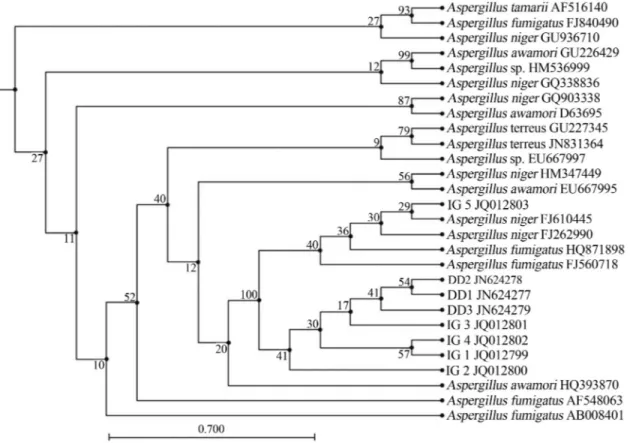
The PCR amplification of 18SrRNAgene was done by using gene specific primers. An amplification product of 1.5kb was obtained for all the isolates as shown in Figure 2. The nuclear small subunit ribosomal DNA (18SrDNA) was selected for characterization and identification of fungi firstly, because established universal fungal primers are available based on the conserved regions of 18SrDNA, mak-ing it possible to obtain the PCR products from most of the fungi. Secondly, the large numbers of 18SrDNAsequences are available in GenBank which makes similarity searches convenient. Several workers have also reported characteriza-tion of fungi based on 18SrRNAgene sequence analysis (Smitet al., 1999; Borneman and Hartin, 2000).
On the basis of 18SrRNAgene similarity isolates IG 1, IG 5 and DD 2 showed 99% sequence homology with
Aspergillus niger(FJ262990) (Yanget al., 2012), IG 2 and DD3 showed 98% sequence homology with Aspergillus fumigatus(AF648063) (Wuet al., 2003). The 18SrRNA
partial sequence placed the isolates IG 3 and DD1 within
320 Gontia-Mishraet al.
Figure 1- Phytase activities for isolates IG 1 (A. niger) and IG 3 (A. awamori) at different pH (a) and temperature (b).
fungi
321
Isolate Media Colony diam (mm) Colony characters Zonation Sporulation Reaction on CREA Identified as Reference
Texture Surface colour Reverse colour
IG 1 CDA 47.33±3.05 Velvety White with dusty yellow sporulating area
White Slightly Radially furrowed
Moderate Moderate growth and good acid pro-duction (large yel-low halo around colony)
Aspergillus niger Sharma and Pandey, 2010
CYA 47.00±1.00 Powdery White periphery with black spores
Yellowish Radially furrowed Heavy
MYA 59.00±3.60 Powdery White periphery with black spores
Cream Radially furrowed at the centre
Moderate
CY2S 71.00±2.65 Powdery White periphery with black spores
Cream Heavily wrinkled Heavy
IG 2 CDA 34.33±1.15 Velvety White with grayish green concentric sporulating rings
Cream Concentric zones Poor Poor growth and low acid produc-tion
A. fumigatus McClenny, 2005
CYA 52.00±1.73 Powdery Cream periphery grayish green sporulating area
Yellowish Radially furrowed Heavy
MYA 47.33±2.08 Powdery Cream with green periphery and grayish green sporulating area
Yellowish Radially furrowed Moderate
CY2S 73.66±3.21 Powdery Cream periphery grayish green sporulating area
Cream Wrinkled and radi-ally furrowed
Heavy
IG 3 CDA 55.66±0.58 Velvety White with brownish sporulating area
White irregularly fur-rowed
Moderate Moderate growth and good acid pro-duction (large yel-low halo around colony)
A. awamori Perroneet al., 2011
CYA 43.33±1.15 Powdery White periphery with dark brown sporulating area
Cream Heavily radially furrowed
Heavy
MYA 57.66±2.08 Velvety White periphery with dark brown sporulating area
Cream Heavily radially furrowed
Moderate
CY2S 67.33±1.53 Velvety White periphery with dark brown sporulating area
Cream Wrinkled Moderate
IG 4 CDA 45.33±1.15 Floccose Green periphery with green sporulating area
Yellow Radially furrowed Poor Moderate growth and good acid pro-duction (large yel-low halo around colony)
A. terreus Zainet al., 2009
CYA 44.33±1.53 Velvety White with dark green sporulating area
Light orange Radially furrowed Poor
MYA 55.66±2.31 Powdery Green periphery with dark green sporulating area
Peach Radially furrowed Moderate
CY2S 75.00±1.00 Powdery Green periphery with dark green sporulating area
Light orange Irregularly fur-rowed
Aspergillus genera with 99% sequence homology with
Aspergillus awamori (HQ393870) (Srinivasan et al., 2012).Similarly, isolate IG 4 showed maximum sequence similarity withAspergillus terreus(JN831364) (Yinet al., 2012). Partial 18SrRNAsequences of all the isolates were submitted to NCBI Genbank under the following accession numbers: IG 1, JQ012799; IG 2, JQ012800; IG 3, JQ012801; IG 4, JQ012802, IG 5, JQ012803; DD1, JN624277; DD2, JN624278 and DD3, JN624279. Phylo-genetic relationship of the fungal isolates with other fungi is shown in Figure 3.
A. nigeris well known for its phytase activity. Phy-tase activity fromA. nigerhas been extensively studied and reported by several workers (Vats and Banerjee, 2005; Gunashree and Venkateswaran, 2009; Soniet al., 2010). Similarly, phytase activity fromAspergillus fumigatushas previously been reported (Mullaneyet al., 2000; Rodriguez
et al., 2000). Isolate IG 3 showed close similarity withA. awamori and has considerably high phytase activity. A. awamori and A. terreus are less extensively studied for phytase activity. To best of our knowledge this is the sec-ond report of phytase production fromA. awamoriandA. terreus.Report of phytase production fromA. awamoriand
A. terreuswith further isolation and characterization ofphy
gene from theseAspergillusstrains will add new informa-tion to the database ofphygenes.
Conclusion
Our finding suggests that phytase production by A. nigerstrain IG 1 andA. awamoristrain IG 3 have signifi-cant values which can be exploited for industrial produc-tion of phytase. Moreover, this enzyme can be used in the animal feed industry for improving the nutritional status of feed. The native fungal communities of soil which degrade phytate from manures and soil with subsequent release of orthophosphate making it available to plants, thus enhance the benefits of manure-derived fertilizer and in combating environmental pollution due to phytate. Additionally,phy
gene from these strains could be used to develop transgenic plants (maize, sorghum and oat) which would in turn uti-lized as animal feed.
Acknowledgments
IG is grateful to Department of Biotechnology, Min-istry of Science and Technology, Government of India, New Delhi for financial assistance.
References
Bae HD, Yanke LJ, Cheng KJ, Selinger LB (1999) A novel stain-ing method for detectstain-ing phytase activity. J Microbiol Me-thods 39:17-22.
322 Gontia-Mishraet al.
Balajee SA (2009) Aspergillus terreus complex. Med Mycol 47:S42-S46.
Borneman J, Hartin RJ (2000) PCR primers that amplify fungal
rRNA genes from environmental samples. Appl Environ Microbiol 66:4356-4360.
Brinch-Pedersen H, Sorensen LD, Holm PB (2002) Engineering crop plants: Getting a handle on phosphate. Trends Plant Sci 7:118-125.
Chadha BS, Gulati H, Minhas M, Saini HS, Singh N (2004) Phytase production by the thermophilic fungusRhizomucor pusillus. World J Microbiol Biotechnol 20:105-109. Engelen AJ, van der Heeft FC, Randsdorp PH, Smit EL (1994)
Simple and rapid determination of phytase activity. J AOAC Int 77:760-764.
Farhat A, Chouayekh H, Farhat MB, Bouchaala K, Bejar S (2008) Gene cloning and characterization of a thermostable phytase fromBacillus subtilisUS417 and assessment of its potential as a feed additive in comparison with a commercial enzyme. Mol Biotechnol 40:127-135.
Fredrikson M, Andlid T, Haikara A, Sandberg AS (2002) Phytate degradation by micro-organisms in synthetic media and pea flour. J Appl Microbiol 93:197-204.
Gunashree BS, Venkateswaran G (2009) Screening of asporo-genic mutants of phytase-producingAspergillus nigerCFR 335 strain. Microb Ecol Health Dis 21:57-63.
Hamada A, Yamaguchi K, Ohnishi N, Harada M, Nikumaru S, Honda H (2005) High-level production of yeast (Schwanniomyces occidentalis) phytase in transgenic rice plants by a combination of signal sequence and codon modi-fication of the phytase gene. Plant Biotechnol J 3:43-55. Hosseinkhani B, Emtiazi G, Nahvi I (2009) Analysis of phytase
producing bacteria (Pseudomonassp.) from poultry faeces and optimization of this enzyme production. Afr J Bio-technol 8:4229-4232.
Hunt J, Boddy L, Randerson PF, Rogers HJ (2004) An evaluation of 18SrDNAapproaches for the study of fungal diversity in grassland soils. Microb Ecol 47:385-395.
Marlida Y, Delfita R, Adnadi P, Ciptaan G (2010) Isolation, char-acterization and production of phytase from endophytic fun-gus its application for feed. Pak J Nutr 9:471-474.
McClenny N (2005) Laboratory detection and identification of
Aspergillusspecies by microscopic observation and culture: The traditional approach. Med Mycol Suppl 43:S125-S128. Mullaney EJ, Daly CB, Sethumadhavan K, Rodriquez E, Lei XG,
Ullah AH (2000) Phytase activity inAspergillus fumigatus
isolates. Biochem Biophys Res Commun 275:759-763. Perrone G, Stea G, Epifani F, Varga J, Frisvad JC, Samson RA
(2011)Aspergillus nigercontains the cryptic phylogenetic speciesA. awamori. Fungal Biol 115:1138-1150.
Rodriguez E, Mullaney EJ, Lei XG (2000) Expression of the
Aspergillus fumigatusphytase gene inPichia pastorisand characterization of the recombinant enzyme. Biochem Bio-phys Res Commun 268:373-378.
Saitou N, Nei M (1987) The neighbor-joining method: A new method for reconstructing phylogenetic trees. Mol Biol Evol 4:406-425.
Samson RA, Noonim P, Meijer M, Houbraken J, Frisvad JC, Varga J (2007) Diagnostic tools to identify black aspergilli. Stud Mycol 59:129-145.
Sharma CB, Gole M (1978)Myo-inositol hexaphos-phytate as a potential inhibitor of amylases of different origin. Phyto-chemistry 17:203-204.
Sharma G, Pandey RR (2010) Influence of culture media on growth, colony character and sporulation of fungi isolated from decaying vegetable wastes. J Yeast Fungal Res 1:157-164.
Simon O, Igbasan F (2002)In vitroproperties of phytases from various microbial origins. Int J Food Sci Technol 37:813-822.
Singh B, Satyanarayana T (2010) Application of phytases of thermophilic mouldSporotrichum thrmophile: A review. J Sci Ind Res 69:411-414.
Smit E, Leeflang P, Glandorf B, van Elsas JD, Wernars K (1999) Analysis of fungal diversity in the wheat rhizosphere by se-quencing of cloned PCR-amplified genes encoding 18S
rRNA and temperature gradient gel electrophoresis. Appl Environ Microbiol 65:2614-2621.
Soni SK, Magdum A, Khire JM (2010) Purification and character-ization of two distinct acidic phytases with broad pH stabil-ity fromAspergillus nigerNCIM 563. World J Microbiol Biotechnol 26:2009-2018.
Srinivasan R, Alagawadi AR, Yandigeri MS, Meena KK, Saxena AK (2012) Characterization of phosphate-solubilizing mi-croorganisms from salt-affected soils of India and their ef-fect on growth of sorghum plants Sorghum bicolor (L.) Moench. Ann Microbiol 62:93-105.
Tamura K, Nei M, Kumar S (2004) Prospects for inferring very large phylogenies by using the neighbor-joining method. Proc Natl Acad Sci 101:11030-11035.
Urbano G, Lopez-Jurado M, Aranda P, Vidal-Valverde C, Teno-rio E, Porres J (2000) The role of phytic acid in legumes: Antinutrient or beneficial function? J Physiol Biochem 56:283-294.
Vats P, Banerjee U (2005) Biochemical characterization of extra-cellular phytase (myoinositol hexakisphosphate phospho-hydrolase) from a hyper-producing strain of Aspergillus nigervan Teighem. J Ind Microbiol Biotechnol 32:141-147. Wang Y, Gao X, Su Q, Wu W, An L (2007) Expression of heat
stable phytase from Aspergillus fumigatus in tobacco (Nicotiana tabacumL. cv. NC89). Ind J Biochem Biophys 44:26-30.
Wodzinski RJ, Ullah AH (1996) Phytase. Adv Appl Microbiol 42:263-302.
Wu Z, Tsumura Y, Blomquist G, Wang XR (2003) 18SrRNA
gene variation among common airborne fungi, and develop-ment of specific oligonucleotide probes for the detection of fungal isolates. Appl Environ Microbiol 69:5389-5397. Yang X, Sun JY, Guo JL, Weng XY (2012) Identification and
proteomic analysis of a novel gossypol-degrading fungal strain. J Sci Food Agri 92:943-951.
Yin Y, Gao Q, Zhang F, Li Z (2012) Medium optimization for the high yield production of single (+)-terrein by Aspergillus terreusstrain PF26 derived from marine spongePhakellia fusca.Process Biochem 47:887-891.
Zain ME, Razak AA, El-Sheikh HH, Soliman HG, Khalil AM (2009) Influence of growth medium on diagnostic characters ofAspergillusandPenicilliumspecies. Afr J Microbiol Res 3:280-286.