PEARLS
SOD Enzymes and Microbial Pathogens:
Surviving the Oxidative Storm of Infection
Chynna N. Broxton, Valeria C. Culotta*
Department of Biochemistry and Molecular Biology, Johns Hopkins University Bloomberg School of Public Health, Baltimore, Maryland, United States of America
Living with ROS through Superoxide Dismutase Enzymes
Since oxygen appeared in the biosphere some 3–5 billion years ago, all organisms have had to deal with the hazards of potentially damaging reactive oxygen species (ROS), such as superox-ide, hydrogen peroxsuperox-ide, and hydroxyl radical. Like all organisms, pathogenic microbes produce ROS as byproducts of aerobic metabolism, but the burden of ROS is magnified when these microbes confront the oxidative burst of the host. As part of the innate immune response, mac-rophages and neutrophils attack invading microbes with toxic superoxide [1]. To counteract this attack, some microbial pathogens express superoxide dismutase enzymes (SOD).
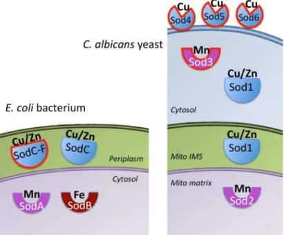
SODs are metalloenzymes that use a redox-active metal to disproportionate two molecules of superoxide to oxygen and hydrogen peroxide, the latter of which is removed by catalase and peroxidase enzymes. SODs have evolved on three separate occasions, yielding a family of Mn and Fe SODs (that use either metal as co-factor), a Cu/Zn SOD family that uses Cu for cataly-sis, and a rare family of Ni SODs [2]. Why so many SODs? This is best answered in terms of metal bioavailability. In a typical gram-negative bacteria, such asEscherichia coli, the cytosol can have ample Mn and/or Fe, but Cu is extruded into the periplasmic/extracellular space [3]. As a result, Mn and Fe SODs are generally intracellular/cytosolic while Cu/Zn SODs are extra-cellular/periplasmic (Fig 1). Consistent with the endosymbiosis theory of mitochondrial evolu-tion, this partitioning of SOD enzymes has been retained in eukaryotic mitochondria: The mitochondrial matrix (equivalent to bacterial cytosol) harbors a Mn SOD, while Cu/Zn SOD is in the mitochondrial intermembrane space and cytosol (equivalent to bacterial periplasmic/ extracellular) (Fig 1).
Custom SOD Enzymes for Bacterial Pathogenesis
The accepted nomenclature for bacterial SODs is SodA, SodB, and SodC for the Mn, Fe, and Cu/Zn SODs, respectively. Because superoxide does not generally cross biological membranes, the intracellular SodA and SodB largely remove intracellular or metabolic sources of superox-ide while the periplasmic/extracellular SodC directly combats superoxsuperox-ide from the animal host. Not all bacterial pathogens contain extracellular SodC; for example, the Lyme disease pathogen
Borrelia burgdorfericontains a single Mn-SodA. This pathogen also does not express Fe-SodB, representing a clever adaptation to limiting Fe supplies in the host [4,5].
The roles of SodA and SodB in bacterial survival and pathogenesis vary greatly depending on the species. In some cases (e.g.,Salmonella typhimurium), loss of SodA has no impact on virulence [6], whereas in other instances (e.g.,Streptococcus agalactiaeorB.burgdorferi), loss of the intracellular SOD attenuates virulence, in spite of there being no growth inhibition in laboratory cultures [7,8]. How could an intracellular SOD promote virulence when host
OPEN ACCESS
Citation:Broxton CN, Culotta VC (2016) SOD Enzymes and Microbial Pathogens: Surviving the Oxidative Storm of Infection. PLoS Pathog 12(1): e1005295. doi:10.1371/journal.ppat.1005295
Editor:Donald C Sheppard, McGill University, CANADA
Published:January 7, 2016
Copyright:© 2016 Broxton, Culotta. This is an open access article distributed under the terms of the Creative Commons Attribution License, which permits unrestricted use, distribution, and reproduction in any medium, provided the original author and source are credited.
Funding:The preparation of this review was supported by NIH funding from R37 GM50016, RO1 AI119949, T32 CA009110, and F31 GM113637. The funders had no role in study design, data collection and analysis, decision to publish, or preparation of the manuscript.
superoxide is extracellular? Although superoxide does not typically cross biological mem-branes, it can do so when protonated. In low pH environments, such as that of a macrophage phagolysosome, the protonated superoxide might enter the bacteria and serve as substrate for intracellular SodA and SodB [9]. Alternatively, the intracellular SODs may exclusively prevent damage from bacteria-derived superoxide that somehow promotes microbial fitness during infection.
Compared to SodA and SodB, the extracellular Cu/Zn SodC is well known to react with host superoxide and is a documented virulence factor for many bacteria [10]. In certain microbes, nature seems to have improved on the Cu/Zn SOD template to support pathogene-sis. SodC was originally identified by Ludmil T. Benov and Irwin Fridovich in non-pathogenic laboratory strains ofE.coli[11], but pathogenicE.coliincluding the highly virulent entero-pathogenic O157:H7 serotype has acquired additional SodC genes through horizontal pro-phage gene transfer (Fig 1). This so-called SodC-F appears superior to chromosomal SodC in terms of protease resistance and enhanced stability for Cu binding [12]. Interestingly, this same pattern is seen withSalmonella, where all strains have a chromosomal SodCII, but highly pathogenic serotypes also carry prophage-derived SodC1 that is resistant to degradation by host proteases and exhibits superior Cu binding stability [13]. The prophage SodCII is more crucial forSalmonellavirulence than the chromosomal SodC [14]. The enhanced biochemical stability of these prophage-derived SODs should prove advantageous to the pathogen in the harsh environment of the host.
Fig 1. The family of SOD enzymes in microbial pathogens.In a gram-negative bacteria such asE.coli
(left), the Mn-SodA and Fe-SodA are intracellular/cytosolic where Mn and Fe metals ions are generally bioavailable. Cu is extruded into the periplasmic space, driving the evolution of Cu/Zn-containing SodC in this extra-cytosolic compartment. In eukaryotes (right), the mitochondria thought to evolve from a gram-negative bacteria exhibits the equivalent partitioning of a Mn-Sod2 to the mitochondrial matrix and Cu/Zn Sod1 to the mitochondrial IMS and cytosol. Some pathogenic microbes have acquired additional SODs (lined in red), including the highly stable prophage-derived SodC-F in pathogenicE.coli(left) and inCandida albicans
(right), the cytosolic Mn-Sod3 as a backup for Cu/Zn Sod1, and the Cu-only extracellular Sod4, Sod5, and Sod6.
Up until 2004, all Cu-containing SODs were believed to also contain a Zn co-factor. Zn does not participate directly in enzyme catalysis, but stabilizes the polypeptide and fine-tunes the redox properties of the catalytic Cu. Surprisingly, SodC ofMycobacterium tuberculosisand of the closely relatedM.lepraeandM.avianspecies is an enzymatically active Cu-SOD that lacks Zn [15]. This Cu-only SOD is well suited to function under Zn-limited conditions. As part of a nutritional immunity response, the host attempts to starve pathogens of nutrient Zn [16], which would have no consequence on a Cu-only SOD.
Fungal Adaptations to Pathogenesis through Specialized SOD
Enzymes
As with other eukaryotes, pathogenic fungi express a largely cytosolic Cu/Zn Sod1 and a dis-tinct Mn-containing Sod2 in the mitochondrial matrix (Fig 1). Cu/Zn Sod1 is a documented virulence factor forCryptococcus neoformansandCandida albicans[17,18]. Activity of fungal Sod1 is limited by the availability of its Cu co-factor [19], and Cu inside the host can vary tre-mendously. Cu can become very high in activated macrophages [20], and consistent with this,
C.albicansmutants defective in Cu detoxification show impairments in macrophage invasion [21]. Cu can also become high in specific host niches, such as in lungs infected withC. neofor-mansand in the bloodstream duringC.albicansandC.neoformansinvasion [22,23]. However, in tissues that are targeted byC.neoformansandC.albicans(such as brain and kidney tissues), Cu availability can become very low [23,24]. We have recently shown thatC.albicansadapts to such variations in host Cu by adjusting its metal co-factor selection for SODs. When host Cu is high, the yeast expresses Cu/Zn Sod1, but when host Cu is low,C.albicanswill switch to a non-Cu alternative, namely a cytosolic Mn-Sod3 (Fig 1) [23]. Cytosolic Mn SODs are extremely rare in biology, and the unusual expression of Mn-Sod3 in theC.albicanscytosol endows this pathogen with uninterrupted SOD activity irrespective of host Cu [23].
In addition to this adaptation with intracellular SODs, certain fungal pathogens possess unusual extracellular Cu/Zn-like SODs that appear tailor-made for host invasion. InC. albi-cans, three extracellular Cu/Zn-like SODs (Sod4, Sod5, and Sod6) are attached to the cell wall through glycosylphosphatidylinositol (GPI) anchors (Fig 1). Of these, Sod5 has been well char-acterized and is known to directly remove host cell-derived superoxide and to promote viru-lence in animal models [25,26]. Upon close inspection, we noted that Sod4, Sod5, and Sod6 can
Fig 2. Model for Cu-only fungal SOD5 at the macrophage–pathogen interface.Activated macrophages
may attack invading microbes with toxic levels of Cu and superoxide (O2-). With its open Cu site, Sod5 on the surface of the fungal pathogen may be able to capture this host Cu, fueling the SOD enzyme to remove the superoxide in counterattack.
only bind Cu (not Zn), making these SODs akin to Cu-only SodC ofM.tuberculosis[15,27]. However, unlike Cu-only SodC, the fungal SODs also lack a region of the SOD protein known as the“electrostatic loop”(ESL), named for its role in electrostatically guiding superoxide to the active site [28]. In spite of having no ESL and no Zn,C.albicansSod5 is a very active SOD [27]. With no ESL, the Cu site of Sod5 is surface-exposed, as opposed to the buried Cu ion of Cu/Zn SODs andMycobacteriumCu-only SodC [27]. We propose that this open Cu site allows the fungal SOD to avidly capture Cu from the host (Fig 2). In particular niches, host Cu can become very high, and Sod5-like SODs may use this Cu to mount a powerful defense against the superoxide attack (Fig 2). Sod5-like SODs (Cu-only, no ESL, extracellular GPI-anchored) can be found throughout Basidiomycota and Ascomycota fungi [27], and they are essential for virulence of other fungi, including the pulmonary pathogenHistoplasma capsulatum[29]. Not all fungal pathogens express extracellular Cu-SODs; an example isC.neoformans. Additionally, there are examples of fungi predicted to express Sod5-like SODs that are not established patho-gens, such asPodospora anserine[27]. Interestingly,P.anserineproduces ROS as part of a dif-ferentiation process [30] and may employ its Sod5-like SOD to handle fungal-derived, rather than host-derived, superoxide.
Concluding Remarks
While all aerobic organisms express SODs for endogenous superoxide, many pathogens have been armed with additional SODs designed to function in the hostile climate of the host– path-ogen interface. This is particularly true for extracellular Cu-SODs of bacteria and fungi that lie in the direct line of fire from host superoxide. The phage-acquired SodCs of pathogenicE.coli
andSalmonellaare resilient towards host proteases and will not readily surrender their Cu co-factor to the host. Additionally, the Cu-only SODs ofMycobacteriumand of fungal pathogens appear optimally designed to function in host environments of low Zn and high Cu. As a final thought, it is worth mentioning that the host cell itself must endure the oxidative insult of its own doing. Like the invading microbe, host cells secrete Cu/Zn SODs to manage extracellular superoxide, but how well this host SOD has evolved to endure the infection battleground remains to be determined.
References
1. Fenlon LA, Slauch JM. Phagocyte roulette in Salmonella killing. Cell Host Microbe. 2014; 15(1):7–8.
Epub 2014/01/21. doi:10.1016/j.chom.2014.01.001 S1931-3128(14)00002-X[pii]. PMID:24439894; PubMed Central PMCID: PMC3936476.
2. Sheng Y, Abreu IA, Cabelli DE, Maroney MJ, Miller AF, Teixeira M, et al. Superoxide dismutases and superoxide reductases. Chem Rev. 2014; 114(7):3854–918. Epub 2014/04/02. doi:10.1021/ cr4005296PMID:24684599.
3. Fu Y, Chang FM, Giedroc DP. Copper Transport and Trafficking at the Host-Bacterial Pathogen Inter-face. Acc Chem Res. 2014; 47(12):3605–13. Epub 2014/10/14. doi:10.1021/ar500300nPMID: 25310275.
4. Troxell B, Xu H, Yang XF.Borrelia burgdorferi, a pathogen that lacks iron, encodes manganese-depen-dent superoxide dismutase essential for resistance to streptonigrin. J Biol Chem. 2012; 287
(23):19284–93. Epub 2012/04/14. M112.344903 [pii] doi:10.1074/jbc.M112.344903PMID:22500025;
PubMed Central PMCID: PMC3365960.
5. Aguirre JD, Clark HM, McIlvin M, Vazquez C, Palmere SL, Grab D, et al. A Manganese-Rich Environ-ment Supports Superoxide Dismutase Activity in a Lyme Disease Pathogen,Borrelia burgdorferi. J Biol Chem. 2013; 288:8468–78. Epub 2013/02/05. M112.433540 [pii] doi:10.1074/jbc.M112.433540PMID: 23376276.
6. Tsolis RM, Baumler AJ, Heffron F. Role ofSalmonella typhimuriumMn-superoxide dismutase (SodA) in protection against early killing by J774 macrophages. Infect Immun. 1995; 63(5):1739–44. Epub
7. Poyart C, Pellegrini E, Gaillot O, Boumaila C, Baptista M, Trieu-Cuot P. Contribution of Mn-cofactored superoxide dismutase (SodA) to the virulence ofStreptococcus agalactiae. Infect Immun. 2001; 69 (8):5098–106. Epub 2001/07/12. doi:10.1128/IAI.69.8.5098–5106.2001PMID:11447191; PubMed
Central PMCID: PMC98605.
8. Esteve-Gassent MD, Elliott NL, Seshu J. sodA is essential for virulence ofBorrelia burgdorferiin the murine model of Lyme disease. Mol Microbiol. 2009; 71(3):594–612. Epub 2008/12/02. MMI6549 [pii]
doi:10.1111/j.1365-2958.2008.06549.xPMID:19040638.
9. Korshunov SS, Imlay JA. A potential role for periplasmic superoxide dismutase in blocking the penetra-tion of external superoxide into the cytosol of Gram-negative bacteria. Mol Microbiol. 2002; 43(1):95–
106. Epub 2002/02/19. 2719 [pii]. PMID:11849539.
10. Battistoni A. Role of prokaryotic Cu,Zn superoxide dismutase in pathogenesis. Biochem Soc Trans. 2003; 31(Pt 6):1326–9. PMID:14641055.
11. Benov LT, Fridovich I.Escherichia coliexpresses a copper- and zinc-containing superoxide dismutase. J Biol Chem. 1994; 269(41):25310–4. Epub 1994/10/14. PMID:7929223.
12. D'Orazio M, Scotti R, Nicolini L, Cervoni L, Rotilio G, Battistoni A, et al. Regulatory and structural prop-erties differentiating the chromosomal and the bacteriophage-associatedEscherichia coliO157:H7 Cu, Zn superoxide dismutases. BMC Microbiol. 2008; 8:166. Epub 2008/10/03. doi: 10.1186/1471-2180-8-166 1471-2180-8-10.1186/1471-2180-8-166[pii]. PMID:18828904; PubMed Central PMCID: PMC2569942.
13. Fang FC, DeGroote MA, Foster JW, Baumler AJ, Ochsner U, Testerman T, et al. VirulentSalmonella typhimuriumhas two periplasmic Cu, Zn-superoxide dismutases. Proc Natl Acad Sci U S A. 1999; 96 (13):7502–7. Epub 1999/06/23. PMID:10377444; PubMed Central PMCID: PMC22115.
14. Ammendola S, Pasquali P, Pacello F, Rotilio G, Castor M, Libby SJ, et al. Regulatory and structural dif-ferences in the Cu,Zn-superoxide dismutases ofSalmonella entericaand their significance for viru-lence. J Biol Chem. 2008; 283(20):13688–99. Epub 2008/03/26. doi:10.1074/jbc.M710499200[pii].
PMID:18362154; PubMed Central PMCID: PMC2376220.
15. Spagnolo L, Toro I, D'Orazio M, O'Neill P, Pedersen JZ, Carugo O, et al. Unique features of the sodC-encoded superoxide dismutase fromMycobacterium tuberculosis, a fully functional copper-containing enzyme lacking zinc in the active site. J Biol Chem. 2004; 279(32):33447–55. Epub 2004/05/25. doi: 10.1074/jbc.M404699200[pii]. PMID:15155722.
16. Kehl-Fie TE, Skaar EP. Nutritional immunity beyond iron: a role for manganese and zinc. Curr Opin Chem Biol. 2010; 14(2):218–24. PMID:20015678. doi:10.1016/j.cbpa.2009.11.008
17. Cox GM, Harrison TS, McDade HC, Taborda CP, Heinrich G, Casadevall A, et al. Superoxide dismut-ase influences the virulence ofCryptococcus neoformansby affecting growth within macrophages. Infect Immun. 2003; 71(1):173–80. Epub 2002/12/24. PMID:12496163; PubMed Central PMCID:
PMC143417.
18. Hwang CS, Rhie GE, Oh JH, Huh WK, Yim HS, Kang SO. Copper- and zinc-containing superoxide dis-mutase (Cu/ZnSOD) is required for the protection ofCandida albicansagainst oxidative stresses and the expression of its full virulence. Microbiology. 2002; 148(Pt 11):3705–13. PMID:12427960.
19. Gleason JE, Li CX, Odeh HM, Culotta VC. Species-specific activation of Cu/Zn SOD by its CCS copper chaperone in the pathogenic yeastCandida albicans. J Biol Inorg Chem. 2014; 19(4–5):595–603. Epub
2013/09/18. doi:10.1007/s00775-013-1045-xPMID:24043471.
20. White C, Lee J, Kambe T, Fritsche K, Petris MJ. A role for the ATP7A copper-transporting ATPase in macrophage bactericidal activity. J Biol Chem. 2009; 284(49):33949–56. PMID:19808669. doi:10. 1074/jbc.M109.070201
21. Douglas LM, Wang HX, Keppler-Ross S, Dean N, Konopka JB. Sur7 promotes plasma membrane organization and is needed for resistance to stressful conditions and to the invasive growth and viru-lence ofCandida albicans. MBio. 2012; 3(1). Epub 2011/12/29. doi:10.1128/mBio.00254-11 e00254-11 [pii] mBio.00254-e00254-11 [pii]. PMID:22202230; PubMed Central PMCID: PMC3244266.
22. Ding C, Festa RA, Chen YL, Espart A, Palacios O, Espin J, et al.Cryptococcus neoformanscopper detoxification machinery is critical for fungal virulence. Cell Host Microbe. 2013; 13(3):265–76. Epub
2013/03/19. doi:10.1016/j.chom.2013.02.002 S1931-3128(13)00068-1[pii]. PMID:23498952; PubMed Central PMCID: PMC3668348.
23. Li CX, Gleason JE, Zhang SX, Bruno VM, Cormack BP, Culotta VC.Candida albicansadapts to host copper during infection by swapping metal cofactors for superoxide dismutase. Proc Natl Acad Sci U S A. 2015; 112(38):E5336–42. Epub 2015/09/10. doi:10.1073/pnas.1513447112PMID:26351691.
25. Frohner IE, Bourgeois C, Yatsyk K, Majer O, Kuchler K.Candida albicanscell surface superoxide dis-mutases degrade host-derived reactive oxygen species to escape innate immune surveillance. Mol Microbiol. 2009; 71(1):240–52. PMID:19019164. doi:10.1111/j.1365-2958.2008.06528.x
26. Fradin C, De Groot P, MacCallum D, Schaller M, Klis F, Odds FC, et al. Granulocytes govern the tran-scriptional response, morphology and proliferation ofCandida albicansin human blood. Mol Microbiol. 2005; 56(2):397–415. PMID:15813733.
27. Gleason JE, Galaleldeen A, Peterson RL, Taylor AB, Holloway SP, Waninger-Saroni J, et al.Candida albicansSOD5 represents the prototype of an unprecedented class of Cu-only superoxide dismutases required for pathogen defense. Proc Natl Acad Sci U S A. 2014; 111(16):5866–71. Epub 2014/04/09.
doi:10.1073/pnas.1400137111[pii]. PMID:24711423.
28. Getzoff ED, Cabelli DE, Fisher CL, Parge HE, Viezzoli MS, Banci L, et al. Faster superoxide dismutase mutants designed by enhancing electrostatic guidance. Nature. 1992; 358(6384):347–51. PMID: 1353610.
29. Youseff BH, Holbrook ED, Smolnycki KA, Rappleye CA. Extracellular superoxide dismutase protects histoplasma yeast cells from host-derived oxidative stress. PLoS Pathog. 2012; 8(5):e1002713. Epub 2012/05/23. doi:10.1371/journal.ppat.1002713PPATHOGENS-D-11-02651[pii]. PMID:22615571; PubMed Central PMCID: PMC3355102.
30. Aguirre J, Rios-Momberg M, Hewitt D, Hansberg W. Reactive oxygen species and development in microbial eukaryotes. Trends Microbiol. 2005; 13(3):111–8. Epub 2005/03/02.