Calcium and Superoxide-Mediated Pathways
Converge to Induce Nitric Oxide-Dependent
Apoptosis in
Mycobacterium
fortuitum-Infected Fish Macrophages
Debika Datta1, Preeti Khatri1, Chaitali Banerjee1, Ambika Singh2, Ramavatar Meena3, Dhira Rani Saha4, Rajagopal Raman2, Paulraj Rajamani3, Abhijit Mitra5, Shibnath Mazumder1*
1Immunobiology Laboratory, Department of Zoology, University of Delhi, Delhi, India,2Gut Biology Laboratory, Department of Zoology, University of Delhi, Delhi, India,3School of Environmental Sciences, Jawaharlal Nehru University, Delhi, India,4Microscopy Laboratory, National Institute of Cholera and Enteric Diseases, Kolkata, India,5Genome Analysis Laboratory, Animal Division, Indian Veterinary Research Institute, Izatnagar, Bareilly, India
Abstract
Mycobacterium fortuitumcauses‘mycobacteriosis’in wide range of hosts although the mechanisms remain largely unknown. Here we demonstrate the role of calcium (Ca+2 )-sig-nalling cascade onM.fortuitum-induced apoptosis in headkidney macrophages (HKM) of Clariassp.M.fortuitumcould trigger intracellular-Ca+2influx leading to the activation of cal-modulin (CaM), protein kinase C alpha (PKCα) and Calmodulin kinase II gamma (CaMKIIg). Gene silencing and inhibitor studies established the role of CaM inM.fortuitum pathogene-sis. We noted that CaMKIIgactivation is regulated by CaM as well as PKCα-dependent superoxide anions. This is altogether first report of oxidised CaMKIIgin mycobacterial infec-tions. Our studies with targeted-siRNA and pharmacological inhibitors implicate CaMKIIgto be pro-apoptotic and critical for the activation of extra-cellular signal regulated kinase 1/2 (ERK1/2). Inhibiting the ERK1/2 pathway attenuated nitric oxide synthase 2 (NOS2)-induced nitric oxide (NO) production. Conversely, inhibiting the NOS2-NO axis by specific-siRNA and inhibitors down-regulated ERK1/2 activation suggesting the crosstalk between ERK1/2 and NO is essential for pathogenesis induced by the bacterium. Silencing the NOS2-NO axis enhanced intracellular bacterial survival and attenuated caspase-8 medi-ated activation of caspase-3 in the infected HKM. Our findings unveil hitherto unknown mechanism ofM.fortuitumpathogenesis. We propose thatM.fortuitumtriggers intracellu-lar Ca+2elevations resulting in CaM activation and PKCα-mediated superoxide generation. The cascade converges in common pathway mediated by CaMKIIgresulting in the activa-tion of ERK1/2-NOS2 axis. The crosstalk between ERK1/2 and NO shifts the balance in favour of caspase dependent apoptosis ofM.fortuitum-infected HKM.
OPEN ACCESS
Citation:Datta D, Khatri P, Banerjee C, Singh A,
Meena R, Saha DR, et al. (2016) Calcium and Superoxide-Mediated Pathways Converge to Induce Nitric Oxide-Dependent Apoptosis inMycobacterium fortuitum-Infected Fish Macrophages. PLoS ONE 11 (1): e0146554. doi:10.1371/journal.pone.0146554
Editor:Srinivasa M Srinivasula, IISER-TVM, INDIA
Received:June 16, 2015
Accepted:December 19, 2015
Published:January 11, 2016
Copyright:© 2016 Datta et al. This is an open
access article distributed under the terms of the
Creative Commons Attribution License, which permits unrestricted use, distribution, and reproduction in any medium, provided the original author and source are credited.
Data Availability Statement:All relevant data are
within the paper and its Supporting Information files.
Funding:This work was supported by National
Introduction
Mycobacterium fortuitum, a rapidly growing and atypical mycobacteria, is one of the causative agents of piscine-mycobacteriosis.M.fortuitumis pathogen of concern not only because of its impact on aquaculture and zoonosis [1] but also due to increased reports from immuno-com-promised individuals [2] and occurrence of multidrug resistant strains [3]. Despite its wide range of infectivity, reports detailing the molecular pathogenesis and virulent attributes ofM.
fortuitumare obscure.
Calcium (Ca+2) is a versatile intracellular messenger that regulates different cellular func-tions. An increase in cytosolic Ca+2influxes can trigger apoptosis in several cell systems.M.
tuberculosis-induced apoptosis through persistent Ca+2influx in infected macrophages is reported earlier [4]. The major effect of cytosolic Ca+2influx is the increased expression of 17-kDa protein calmodulin (CaM) that functions as the predominant Ca+2sensor in eukaryotic cells. CaM has four distinct structural motifs known as the‘E-F hand’. Each E-F hand consists of an N-terminal helix (the E helix), a centrally located Ca+2coordinating loop and a C-termi-nal helix (the F helix). The binding of Ca+2induces conformational changes in CaM releasing considerable free energy and exposing critical hydrophobic patches on the surface of CaM that binds to complementary sites present on numerous effector proteins [5]. The involvement of Ca+2-CaM pathway inM.bovisBCG infection has been reported [6]. An important down-stream effector is calmodulin-dependent protein kinase II (CaMKII), a multifunctional Ser/ Thr kinase. Binding of Ca+2-CaM relieves auto inhibition, resulting in inter subunit phosphor-ylation and activation of CaMKII. The Ca+2-CaM-CaMKII pathway has been implicated in the activation of other signalling pathways including mitogen activated protein kinase (MAPK) during mycobacterial pathogenesis [7]. There are several isoforms of CaMKII and the pro-apo-ptotic role of the gamma-isoform (CaMKIIg) on macrophage apoptosis has been increasingly reported [8].
Protein kinase C (PKC) comprises a family of Ser/Thr kinases that perform myriad of func-tions including immune signalling [9]. On the basis of structure and cofactor requirement PKC are classified into conventional-PKC (cPKC), novel-PKC (nPKC) and atypical-PKC (aPKC). PKC activation as host containment or defence phenomenon is well implicated in mycobacte-rial infections [10]. It has been suggested that cPKC signalling is integral to ROS generation and the activation of MAPK family proteins, though the mechanisms remain poorly under-stood [11]. The generation of ROS constitutes an important macrophagic response to myco-bacteria by impacting several intracellular signalling pathways with pathophysiological implications [12]. Recent reports suggest that mycobacterial virulence factors alter intracellular Ca+2-levels leading to enhanced ROS production and subsequent macrophage apoptosis [13].
Mitogen activated protein kinase (MAPK) represents an evolutionarily conserved kinase cascade which acts as focal point in the regulation of diverse cellular functions including cell proliferation or death [14]. Extra cellular signal regulated kinase (ERK 1/2) is an important component of the MAPK family and there are reports suggesting the kinase contributing to pathogenesis induced by several pathogenic mycobacteria [6,14,15]. Among the several important genes that are up-regulated by ERK 1/2 in mycobacterial infections NOS2 is impor-tant [16]. Nitric oxide inducedviathe NOS2 pathway inhibits the growth of mycobacteria and is reported to be critical for clearing the pathogen from infected mice [17,18]. However, the role of NO in case of atypical mycobacterial pathogenesis is inconclusive [19]. NO induces its pro-apoptotic effect through the activation of caspase-8 [20]. Pathological conditions lead to different outcomes, of which apoptosis has been greatly studied with respect to mycobacterial infections [21]. Although, caspase-mediated apoptosis is considered to be the classical pathway there are reports suggesting the initiation of the death program could also be
caspase-Commission Fellowship (AS). The funders had no role in study design, data collection and analysis, decision to publish, or preparation of the manuscript.
Competing Interests:The authors have declared
independent in mycobacterial infection [22,23]. Caspase-mediated apoptosis occurs through two distinct pathways, the extrinsic or caspase-8 and intrinsic or caspase-9 pathway which often cross-talk and have been implicated in mycobacterial infections [21]. The final step in the caspase cascade is the activation of executioner caspase or caspase-3. The implication of apo-ptosis in mycobacterial pathogenesis is a matter of speculation. On one hand, there are studies documenting apoptosis limits mycobacterial spread and infection [24,25]. Results from other groups [26,27] also suggest that the apoptosing macrophages might act as Trojan horse in the dissemination of mycobacteria to unsuspecting macrophages. It has also been suggested that virulent mycobacteria induce necrosis [28] or necroptosis [29] rather than apoptosis of infected macrophages.
It is important to note that information pertaining to mycobacterial pathogenesis is primar-ily based on mammalian models against typical mycobacterial pathogens. There is little infor-mation on the pathogenicity induced by atypical mycobacteria likeM.fortuitum. Fish serves as natural hosts forM.fortuitum. They possess an elaborate innate immune arm and fish macro-phages are critical for countering different pathogens [30]. In the present study, we explored the mechanisms ofM.fortuitumpathology using macrophages isolated from head kidney (HK) or anterior kidney fromClariassp. The HK is an important lymphoid organ in fish and rich source of macrophages [31]. In recent years, work from our laboratory has successfully established that HKM are inherently phagocytic and serve as an alternate model to study bacte-rial infection [8,30,32].
Here, we sought to study the role of Ca+2-dependent signalling molecules onM.
fortuitum-induced pathology. Our study demonstrated the primal role of CaM and PKCα-mediated ROS with CaMKIIgacting as platform wherein the two pathways integrate initiating cascade of events leading to extrinsic pathway mediated apoptosis ofM.fortuitum-infected HKM.
Material and Methods
Ethics Statement
Animal experiments described in this study were approved by the Animal Ethics Committee, University of Delhi (DU/ZOOL/IAEC-R/2013/34) and carried out in accordance with the pro-tocols approved by The Prevention of Cruelty to Animals Act, Govt. of India.
Bacterial strains and growth conditions
Mycobacterium fortuitum(Strain 993) was purchased from Microbial Type Culture Collection and Gene Bank (MTCC), Chandigarh, India. For infection studies, the bacteria were grown to midlog phase (120 h) in Middlebrook 7H9 broth (Himedia) at 30°C supplemented with 0.05% Tween-80, 0.50% glycerol and 100μg/mL ampicillin. The stocks were maintained at -80°C in
10% glycerol as well as Lowenstein Jensen media (Himedia) at 4°C for further use. The anti-microbial profile suggested the strain to be amikacin sensitive.
Isolation of HKM and infection with
M.
fortuitum
infection (MOI) of 1: 10 (HKM: bacteria). The number of HKM used for different experiments are mentioned in corresponding section. A short spin of 5 min was given to facilitate bacteria-HKM interactions, the cells distributed in 6 well tissue culture plates and incubated for 4 h at 30°C. Subsequently, amikacin (50μg/mL, HiMedia) was added and the cells further incubated
for 1 h to kill the cellular bacteria. The concentration of amikacin effectively killed extra-cellular bacteria without affecting HKM viability (data not shown). Finally, the infected HKM were washed and re-suspended in complete RPMI containing amikacin (5μg/mL) and
incu-bated at 30°C for further studies.
Inhibitors used
Intracellular Ca+2chelator [1, 2-Bis (2-aminophenoxy) ethane-N,N,N0,N0-tetraacetic acid
tet-rakis (acetoxymethyl ester), BAPTA/AM, 5μM)], NADPH Oxidase inhibitor
(Diphenyleneio-donium chloride, DPI, 10μM), calmodulin inhibitor (Calmidazolium chloride, CMZ, 10 nM),
pan-PKC inhibitor (Chelerythrin chloride, CC, 5μM), PKCαinhibitor (Gö6976, 5μM), Raf-1
kinase inhibitor (GW5074, 10μM), inducible nitric oxide synthase inhibitor (aminoguanidine
hemisulfate, AMG, 1 mM andNω-Nitro-L-arginine methyl ester hydrochloride, L-NAME, 1
mM), were purchased from Sigma. NADPH Oxidase inhibitor (Apocynin, APO, 100μM) was
purchased from Calbiochem. Competitive inhibitor of CaMKII (KN-93, 10μM) and its
struc-tural analogue (KN-92, 10μM) were purchased from Cayman and Calbiochem respectively.
MEK1/2 inhibitor (PD98059, 10μM) was purchased from Selleck chemicals. The MEK-ERK1/
2 inhibitor (U0126, 20μM) was from Promega. Pan-caspase inhibitor (Z-VAD-FMK, 10μM),
caspase-8 inhibitor (Z-IETD-FMK, 10μM,) and caspase-3 inhibitor (Z-DEVD-FMK, 10μM)
were purchased from Biovision. The HKM were pre-treated with the specific inhibitors for 1 h prior to infection withM.fortuitum. The concentration of different inhibitors was based on their specificity and cytotoxicity. The HKM treated with the indicated concentrations of the inhibitors remained as viable as control HKM at all time points as determined by the trypan blue (0.4%) dye exclusion method (data not shown) and were maintained during the entire course of the experiment.
siRNA Transfection
The siRNA transfection was carried out using HiPerFect Transfection Reagent (Qiagen) as described earlier [8]. Briefly, the siRNA-HiPerFect complex (5μl each) was added gently to
Opti-MEM (Invitrogen), incubated for 20 min then added to the HKM cultures maintained in Opti-MEM. The HKM-siRNA complex was incubated overnight at 30°C with 5% CO2,washed,
placed in complete-RPMI and infected withM.fortuitumas mentioned above. Targeted gene knock down was confirmed by Real-Time PCR, protein and apoptosis assays. Five nano mole each of targeted [(CaM, SENSE-5’-CCAUUACGACCAAAGAGUU-3’& ANTISENSE-5’-AA
CUCUUUGGUCGUAAUGG-3’); (CaMKIIg, SENSE-5’- GGACAUUUGGGCUUGUGGA-3’
& ANTISENSE-5’-UCCACAAGCCCAAAUGUCC-3’) [8], (NOS2, SENSE-5’-CGCUACAAC
AUUCUUGAGA-3’& ANTISENSE-5’-UCUCAAGAAUGUUGUAGCG-3’)] and siRNA
Uni-versal negative CONTROL (Sigma) were used for this study.
Apoptosis study
HKM pre-treated or transfected with or without indicated concentrations of respective inhibi-tors and targeted or scrambled siRNAs were infected with or withoutM.fortuitumand apopto-tic changes studied at 24 h p.i.
was performed usingIn situapoptosis detection kit (Apop Tag Plus Fluorescein In Situ Detec-tion Kit, Chemicon, Millipore) according to the manufactures protocol. In brief, the TdT enzyme was added to the samples and incubated for 30 mins at 30°C following which the stop buffer was added. After washing with PBS anti-digoxigenin-fluorescein conjugate was added to the samples and incubated at 30°C in dark. Nuclear stain was done with DAPI (1μg/mL,
Sigma) and the cells were visualized by confocal microscope (× 40 oil immersion, Nikon Eclipse A1Rsi-TiE-300). HKM were incubated with apoptosis inducer staurosporine (STS, 1μM, Sigma) for 6 h as positive control for the assay.
(ii)Hoechst 33342. Control and infected HKM (1×10⁶) were washed, fixed with 3.7% paraformaldehyde solution at room temperature, stained with Hoechst 33342 (2μg / mL,
Sigma) and visualized under fluorescence microscope (×40, Nikon Eclipse 400) within 30 mins of adding the stain. Total 100 cells were studied in each field and three fields were observed to determine the percentage of apoptotic HKM [8]. HKM were incubated with apoptosis inducer staurosporine (STS, 1μM, Sigma) for 6 h as positive control for the assay.
(iii)AnnexinVFITC& Propidium Iodide (AV-PI). The AVFITC-PI staining was performed following the manufacturer’s instructions (BD-Pharmingen). Briefly, control and infected HKM (1×10⁶) were washed and stained with AVFITC-PI mixture and analysed under fluores-cence microscope (× 40, Nikon Eclipse 400) within 30 mins of adding the dyes. Hundred HKM were studied in each field and three such fields were included to determine the percentage of apoptotic HKM [8]. HKM were incubated with apoptosis inducer staurosporine (STS, 1μM,
Sigma) for 6 h as positive control for the assay.
(iv)TEM. Control and infected HKM (1×10⁷) were washed, fixed with 2.5% glutaralde-hyde (Polaron, Biorad) in 0.1 M phosphate buffer (pH 7.4). The fixed HKM were treated with 1% phosphate buffered OsO4 (Sigma), dehydrated through graded series of ethanol and pro-pylene oxide (Merck) and embedded in Epon 812 (TAAB). thin sections (60 nm, Ultra-microtome, Leica) were placed on nickel grids (Sigma), stained with uranyl acetate (BDH) and lead citrate (Polaron) and examined under Tecnai 12 Bio-twin transmission electron micro-scope (FEI, 80 kV) [33].
Measurement of intracellular-Ca
+2levels
The intracellular-Ca+2was measured using the Fluo-4 (Fluo-4 Direct Calcium Assay Kit, Life Technologies) following the manufacturer’s protocol. Briefly, the HKM (1×10⁵) pre-treated with or without indicated inhibitors were infected withM.fortuitumfor the indicated time intervals. The HKM were washed, loaded with Fluo-4 and changes in the fluorescent intensity recorded using microplate reader at 485 nm excitation and 520 nm emissions respectively (Bio-Tek, Synergy HT) [34].
Measurement of superoxide anion levels
The production of ROS (superoxide radicals) was detected and quantified by fluorimetric assays using DHE (Invitrogen, 5μM). Briefly, the HKM (1×10⁶) uninfected or infected withM.
fortuitumfor indicated time intervals were washed and incubated with DHE for 15 mins at 30°C. The changes in fluorescence intensity were quantified by microplate reader at excitation and emission at 520 and 580 nm respectively.
RNA extraction and cDNA synthesis
The RNA was isolated from HKM (2×10⁷) control or transfected separately with or without targeted or scrambled siRNA and infected withM.fortuitumat indicated time point p.i. using TRIZOL (Sigma). From 1μg of DNase treated (RNase-free) RNA the cDNA was prepared
using first strand cDNA synthesis kit as per manufacturer’s instructions (MBI Fermentas).
Cloning, Amplification, Sequencing of NOS2
To identify the NOS2 gene inClariassp the degenerate primers were designed (Table 1). The cDNA was amplified; the product extracted using HiPura gel extraction kit (Himedia), cloned into pGEM-T EASY vector (Promega) and sequenced (Macrogen). The sequence obtained
(Table 2) were aligned to nBLAST and submitted to NCBI database (Accession No.
KF956810).
Real-time PCR
The quantification of CaM, CaMKIIgand NOS2 were done using SYBR green PCR Master Mix (Applied Biosystems) by Real-Time PCR (ABI ViiA, Applied Biosystems) as described earlier [8]. The gene specific real-time primers have been listed inTable 3. The expression of different genes were analysed by comparativeΔΔCTmethod whereinβ-actin was taken as the
internal control (endogenous control) and uninfected HKM (0h) was used as the calibrator.
PKC activity assay
The PKC activity was quantified using PKC kinase activity assay kit (Enzo Life Sciences) fol-lowing the manufacturer’s protocol with chemicals supplied with the kit. The wells provided with the kit were pre-coated with PKC substrate which gets phosphorylated by the active PKC. The HKM (1×10⁶) pre-treated with or without inhibitors were infected withM.fortuitum, col-lected at indicated time p.i. and re-suspended in chilled lysis buffer containing protease inhibi-tors. The lysates were added to the appropriate wells followed by the addition of ATP to initiate the reaction. The assay was developed with tetramethylbenzidine substrate (TMB) and the relative change in PKC phosphotransferase activity plotted from the OD (450 nm) values obtained.
Table 1. Degenerate primers for NOS2.
NOS2-Degenerate primers
Forward Primers Reverse Primers
FP1 5’-TAYGCTGGCTAYCAGATG—3’ RP1 5’-CTGYTGCCAGAARCTKCGGAA- 3’
FP2 5’-GGYTGGTACATGGGCACMGA—3’ RP2 5’-ATGRGCAAAGGCRCAGAACYG- 3’
RP3 5’-CATCTCCTGGTGRAASACRGG- 3’
R = A/G; Y = C/T; K = G/T, M = A/C
doi:10.1371/journal.pone.0146554.t001
Table 2. NOS2 gene sequence.
>NOS2 (Accession No. KF956810)
GGTTGGTACATGGGCACAGAGATTGGAGCAAGGGACTTCTGTGATCCTCAGCGCTACAACATTCTTGA GAAAGTTGGACGCTGTATGGGGTTGGATACACACAAGCTTTCATCGCTATGGAAGGATGAAGCTCTAG TTGCTGTCAATGTTGCAGTGATTCACAGTTTTCAGAAAAATAAAGTGACCATCACAGACCACCACACTG CCACAGAGTCCTTCATGAAGTACATGGAGACAGAATTGCGCCTGCGTGGTGGCTGTCCTGCCGACTG GGTTTGGCTGGTACCTCCTATGTCTGGCTCTCTGACCCCCGTCTTCCACCAGGAGATG
CaM & CaMKIIg
Assay
CaM and CaMKIIgassays were performed following the manufacturer’s protocol (USCN Life Science Inc.) with chemicals supplied with the kit. Briefly, HKM (1×10⁶) pre-incubated or transfected with or without inhibitors or siRNAs were infected with or withoutM.fortuitum. The HKM were collected at indicated time p.i. and re-suspended in chilled lysis buffer contain-ing protease inhibitors. For the quantitative detection of CaM and CaMKIIg, the cell lysates were harvested and the assays performed in plates pre-coated with biotin-conjugated antibody specific to CaM or CaMKIIg[8]. The amount of CaM and CaMKIIgwere interpolated from their respective standard curves obtained by plotting the O.D. (450 nm) of the standards.
ERK 1 /2 Assay
The total ERK 1/2 and pERK 1/2 were measured using specific kits (Enzo Life Sciences). The HKM (1×10⁶) pre-treated or transfected with or without indicated inhibitors or siRNAs respectively followed byM.fortuituminfection. At 24 h p.i. the cell lysates collected and the assays performed in microtiter plates pre-coated with antibody specific to total ERK 1/2 and pERK 1/2 respectively [8]. The assays were developed with TMB substrate, reaction terminated by addition of stop solution and absorbance read at 450 nm using a microplate reader. The amount of total and pERK 1/2 were interpolated from their respective standard curves obtained by plotting the O.D. of the standards.
Immunocytochemistry of NOS2
To detect NOS2 expression, the HKM (5×10⁶) were pre-incubated with or without different inhibitors or transfected with specific siRNA and infected withM.fortuitumas mentioned ear-lier. At 24 h p.i. the cells were fixed in 3% paraformaldehyde at room temperature for 30 min and subsequently incubated with PBS containing BSA (2 mg/mL, blocking solution) and saponin (0.2 mg/mL, permeabilization solution) for 1 h at room temperature. After washing the HKM were incubated with mouse anti-NOS2 primary antibody (1: 200) overnight at 4°C. Next day, the HKM were washed and stained with FITC-conjugated anti-mouse secondary antibody (1: 250) for 3 h at 30°C. The nuclear staining was done by adding DAPI (1μg/mL, Sigma). The cells were
visualized by confocal microscope (× 40 oil immersion, Nikon Eclipse A1Rsi-TiE-300).
Nitric oxide assay
The HKM (1×10⁶) pre-treated separately with or without indicated concentrations of inhibi-tors, siRNA (targeted/scrambled) for indicated time periods were infected with or withoutM.
fortuitum. Sodium nitroprusside (SNP, 2 mM, Enzo Life Sc) was used as positive control in the
Table 3. Real-time primers for qPCR.
Gene Real-Time Primers Size Accession No.
CaM FP: 5’-AAGATGGAGATGGCACCATTA-3’ 150 KF892532
RP: 5’-TGGTCAGGAACTCTGGGAAG-3’
CaMKIIg FP: 5’-TTGTTGACATCTGGGCATGT-3’ 111 KF892533
RP: 5’- CATAAGCTCCGCTTTGATCT -3’
NOS2 FP: 5’-GACCATCACAGACCACCACA-3’ 114 KF956810
RP: 5’-GACATAGGAGGTACCAGCCAA-3’
β-actin FP: 5’- CGAGCAGGAGATGGGAACC-3’ 102 AF057040
RP: 5’-CAACGGAAACGCTCATTGC-3’
assay. The cell free supernatants were collected at indicated time p.i., mixed with equal volume of Griess reagent (Invitrogen-Molecular Probes) and incubated at 30°C for 10 min. The absor-bance was read at 540 nm in microplate reader and the amount of nitrite generated was calcu-lated from the NaNO2standard curve.
Caspase-8 and Caspase-3 assay
The caspase-3 and -8 activities were detected using specific assay kits from Biovision and Gen-Script respectively following the manufacturer’s instruction using chemicals supplied with the kit. The HKM (1×10⁶) pre-incubated with or without inhibitors were infected withM. fortui-tumas described earlier were collected 24 h p.i., lysed and the supernatant obtained (50μl)
incubated with caspase-8 or -3 specific substrates in reaction buffer for 5 h at 30°C. ThepNA light emission was quantified using a microtiter plate reader at 405 nm and the relative fold changes of caspase-8, caspase-3 plotted.
Quantification of intracellular
M.
fortuitum
growth
The HKM (1×10⁶) pre-treated with or without indicated inhibitors or transfected with specific siRNA were infected withM.fortuitumas described earlier. The cells were harvested at indi-cated time intervals lysed with 0.1% Triton X-100, serially diluted and plated on middlebrook 7H11 agar containing ampicillin (100μg/ mL). The number of viable bacteria (CFU) was
enu-merated following incubation at 30°C.
Statistical analysis
Paired t- test was done to calculate the statistical analysis of the data in between the groups of uninfected, infected and infected followed by pre-treatment with indicated inhibitors or trans-fection with specific siRNA. p<0.05 was considered as the minimum significant level.
Results
M.
fortuitum-induced HKM apoptosis is caspase dependent
The HKM were infected withM.fortuitumat MOI 1: 10 and 24 h post infection (p.i.) we noted significant cell death as evident from trypan blue staining. Microscopic observations suggested characteristic changes in HKMviz., cell shrinkage, increased granularity, cell rounding, cell aggregation and detachment from the culture plates unlike the uniform monolayer with elon-gated morphology of uninfected HKM (data not shown).
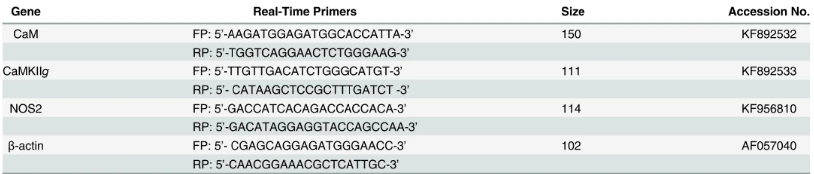
We were interested to determine the nature of cell death induced byM.fortuitum. Terminal deoxynucleotidyl transferase (TdT) dUTP nick-End labeling (TUNEL) assay has been designed to detect apoptotic cells that undergo extensive DNA degradation. We observed significant TUNEL positive HKM (45.23% ± 1.5, p<0.05) due to infection withM.fortuitumcompared to the uninfected control cells 5.01% ± 2.1 (Fig 1A) which suggested thatM.fortuituminfection lead to the macrophage apoptosis. Significant number of TUNEL positive HKM (53.55 ± 2.1; p<0.05) were noted following treatment with STS (positive control) for 6 h.
In parallel study HKM were collected 24 h p.i., stained with AV/PI and analysed by fluores-cence microscopy (Fig 2BandS1 Fig). We observed significant number of AV+PI-M. fortui-tum-infected HKM (34.33% ± 1.37 p<0.05) compared to the uninfected control HKM (6.11% ± 1.91) which further emphasised apoptosis as the outcome ofM.fortuitum-induced pathoge-nicity in HKM. STS (positive control) treated HKM for 6 h showed significant presence of AV+PI-cells (40.03% ± 1.6, p<0.05).
Fig 1.M.fortuituminduces caspase dependent HKM apoptosis.(A) TUNEL assay of uninfected, STS treated (positive control),M.fortuitum-infected with or without Z-VAD-FMK pre-treated HKM. The HKM were observed under confocal microscope (×40). (B) TEM analysis of uninfected andM. fortuitum-infected HKM. All the experiments were performed 24 h p.i. STS treated HKM were analysed 6 h post incubation. The images are representative of three independent experiments. N, nucleus; M, mitochondrion; FN, fragmented nucleus; B, bacteria; AB, apoptotic bodies. HKM, control head kidney macrophage; HKM+STS, HKM treated with STS; HKM+MF, HKM infected withM.fortuitum; HKM+MF+Z-VAD-FMK, HKM pre-treated with Z-VAD-FMK followed byM. fortuituminfection.
Finally, we used TEM to studyM.fortuitum-induced ultra-structural changes. TEM demon-strated distinct morphological changes that included pseudopodial retraction, plasma mem-brane blebbing, nuclear fragmentation, disintegration of mitochondria, condensation of chromatin and vacuolization, characteristic of apoptosis. Control HKM exhibited extended pseudopodia, regular nucleus with diffused chromatin (Fig 1B). Collectively, our results con-firmed that HKM-cytopathy induced byM.fortuitumis apoptotic in nature.
Macrophage apoptosis induced by mycobacterial pathogens has been observed to be both caspase-dependent and -independent. To address this, we used the pan caspase inhibitor Z-VAD-FMK and observed that pre-treatment with the inhibitor significantly reducedM. for-tuitum-induced HKM apoptosis (Figs1A,2A and 2B) confirming the role of caspase inM. for-tuitumpathogenesis. The next step was to assay executioner caspase activation and we noted significant increase in caspase-3 activity inM.fortuitum-infected HKM (Fig 2C). Pre-treatment with the caspase-3 inhibitor Z-DEVD-FMK resulted in significant inhibition in caspase-3 activity (Fig 2C), with concomitant reduction in HKM apoptosis (Fig 2A and 2B). Treatment with STS (positive control) induced significant caspase-3 activity in HKM (Fig 2C).
The role of caspase inhibitors on intracellular growth ofM.fortuitumwas studied by dilution plating of HKM pre-treated with or without Z-VAD-FMK and Z-DEVD-FMK. We observed that the intracellular survival ofM.fortuitumwas significantly improved in presence of both Z-VAD-FMK and Z-DEVD-FMK (S2 Fig). The caspase inhibitors did not influence bacterial growthper sewhen added to 7H9 broth or complete-RPMI (data not shown). Together, these observations confirmed thatM.fortuitum-induced HKM apoptosis is caspase-dependent.
M.
fortuitum
induces intracellular Ca
+2-surge in HKM
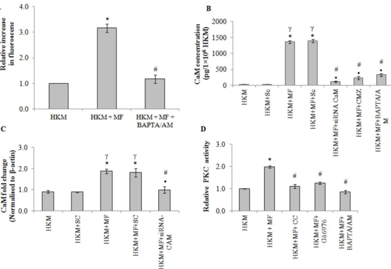
The role of Ca+2has been implicated in the pathogenicity of mycobacterial infections. We were interested to studyM.fortuitum-induced alterations in cytosolic Ca+2levels and determine the role of altered Ca+2homeostasis on HKM apoptosis. As a first step, Fluo-4 was used to study
M.fortuitum-induced alterations in cytosolic Ca+2levels. It was observed thatM.fortuitum -infection led to gradual increase in Ca+2levels as early as 15 mins of infection reaching its peak by 60 min; thereafter the levels gradually declined reaching basal levels 2 h p.i. (S3 Fig). Pre-treatment with BAPTA/AM down regulated the Ca+2surge (Fig 3A), inhibited caspase-3 acti-vation (Fig 2C) and attenuated HKM apoptosis (Fig 2A and 2B). In continuation to this, we noted that pre-treatment with BAPTA/AM enhanced intra-cellular bacterial survival (S2 Fig) Together these results highlighted the critical role of intracellular Ca+2onM.fortuitum -induced HKM apoptosis.
Cytosolic Ca
+2elevation lead to CaM over-expression
On establishing the key role of Ca+2we set out to identify the Ca+2-dependent pathways acti-vated consequent toM.fortuituminfection. Calmodulin (CaM) being an evolutionary
Fig 2. The involvement of pro-apoptotic intermediates inM.fortuituminfection.HKM were pre-treated separately with indicated inhibitors or
transfected with specific siRNAs prior toM.fortuituminfection and at 24 h p.i. apoptosis measured by (A) Hoechst 33342 staining (B) AV/PI staining and (C) relative caspase-3 activity. STS treated HKM were analysed 6 h post incubation. Vertical bars represent mean±SE (n = 3).*P<0.05, compared to HKM;
γP<
0.05, compared to HKM+Sc;#P<0.05, compared to HKM+MF;•P<0.05, compared to HKM+MF+Sc. HKM, control head kidney macrophage; HKM+STS,
HKM treated with STS (positive control); HKM+Sc, HKM transfected with scrambled siRNA; HKM+MF, HKM infected withM.fortuitum; HKM+MF+Sc, HKM transfected with scrambled siRNA infected withM.fortuitum; HKM+MF+siRNA-CaM, HKM transfected with siRNA-CaM infected withM.fortuitum; HKM+MF +siRNA-CaMKIIg, HKM transfected with siRNA-CaMKIIginfected withM.fortuitum; HKM+MF+siRNA-NOS2, HKM transfected with siRNA-NOS2 infected withM.fortuitum; HKM+MF+BAPTA/AM, HKM+MF+CMZ, HKM+MF+CC, HKM+MF+Gö6976, HKM+MF+APO, HKM+MF+DPI, HKM+MF+KN-93, HKM +MF+KN-92, HKM+MF+GW5074, HKM+MF+PD98059, HKM+MF+U0126, HKM+MF+LNAME, HKM+MF+AMG, HKM+MF+Z-VAD-FMK, HKM+MF +Z-IETD-FMK, HKM+MF+Z-DEVD-FMK, HKM pre-treated with BAPTA/AM, CMZ, CC, Gö6976, APO, DPI, KN-93, KN-92, GW5074, PD98059, U0126, LNAME, AMG, Z-VAD-FMK, Z-IETD-FMK or Z-DEVD-FMK respectively and infected withM.fortuitum.
conserved Ca+2sensor was our prime target.M.fortuituminfection led to significant increase in CaM mRNA expression with maximum levels recorded 4 h p.i. (S3 Fig) thereafter, gradually declined reaching the basal levels by 24 h p.i. Maximum CaM protein was also detected 4 h p.i.
(S3 Fig). Hence, we selected 4 h p.i. for subsequent studies on CaM. We measured CaM level in
presence of CMZ and BAPTA/AM. Pre-treatment with CMZ inhibited CaM expression (Fig 3B) implicating the role of CaM onM.fortuitum-induced HKM pathology. The inhibitory role of BAPTA/AM (Fig 3B) further proved CaM expression due to modulation of Ca+2levels in the infected HKM.
The next step in this direction was to correlate CaM expression with HKM apoptosis. We observed that pre-treatment with CMZ attenuated caspase-3 activity (Fig 2C) and inhibited HKM apoptosis (Fig 2A and 2B) suggesting the role of CaM onM.fortuitumpathogenesis. Finally, we used CaM-siRNA to validate these results and observed that transfection with
Fig 3.M.fortuitumalters intracellular Ca+2homeostasis leading to activation of CaM and PKC-α.(A) HKM were pre-treated with or without BAPTA/AM followed byM.fortuituminfection and cytosolic Ca+2elevation was measured using Fluo-4 at 1 h of infection. (B) HKM transfected with indicated siRNA or pre-treated separately with indicated inhibitors and at 4 h p.i. CaM-protein expression detected by EIA. (C) Fold changes in CaM-mRNA expression was determined in HKM transfected with specific siRNA or non-targeted siRNA followed byM.fortuituminfection 4 h p.i by qPCR. (D) HKM were pre-treated separately with or without indicated inhibitors and PKC activity determined at 2 h p.i. Vertical bars represent mean±SE (n = 3).*P<0.05, compared to HKM;
γP<
0.05, compared to HKM+Sc;#P<0.05, compared to HKM+MF;•P<0.05, compared to HKM+MF+Sc. HKM, control head kidney macrophage; HKM+Sc,
HKM transfected with scrambled siRNA; HKM+MF, HKM infected withM.fortuitum; HKM+MF+Sc, HKM transfected with scrambled siRNA infected withM. fortuitum; HKM+MF+siRNA-CaM, HKM transfected with siRNA-CaM infected withM.fortuitum; HKM+MF+BAPTA/AM, HKM+MF+CMZ, HKM+MF+CC, HKM+MF+Gö6976, HKM were pre treated with BAPTA/AM, CMZ, CC, Gö6979 respectively followed by infection withM.fortuitum.
CaM-siRNA down-regulated CaM expression at mRNA (Fig 3C) and protein levels (Fig 3B) and attenuatedM.fortuitum-induced HKM apoptosis (Fig 2A and 2B). Collectively, our results clearly establish the role of CaM in the cascade of events leading toM.fortuitum-induced HKM apoptosis.
Activation of PKC
α
is critical for
M.
fortuitum
pathogenesis
PKC is an important intermediate in Ca+2-signalling and implicated in apoptosis. Infection withM.fortuituminduced significant PKC activation with peak activity recorded 2 h p.i. (S3 Fig). Hence, this time interval was selected for subsequent PKC studies. The HKM were pre-treated with the pan-PKC inhibitor, CC and checked for enzyme activity and apoptosis respec-tively. The inhibitor binds with the catalytic domain of PKC and affects the translocation of the kinase from cytosol to plasma membrane [35]. CC down-regulated PKC activation (Fig 3D) and inhibited HKM apoptosis (Fig 2A and 2B) implicating PKC as pro-apoptotic molecule in
M.fortuitumpathology. BAPTA/AM pre-treatment also down-regulated PKC activity (Fig 3D) confirming the involvement of Ca+2-dependent PKC inM.fortuitum-infected HKM.
Amongst the Ca+2-dependent PKC sub-family the role of PKCαhas been reported in myco-bacterial pathogenesis [10,36,37]. In this direction, the HKM were pre-treated with the PKCα
inhibitor, Gö6976 and apoptosis assayed 24 h p.i. Gö6976 is a selective inhibitor of PKCα, though the mechanisms are not well understood [10,11,36]. It was observed that pre-treat-ment with Gö6976 down-regulated PKC activity (Fig 3D) and HKM apoptosis (Fig 2A and 2B). Based on these results we conclude that PKCαplays an important role in the pathogenicity ofM.fortuitum.
PKC
α
promotes superoxide production in
M.
fortuitum-infected HKM
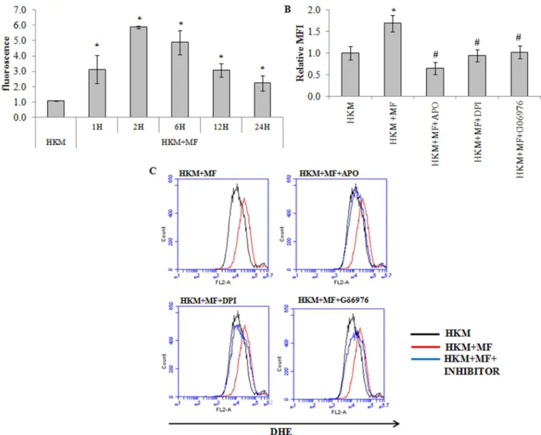
Reactive oxygen species (ROS) play a crucial role in host immunity to mycobacterial patho-gens. However, the role of ROS in the pathogenesis ofM.fortuitumis poorly understood. We used specific dye DHE and noted significant superoxide anion production in theM.fortuitum -infected HKM with maximum levels recorded 2 h p.i. though the levels remained significantly elevated till 24 h p.i. (Fig 4A). To correlate superoxide generation withM.fortuitum pathogen-esis the HKM were pre-treated with antioxidants APO and DPI. Pre-treatment with APO and DPI down-regulatedM.fortuitum-induced superoxide anion production (Fig 4B and 4C) and attenuated caspase-3 mediated apoptosis (Fig 2A, 2B and 2C) implicating the role of superox-ide to the pathogenesis induced by the bacterium.
The next step was identifying the upstream events that lead to superoxide anion generation. PKCαdependent ROS production through the activation of NADPH Oxidase is reported ear-lier [11]. We hypothesised a similar role of PKCαin our study. To that end the HKM were pre-treated with Gö6976 andM.fortuitum-induced superoxide anion production studied. We observed that superoxide anion production was significantly reduced in presence of Gö6976
(Fig 4B and 4C) clearly indicating the pro-active role of PKCαon superoxide anion generation
inM.fortuitum-infected HKM.
CaM and superoxide converge to activate CaMKIIg
in
M.
fortuitum
infected HKM
conferred significant protection to the infected HKM from undergoing apoptotic death (Fig
2A, 2B and 2C). Expectedly, HKM apoptosis could not be prevented in presence of KN-92, the
inactive analogue of KN-93 suggesting a prime role of CaMKII on mediatingM.fortuitum -induced HKM apoptosis.
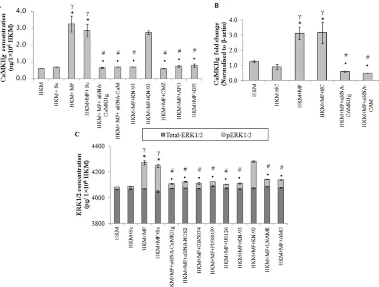
Among the several CaMKII isoforms, CaMKIIglargely modulates macrophage functioning. The CaMKIIgexpression was checked by qPCR and results suggested maximum CaMKIIg -mRNA expression 4 h p.i.; thereafter the levels though declined were significant till 24 h p.i. (S4 Fig). The next step was measuring CaMKIIglevel 24 h p.i. by EIA. We selected this time point because significant CaMKIIgmRNA expression was observed even at 24 h p.i. Significant CaMKIIgexpression was observed inM.fortuitum-infected HKM which was inhibited in
Fig 4.M.fortuituminduces PKCα-mediated superoxide generation.(A) HKM were infected with or withoutM.fortuitumand at indicated time p.i. superoxide generation measured using DHE. HKM were pre-treated separately with or without indicated inhibitors followed byM.fortuituminfection and superoxide generation measured 2 h p.i. by FACS using DHE (B) The relative median fluorescence intensity (MFI) and (C) Histogram. Vertical bars represent mean±SE (n = 3).*P<0.05, compared to HKM;#P<0.05, compared to HKM+MF. HKM, control head kidney macrophage; HKM+MF, HKM infected withM.
fortuitum;HKM+MF+APO, HKM+MF+DPI, HKM+MF+Gö6976, HKM were pre treated with APO, DPI, and Gö6979 respectively followed by infection withM. fortuitum.
presence of KN-93 but not by KN-92 (Fig 5A). Pre-treatment with CMZ also suppressed CaM-KIIglevel (Fig 5A).
Gene silencing was used to further validate our observations. We observed that transfection with CaM-and CaMKIIg-siRNA down-regulated CaMKIIgexpression at mRNA (Fig 5B), pro-tein level (Fig 5A) and attenuatedM.fortuitum-induced HKM apoptosis (Fig 2A and 2B). Based on these observations we conclude that signalling via Ca+2-CaM-CaMKIIgpathway is instrumental for initiatingM.fortuitum-induced apoptotic cascade in HKM.
Fig 5. CaMKIIginduces activation of pro-apoptotic ERK1/2 inM.fortuitumpathogenesis.HKM were transfected or pre-treated with or without indicated siRNAs or inhibitors respectively and CaMKIIgexpression determined by (A) EIA at 24 h p.i. and (B) qPCR at 4 h p.i. (C) HKM were transfected or pre-treated separately with or without indicated siRNAs or inhibitors respectively and at 24 h p.i. checked for total-ERK1/2 and pERK1/2 levels. Vertical bars represent mean±SE (n = 3).*P<0.05, compared to HKM;γ
P<0.05, compared to HKM+Sc;#P<0.05, compared to HKM+MF;•P<0.05, compared to HKM+MF+Sc.
HKM, control head kidney macrophage; HKM+Sc, HKM transfected with scrambled siRNA; HKM+MF, HKM infected withM.fortuitum; HKM+MF+Sc, HKM transfected with scrambled siRNA infected withM.fortuitum; HKM+MF+siRNA-CaM, HKM transfected with siRNA-CaM infected withM.fortuitum; HKM+MF +siRNA-CaMKIIg, HKM transfected with siRNA-CaMKIIginfected withM.fortuitum; HKM+MF+NOS2, HKM transfected with siRNA-NOS2 infected withM. fortuitum; HKM+MF+CMZ, HKM+MF+KN-93, HKM+MF+KN-92, HKM+MF+APO, HKM+MF+DPI, HKM+MF+GW5074, HKM+MF+PD98059, HKM+MF +U0126, HKM+MF+L-NAME, HKM+MF+AMG, HKM were pre treated with CMZ, KN-93, KN-92, APO, DPI, GW5074, PD98059, U0126, LNAME or AMG respectively followed by infection withM.fortuitum.
The NADPH Oxidase pathway has been reported to drive CaMKII activation following decline in Ca+2-CaM signals [38]. Our results with APO and DPI encouraged us to investigate this and we noted that CaMKIIgactivity was significantly inhibited in presence of APO and DPI (Fig 5A) suggesting the role of NADPH Oxidase/superoxide pathway on CaMKIIg activa-tion inM.fortuitum-infected HKM.
ERK1/2 is pro-apoptotic and downstream to CaMKIIg
ERK1/2 activation in mycobacterial pathogenesis is well established [14]. The activation of ERK1/2 depends on upstream Raf-1-MEK1/2 signalling. Hence, the HKM were infected with
M.fortuitumin the presence of the pharmacological inhibitors GW5074, PD98059 and U0126 respectively and the changes in total-ERK1/2 and phosphorylated ERK1/2 (pERK1/2) levels measured 24 h p.i. (Fig 5C). GW5074 is a potent and selective Raf-1 inhibitor with no effect on the activity of MKK/MEK signalling. PD98059 is a non-ATP competitive inhibitor that binds to MKK/MEK1, preventing its activation by upstream protein kinases, such as Raf [39]; it does not directly inhibit ERK1/2. U0126, also a non-ATP competitive inhibitor prevents MEK from phosphorylating downstream ERK1/2 thereby inhibiting ERK1/2 functioning [8,39].M. for-tuituminduced significant increase in pERK1/2 levels which was down-regulated in presence of GW5074, PD98059 and U0126 (Fig 5C). No noticeable change in total-ERK1/2 level was observed in the infected HKM.
The role of CaMKII on the activation of ERK1/2 has been reported in mycobacterial infec-tion [7]. To address this HKM were pre-treated with KN-93 and KN-92, or transfected with CaMKIIg-siRNA andM.fortuitum-induced changes in total-ERK1/2 and pERK1/2 levels stud-ied 24 h p.i. To our expectation, KN-93 and CaMKIIg-siRNA inhibitedM.fortuitum-induced pERK1/2 expression but did not influence the total-ERK1/2 levels in infected or control HKM
(Fig 5C).
Finally, the implication of ERK1/2 activation onM.fortuitum-infected HKM was studied. We observed that HKM apoptosis was attenuated in presence of GW5074, PD98059 and U0126 (Fig 2A and 2B). Based on these results we suggest that ERK1/2 is pro-apoptotic and downstream to CaMKIIginM.fortuituminfections.
ERK1/2 and NOS2-NO crosstalk determines the fate of
M.
fortuitum
infected HKM
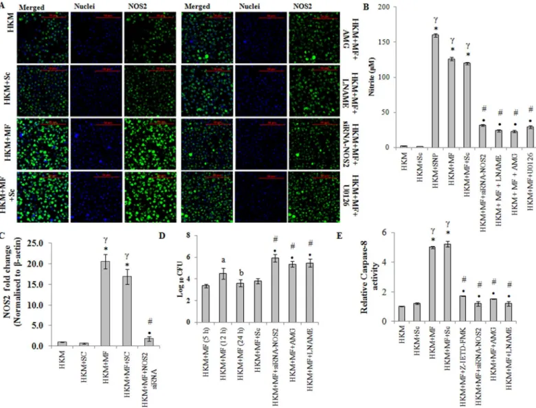
Nitric oxide (NO) production in macrophagesviaNOS2 pathway is an important host defence mechanism against mycobacterial pathogens [17,18]. However, the role of NO onM. fortui-tum-induced pathogenicity is poorly understood. We observed significant NO production in HKM infected withM.fortuitumwith maximum levels recorded 24 h p.i. (S4 Fig). We designed degenerate primers (Table 1) for NOS2 using the homologous stretch across verte-brates as the template and the PCR product was cloned and sequenced. The sequence showed 90% similarity with NOS2-mRNA of channel catfish,Ictalurus punctatus. Based on the sequence (Accession no. KF956810,Table 2) primers for qPCR studies were designed (Table 3) and NOS2-mRNA expression quantified. We observed maximum NOS2-mRNA expression at 12 h p.i. (S4 Fig) which declined at 24 h p.i. but was still significant in the infected HKM. The next step was to confirm NOS2 protein expression by immunofluroscence. As maximum NO production coupled with significant NOS2-mRNA expression was observed 24 h p.i. it was selected for subsequent studies. Our results clearly suggest increased NOS2 protein expression inM.fortuituminfected HKM (Fig 6AandS5 Fig).
NOS2 activation. We observed that pre-treatment with AMG and LNAME (i) inhibited NOS2 expression (Fig 6AandS5 Fig), (ii) NO production (Fig 6B) and (iii) down-regulated caspase-3 activity and attenuatedM.fortuitum-induced HKM apoptosis (Fig 2A, 2B and 2C). Sodium nitroprusside used as the positive control induced significant NO production in uninfected HKM. Together, these results suggested that induction of apoptosis is linked to NO production inM.fortuitum-infected HKM.
Fig 6. NOS2-NO axis induces caspase-8 activation inM.fortuituminfected HKM.(A) HKM were transfected or pre-treated separately with or without specific siRNA and indicated inhibitors respectively and at 24 h p.i. NOS2 protein (FITC conjugated) expression studied by confocal microscopy (×40). The images are representative of three independent experiments. (B) HKM were transfected or pre-treated separately with or without indicated siRNA or inhibitors respectively and at 24 h p.i. NO release measured. HKM treated with SNP was taken as positive control for the assay. (C) Fold change in NOS2 mRNA expression was determined in HKM transfected with specific siRNA or non targeted siRNA followed byM.fortuituminfection 12 h p.i. (D) Intracellular bacterial load was determined in HKM infected withM.fortuitumat indicated time p.i. or HKM transfected or pre-treated with indicated siRNA or inhibitors respectively 24 h p.i. by dilution plating on 7H11 agar plate. (E) Relative caspase-8 activity measured at 24 h p.i. in HKM pre-treated or transfected with indicated inhibitors or siRNA respectively. Vertical bars represent mean±SE (n = 3).*P<0.05, compared to HKM;γP<0.05, compared to HKM+Sc;#P<0.05,
compared to HKM+MF;•P<0.05, compared to HKM+MF+Sc;aP<0.05, compared to HKM+MF (5 h);bP<0.05, compared to HKM+MF (12 h). HKM, control
head kidney macrophage; HKM+Sc, HKM transfected with scrambled siRNA; HKM+MF, HKM infected withM.fortuitum; HKM+MF+Sc, HKM transfected with scrambled siRNA infected withM.fortuitum; HKM+MF+siRNA-NOS2, HKM transfected with siRNA-NOS2 infected withM.fortuitum; HKM+MF+AMG, HKM+MF+LNAME, HKM+MF+U0126, HKM+MF+Z-IETD-FMK, HKM were pre-treated with AMG, LNAME, U0126 or Z-IETD-FMK, respectively followed by M.fortuituminfection.
To further confirm this we used gene silencing approach. We noted that transfection with NOS2-siRNA down regulated NOS2 mRNA expression (Fig 6C), NOS2 protein expression
(Fig 6AandS5 Fig), NO production (Fig 6B) and caspase-3 mediated HKM apoptosis (Fig 2A,
2B and 2C) which clearly established the importance of NOS2 dependent NO generation inM.
fortuitum-induced HKM apoptosis.
Our next aim was studying the effect of NO on bacterial growth. The HKM were transfected or pre-treated separately with NOS2-siRNA or AMG and LNAME respectively and 24 h p.i. lysed, serially diluted and plated on 7H11 agar for enumerating CFU. It was noted that the sur-vival of intra-cellularM.fortuitumwas significantly improved by inactivating the NOS2-NO axis (Fig 6D) which confirmed the bactericidal role of NOS2-NO onM.fortuitum. The addi-tion of AMG and LNAME in the culture medium did not influence bacterial growthper se
(data not shown).
Once the importance of NOS2-NO axis onM.fortuitum-induced pathogenesis was estab-lished we looked for the upstream signals influencing NO release. ERK1/2 has been implicated on the transcriptional activation of NOS2 in mycobacterial pathogenesis [16]. There are also reports documenting the involvement of NO on the activation of ERK1/2 [40]. Hence, the HKM were pre-treated with U0126 to study NOS2 expression and NO production as well the impact of attenuating NOS2-NO signalling on ERK1/2 activation probed. We observed that pre-treatment with U0126 attenuated NOS2 expression (Fig 6A) and inhibited NO production
(Fig 6B). Reversibly, attenuating the NOS2-NO signalling by AMG, LNAME or NOS2-siRNA
inhibited the activation of ERK1/2 (Fig 5C). These results suggests a cross-talk between ERK1/ 2 and NOS2-NO axis inM.fortuitum-infected HKM.
NO activates caspase-8 in
M.
fortuitum
infected HKM
We sought to identify the apical caspases instigating the death process. Significant increase in caspase-8 activity was observed and pre-treatment with the caspase-8 inhibitor, Z-IETD-FMK attenuated its activity (Fig 6E) and reduced HKM apoptosis (Fig 2A and 2B). Concomitantly, Z-IETD-FMK also inhibited caspase-3 activity (Fig 2C) in the infected HKM. Based on these results we suggest the role of caspase-8 as initiator caspase inM.fortuitum-induced HKM apo-ptosis. Furthermore, transfection with NOS2-siRNA or pre-treatment with AMG or LNAME inhibited caspase-8 activation (Fig 6E). We conclude that NO induces it apoptotic influence by activating caspase-8 inM.fortuitum-infected HKM which activated caspase-3 to accomplish the apoptotic cascade.
Discussion
There is little information on pathogenesis by atypical mycobacteria likeM.fortuitum. Our results for the first time elucidate the hierarchy of pro-active signalling molecules inM. fortui-tumpathogenesis.
We observed thatM.fortuituminduces HKM apoptosis as evident by nuclear morphology, extra-membranous presence of phosphatidyl serine, host cell DNA fragmentation, develop-ment of cytoplasmic vacuoles and apoptotic bodies. Macrophagic death has been observed in presence of several intracellular pathogens including mycobacteria although the mechanisms and implications remain unexplained.M.fortuitum-induced apoptosis of human and murine macrophages has been reported earlier [41]. Our study for the first time reports the induction of similar pathological consequences in fish suggesting the pro-apoptotic trait to be a conserved virulence factor ofM.fortuitumto counteract immune responses in different hosts.
pro- and anti-apoptotic effects on macrophages [4,43]. From our results the pro-apoptotic involvement of Ca+2inM.fortuituminfection was distinct. This is the first evidence linking increase in intra-cellular Ca+2levels toM.fortuitum-induced macrophage apoptosis. The increase of Ca+2levels are‘deciphered’by various intracellular Ca+2binding proteins that con-vert the signals into a wide variety of biochemical changes. Among the several downstream Ca+2-binding proteins through which Ca+2induces its varied effects CaM is important. It is one of the most conserved proteins and has also been reported from fish [8]. We found the sig-nificant role of CaM onM.fortuitumpathogenesis. Calmodulin showed parallel expression at transcription and translational level. This kind of observation has been noted with other earlier signalling molecules [44]. A similar trend of CaM expression has recently been reported from our laboratory inA.hydrophila-infected HKM [8]. Interestingly, we observed significant decline in CaM protein levels in presence of Ca+2chelator BAPTA/AM as well as CMZ. How BAPTA/AM and CMZ influences CaM expression is not very clear to us. Earlier studies sug-gested the pro-active role of Ca+2as second messenger in regulating the transcription of several proteins including CaM in plant [45] and mammalian cells [46]. Our results with BAPATA/ AM, extends these observations suggesting a regulatory role of Ca+2on the expression of CaM and CaM-dependent kinases in fish HKM. Regarding the role of CMZ on CaM expression there could be two possibilities. 1. CMZ regulates the cytosolic Ca+2levels [47] thereby affect-ing the transcription of CaM. 2. It may contribute to off target actions that suppress CaM pro-tein expression or stability. We propose that the increase in intracellular Ca+2levels induced by
M.fortuitumacts at two distinct levels. Firstly, at the molecular level it influences transcription and the mode of association of CaM with various downstream target proteins and secondly induces conformational states in CaM leading to target-specific activation and concomitant release of free energy critical for the transduction of Ca+2signals inM.fortuituminfection.
There are several downstream targets for CaM amongst which the involvement of CaMKII is fairly well reported. CaMKII exists in several isoforms and their involvement in macrophage apoptosis is not well reported. We have used CaMKIIgisoform for this study since it largely modulates macrophage functioning [8,48]. Our findings clearly established the pro-apoptotic role of CaMKIIgonM.fortuitum-pathogenesis. An interesting observation was the decrease in CaMKIIgprotein levels in presence of CMZ and KN-93 in the infected HKM. It is well known that activated-CaM can influence the transcription of several genes either directly or by modu-lating the activity of CaM-binding transcription factors (CAMTA) [45]. We hypothesize that the binding of CMZ compromises the transcriptional activity of CaM thereby affecting genes involved in the Ca+2-cascade like CaMKII. The fluctuations in cytosolic Ca+2levels also influ-ence CaMKII transcription [46]. CMZ and KN-93, besides inhibiting intra-cellular Ca+2levels also interferes with cyclic nucleotide metabolism and G protein mediated signalling important for regulating the transcriptional activity of CaMKII [47,49]. Increased CaMKII expression was reported earlier inM.bovis[50] andM.smegmatis[7], and our results extend this toM.
fortuitumsuggesting CaMKIIgas a virulence factor in host though the mechanisms remain to be established. In this context identifying the role of other CaMKII isoforms inM.fortuitum -pathogenesis would be interesting.
Members of the PKC family have been implicated in mycobacterial pathogenesis but their involvement duringM.fortuituminfections remains to be addressed. Our data clearly sug-gested the pro-apoptotic role of PKCαonM.fortuitum-pathology. Earlier studies suggested PKCαto be crucial forM.bovisBCG [36] andM.tuberculosis[37]. Thus, our results confirm earlier observations reporting PKCαmodulation to be a conserved strategy of mycobacterial pathogens to induce pathological changes.
mycobacteria-induced ROS generation to be pro-apoptotic hence a critical host resistance determinant [12]. It has also been proposed that mycobacteria have evolved mechanism to impair or detoxify ROS to facilitate intracellular survival [51]. The NADPH oxidase complex is well characterised and correlated with superoxide generation in fish macrophages against mycobacterial pathogens [52]. We observed increased superoxide generation consequent toM.
fortuituminfection and pre-treatment with APO and DPI significantly reduced superoxide generation and HKM apoptosis. These results are in line with earlier studies reporting the role ofM.fortuitum-induced robust ROS generation in mammalian macrophage apoptosis [53]. The role of PKC on ROS generation through the activation of NADPH Oxidase has been dem-onstrated under various conditions [11]. Only recently, the involvement of different PKC iso-forms on ROS generation is becoming apparent. We observed that inhibiting PKCα
suppressed superoxide generation implicating its role on ROS generation inM.fortuitum
infection. We postulate PKCαmight be involved in the phosphorylation and membrane trans-location of the enzyme components [54].
An unexpected and intriguing observation we made is that significant CaMKIIgactivity was recorded at late hours of infection when both Ca+2and CaM levels had declined significantly. Coupled to that we noted sustained ROS generation inM.fortuituminfected HKM. Each CaM-KII monomer consists of an N-terminal catalytic domain a C-terminal association domain that enables assembly of the holoenzyme and a regulatory domain between the catalytic and associ-ation domains. Under conditions of brief increases in intracellular Ca+2levels CaMKII returns to an inactive conformation after Ca+2/CaM unbinding. Only recently it was observed that ROS can prolong CaMKII activation in absence of Ca+2/CaM signalingviaoxidation of methi-onine residues present in the regulatory subunit [38]. There are no reports on this alternate mechanism of CaMKII activation in microbe-induced pathogenesis. We observed that ROS induced via PKCαcontributes to CaMKIIgactivation in the HKM in absence of Ca+2-CaM sig-nalling. Our results support the mechanism by which CaMKII can integrate Ca+2and ROS sig-nals [38]. We propose that the Ca+2/CaM binding serves as the initial trigger for CaMKII activation inM.fortuitumpathogenesis. Subsequently, the pro-oxidant rich conditions in the infected HKM induce the oxidation of methionine residues in CaMKII which resets the Ca+2 sensitivity of CaMKII so that very low levels of intracellular Ca+2are sufficient for prolonging the kinase activity. The constutitively active Ca+2/CaM autonomous CaMKII promotes core events important for initiating the apoptotic cascade inM.fortuituminfected HKM. It is important to note that oxidized CaMKII is reduced and inactivated by methionine sulfoxide reductase A (MsrA) [5]. We believe that followingM.fortuituminfections the balance between oxidised CaMKII and Met-reduced CaMKII is lost in the HKM. Thus, understanding of the ox-CaMKII/MsrA signaling pathway would provide new insights into how Ca+2 /CaM-ROS-CaMKII axis causes apoptosis ofM.fortuituminfected HKM.
The activation of ERK1/2 plays a critical role in microbial pathogenesis. However, there is little evidence suggesting ERK1/2 activation byM.fortuitum. The role of ERK1/2 as pro-apo-ptotic molecule has been demonstrated againstM.avium[15] andM.bovisBCG [6]. Based on our results and earlier reports we suggest that ERK1/2 activation is an important virulence mechanism inM.fortuitumpathogenesis. Earlier studies have also indicated the role of CaM-KII on activating the ERK1/2 pathway [7]. We observed that inhibiting CaMKIIg down-regu-lated ERK1/2 activation which confirmed that ERK1/2 is downstream to Ca+2-CaM-CaMKIIg
pathway and its presence was reported in fish [56]. For a direct proof we assayed ERK1/2 in presence of the Raf-1 inhibitor, GW5074, and our results proved that Raf-1 plays the role of upstream regulator in CaMKIIginduced ERK1/2 activation. Thus we conclude that CaMKIIg
triggered Raf-1-MEK1/2-ERK1/2 signalling duringM.fortuituminfection.
NOS2-induced NO plays an important role in host defence against microbial pathogens [17,18]. It appears that NO act as pro-and anti-apoptotic factor depending upon experimental conditions [57]. Using a combination of specific inhibitors and specific siRNA we report that NO helps in the containment of the intra-cellular bacteria and acts as pro-apoptotic molecule inM.fortuitumpathology. Our findings contradict earlier studies reportingM.fortuitumdoes not produce NO in mammalian macrophages [18]. The ability of mycobacteria to survive and induce pathology varies among different host species [52,58]. Fish is the natural host forM.
fortuitumwhich could be the reason behind the observed differences with mammalian macrophages.
The role of NO on piscine mycobacteriosis has not been studied in details. In context of the
M.marinum-goldfish and -zebrafish models a decline in NO production was reported which aided in the spread and persistence of the bacterium [52,58]. Looking at these contradictory results it seems likely that the different fish pathogenic mycobacteria alike the mammalian counterparts have evolved multiple mechanisms for countering macrophagic responses in dif-ferent hosts.
Signalling through MAPK pathway influences NO production during mycobacterial infec-tions [16]. We asked whether ERK1/2 has a role on NO production inM.fortuituminfected HKM. Our inhibitor studies clearly demonstratedM.fortuituminduced NO production is linked to MEK1/2-ERK1/2 signalling. These results are in line with earlier studies suggesting a role of ERK1/2 on inducing NO release in macrophages infected with mycobacterial pathogens [16]. It has been proposed that ERK1/2 could modulate NOS2viarelease of pro-inflammatory cytokines like TNF-α[59] with NFκB playing an intermediatory role in the process [60,61]. Further studies are needed to identify the role of other macrophage soluble mediators like TNF-α, IL-1βas well as NFκB and co-relate them with NO-mediated HKM apoptosis in our model. There are several recent reports implicating NO in the activation of ERK1/2 with patho-logical consequences [40]. To the best of our knowledge this has not been reported during host-mycobacterial interactions. This prompted us to examine the interaction between NO and ERK1/2 inM.fortuitum-infected HKM. We noted that inhibiting NO production led to down-regulation of ERK1/2 activation. It has been reported that NO activates ERK1/2 pathway in cGMP/PKG-dependent as well as Ras-dependent but cGMP- independent [40] manner. These mechanisms may be involved in the activation of ERK1/2 by NO inM.fortuitum -infected HKM. Our results for the first time demonstrated ERK1/2-NO cross-talk in mycobac-terial pathogenesis paving way for understanding of the molecular mechanisms of mycobacter-iosis. It is important to note that ERK1/2 and NO-mediated effects depend on the duration of exposure and their levels in the cells [40]. We propose that a positive feedback mechanism operates between ERK1/2 and NO due to which their levels increase significantly and once it crosses the critical threshold it tilts the balance in favour of apoptosis.
further studies are needed to understand this cross-talk. We extended our study by showing that consequent to NOS2-NO inhibition caspase-3 activation was abrogated. We conclude that caspase-8 acts as conduit between NO and caspase-3 inM.fortuitum-infected HKM. Based on our observations we suggestM.fortuituminduces HKM apoptosis through a caspase 8-depen-dent pathway and NO promotes apoptosis by amplifying caspase-8 and -3 activation. In this context it would be interesting to investigate the involvement of caspase-9 and study the cross-talk between the two initiator caspases duringM.fortuituminfection.
Our study for the first time traces the course of events that lead toM.fortuituminduced macrophage apoptosis in fish (Fig 7). We propose thatM.fortuituminfection alters the intra-cellular Ca+2levels. CaM and the robust superoxide levels induced via-PKCα-NADPH Oxidase converges to prolong CaMKIIgactivations thereby sustaining the levels of ERK1/2 critical for NO release. The cross-talk between ERK1/2 and NO‘places checks and balances’eventually tilting in favour of caspase activation and HKM apoptosis. We are currently identifying which mycobacterial molecule (s) mediate this signalling cascade and if specifically modifying this apoptotic programme changes the course of infectionin vivo.
Supporting Information
S1 Fig.M.fortuituminduces HKM apoptosis. (PDF)
S2 Fig. Inhibition of Ca+2and caspase activity enhanced the intracellular bacterial load. (PDF)
S3 Fig.M.fortuitumincreases cytosolic Ca+2leading to the activation of CaM and PKC. (PDF)
S4 Fig.M.fortuituminduces the over expression of CaMKIIgand NOS2/NO in infected HKM.
(PDF)
S5 Fig.M.fortuituminduces NOS2 activation in infected HKM. (PDF)
Acknowledgments
The authors are grateful to S. Das, North Shore University Health system and T. Mazumder, Georgia Regents University for helpful discussions and critically analyzing the manuscript. The NOS2 antibody was kind gift from B. Ghosh, Institute of Genomics and Integrative Biology. We thank U. Rai, University of Delhi for the help in fluorescence microscopy, P. Arora for flowcytometry work and A. K. Pal for maintenance of fish. This work was supported by NFBSFARA/ICAR Project Grant (RNAi-2014) and University of Delhi Doctoral Research Pro-gramme, Dean (R)/R&D/2012/917. DD, PK, CB and AS were supported by ICAR Fellowship (Govt of India), CSIR Fellowship (Govt of India), DST-INSPIRE Fellowship (Govt of India), UGC Fellowship (Govt of India) respectively. The funding bodies had no role in study design, data collection and analysis, decision to publish, or preparation of the manuscript.
Fig 7. Overview of the work.M.fortuitumalters intracellular Ca+2homeostasis leading to activation of CaM
and PKCα. PKCαinduces superoxide anions generationviaNADPH Oxidase. CaM and superoxide together activates CaMKIIg. In downstream, CaMKIIgmodulates ERK1/2 to activate NOS2-NO axis. The ensuing ERK1/2-NO positive feedback loop leads to caspase-8 mediated caspase-3 activation and HKM apoptosis.
Author Contributions
Conceived and designed the experiments: DD RR SM. Performed the experiments: DD PK AS RM DRS. Analyzed the data: DD PK CB DRS RR SM. Contributed reagents/materials/analysis tools: SM PR AM. Wrote the paper: DD CB SM.
References
1. Shukla S, Sharma R, Shukla SK. Detection and identification of globally distributed mycobacterial fish pathogens in some ornamental fish in India. Folia Microbiol. 2013; 58: 429–436.
2. Smith MB, Schnadig VJ, Boyars MC, Woods GL. Clinical and Pathologic Features ofMycobacterium fortuitumInfections: An Emerging Pathogen in Patients with AIDS. Am J Clin Pathol. 2001; 116: 225–
232. PMID:11488069
3. Kashyap VK, Gupta RK, Shrivastava R, Srivastava BS, Srivastava R, Parai MK, et al.In vivoactivity of thiophene-containing trisubstituted methanes against acute and persistent infection of non-tubercular Mycobacterium fortuitumin a murine infection model. J Antimicrob Chemother. 2012; 67: 1188–1197. doi:10.1093/jac/dkr592PMID:22311937
4. Rojas M, Garcı´a LF, Nigou J, Puzo G, Olivier M. Mannosylated Lipoarabinomannan Antagonizes Mycobacterium tuberculosis-Induced Macrophage Apoptosis by Altering Ca+2-Dependent Cell
Signal-ing. J Infect Dis. 2000; 182: 240–51. PMID:10882603
5. Erickson JR, Joiner ML, Guan X, Kutschke W, Yang J, Oddis CV et al. A dynamic pathway for calcium-independent activation of CaMKII by methionine oxidation. Cell. 2008; 133: 462–474. doi:10.1016/j. cell.2008.02.048PMID:18455987
6. Me´ndez-Samperio P, Miranda ATE. Activation of ERK1/2 and TNF-aproduction are mediated by cal-cium/calmodulin, and PKA signalling pathways duringMycobacterium bovisinfection. J Infect. 2006; 52: 147–153. PMID:16442440
7. Yadav M, Roach SK, Schorey JS. Increased Mitogen-Activated Protein Kinase Activity and TNF-Pro-duction Associated withMycobacterium smegmatisbut NotMycobacterium avium-Infected Macro-phages Requires Prolonged Stimulation of the Calmodulin/Calmodulin Kinase and Cyclic AMP/Protein Kinase A Pathways. J Immunol. 2004; 172: 5588–5597. PMID:15100302
8. Banerjee C, Khatri P, Raman R, Bhatia H, Datta M, Mazumder S. Role of Calmodulin-Calmodulin Kinase II, cAMP/Protein Kinase A and ERK 1/2 onAeromonas hydrophila-Induced Apoptosis of Head Kidney Macrophages. PLoS Pathog. 2014; 10(4): e1004018. doi:10.1371/journal.ppat.1004018
PMID:24763432
9. Steinberg SF. Structural Basis of Protein Kinase C Isoform Function. Physiol Rev. 2008; 88: 1341–
1378. doi:10.1152/physrev.00034.2007PMID:18923184
10. Yadav M, Clark L, Schorey JS. Macrophage’s Proinflammatory Response to a Mycobacterial Infection Is Dependent on Sphingosine Kinase-Mediated Activation of Phosphatidylinositol Phospholipase C, Protein Kinase C, ERK1/2, and Phosphatidylinositol 3-Kinase. J Immunol. 2006; 176: 5494–5503. PMID:16622018
11. Thamilselvan V, Menon M, Thamilselvan S. Oxalate-induced activation of PKC-αandδregulates NADPH oxidase-mediated oxidative injury in renal tubular epithelial cells. Am J Physiol Renal Physio. 2009; l297: F1399–F1410.
12. Yang C, Shin D, Lee H, Son JW, Lee SJ, Akira S, et al. ASK1-p38 MAPK-p47phox activation is essen-tial for inflammatory responses during tuberculosis via TLR2-ROS signalling. Cell Microbiol. 2008; 10: 741–754. PMID:18028450
13. Choi JA, Lim YJ, Cho SN, Lee JH, Jeong JA, Kim EJ, et al. Mycobacterial HBHA induces endoplasmic reticulum stress-mediated apoptosis through the generation of reactive oxygen species and cytosolic Ca2+in murine macrophage RAW 264.7 cells. Cell Death Dis. 2013; 4: e957; doi:10.1038/cddis.2013.
489PMID:24336077
14. Schorey JS, Cooper AM. Macrophage signalling upon mycobacterial infection: the MAP kinases lead the way. Cell Microbiol. 2003; 5: 133–142. PMID:12614457
15. Bhattacharyya A, Pathak S, Kundu M, Basu J. Mitogen-activated protein kinases regulate Mycobacte-rium aviuminduced tumor necrosis factor-αrelease from macrophages. FEMS Immunol Med Mic. 2002; 34: 73–80.
16. Chan ED, Morris KR, Belisle JT, Hill P, Remigio LK, Brennan PJ, et al. Induction of Inducible Nitric Oxide Synthase-NO•by Lipoarabinomannan ofMycobacterium tuberculosisis Mediated by MEK1-ERK, MKK7-JNK, and NF-kB Signaling Pathways. Infect Immun. 2001; 69: 2001–2010. PMID:
17. Chan J, Xing Y, Magliozzo RS, Bloom BR. Killing of VirulentMycobacterium tuberculosisby Reactive Nitrogen Intermediates Produced by Activated Murine Macrophages. J Exp Med. 1992; 175: 1111–
1122. PMID:1552282
18. Rojas M, Barrera LF, Puzo G, Garcia LF. Differential Induction of Apoptosis by VirulentMycobacterium tuberculosisin Resistant and Susceptible Murine Macrophages Role of Nitric Oxide and Mycobacterial Products. J Immunol. 1997; 159: 1352–1361. PMID:9233632
19. Grayfer L, Hodgkinson JW, Belosevic M. Antimicrobial responses of teleost phagocytes and innate immune evasion strategies of intracellular bacteria. Dev Comp Immunol. 2014; 43: 223–242. doi:10. 1016/j.dci.2013.08.003PMID:23954721
20. Du C, Guan Q, Diao H, Yin Z, Jevnikar AM. Nitric oxide induces apoptosis in renal tubular epithelial cells through activation of caspase-8. Am J Physiol Renal Physiol. 2006; 290: F1044–F1054. PMID:
16352744
21. Behar SM, Martin CJ, Booty MG, Nishimura T, Zhao X, Gan H, et al. Apoptosis is an innate defence function of macrophages againstMycobacterium tuberculosis. Mucosal Immunol. 2011; 4: 279–287. doi:10.1038/mi.2011.3PMID:21307848
22. Lee J, Remold HG, Ieong MH, Kornfeld H. Macrophage apoptosis in response to high intracellular bur-den ofMycobacterium tuberculosisis mediated by a novel caspase-independent pathway. J Immunol. 2006; 176: 4267–4274. PMID:16547264
23. O’Sullivan MP, O’Leary S, Kelly DM, Keane J. A Caspase-Independent Pathway Mediates Macro-phage Cell Death in Response toMycobacterium tuberculosisinfection. Infect Immun. 2007; 75: 1984–1993. PMID:17283090
24. Molloy A, Laochumroonvorapong P, Kaplan G. Apoptosis, but Not Necrosis, of Infected Monocytes is Coupled with Killing of Intracellular Bacillus Calmette-Gu6rin. J Exp Med. 1994; 180: 1499–1509. PMID:7931080
25. Arcilaa ML, Sa´nchez MD, Ortiz B, Barrera LF, Garcı´a LF, Rojas M. Activation of apoptosis, but not necrosis, duringMycobacterium tuberculosisinfection correlated with decreased bacterial growth: Role of TNF-a, IL-10, caspases and phospholipase A2. Cell Immunol. 2007; 249: 80–93. PMID:
18160064
26. Davis JM, Ramakrishnan L. The role of the granuloma in expansion and dissemination of early tubercu-lous infection. Cell. 2009; 136:37–49. doi:10.1016/j.cell.2008.11.014PMID:19135887
27. Aguilo JI, Alonso H, Uranga S, Marinova D, Arbués A, de Martino A, et al. ESX-1-induced apoptosis is involved in cell-to-cell spread ofMycobacterium tuberculosis. Cell Microbiol. 2013; 15: 1994–2005. doi:10.1111/cmi.12169PMID:23848406
28. Chen M, Gan H, Remold HG. A mechanism of virulence: virulentMycobacterium tuberculosisstrain H37Rv, but not attenuated H37Ra, causes significant mitochondrial inner membrane disruption in mac-rophages leading to necrosis. J Immunol. 2006; 176: 3707–3716. PMID:16517739
29. Roca FJ, Ramakrishnan L. TNF Dually Mediates Resistance and Susceptibility to Mycobacteria Through Mitochondrial Reactive Oxygen Species. Cell. 2013; 153: 521–534. doi:10.1016/j.cell.2013. 03.022PMID:23582643
30. Majumdar T, Ghosh D, Datta S, Sahoo C, Pal J, Mazumder S. An attenuated plasmid-cured strain of Aeromonas hydrophilaelicits protective immunity inClarias batrachusL. Fish Shellfish Immunol. 2007; 23: 222–230. PMID:17208455
31. Sorensen KK, Sveinbjornsson B, Dalmo RA, Smedsrod B, Bertheussen K. Isolation, cultivation and characterization of head kidney macrophages from Atlantic cod,Gadus morhuaL. J Fish Dis. 1997; 20: 93–107.
32. Banerjee C, Singh A, Das TK, Raman R, Shrivastava A, Mazumder S. Ameliorating ER-stress attenu-atesAeromonas hydrophila-induced mitochondrial dysfunctioning and caspase mediated HKM apopto-sis inClarias batrachus. Sci Rep. 2014; doi:10.1038/srep05820
33. Datta S, Saha DR, Ghosh D, Majumdar T, Bhattacharya S, Mazumder S. Sub-lethal concentration of arsenic interferes with the proliferation of hepatocytes and inducesin vivo apoptosis inClarias batrachus L. Comp Biochem Physiol. 2007; 145: 339–349.
34. Spoida K, Masseck OA, Deneris ES, Herlitze S. Gq/5-HT2c receptor signals activate a local GABAergic inhibitory feedback circuit to modulate serotonergic firing and anxiety in mice. Proc Natl Acad Sci. 2014; 111, 6479–6484. doi:10.1073/pnas.1321576111PMID:24733892
35. Herbert JM, Augereau JM, Gleye J, Maffrand JP. Chelerythrine is a potent and specific inhibitor of pro-tein kinase C. Biochem Biophys Res Commun. 1990; 172: 993–999. PMID:2244923