J of Evolution of Med and Dent Sci/ eISSN- 2278-4802, pISSN- 2278-4748/ Vol. 4/ Issue 27/ Apr 02, 2015 Page 4750
A RARE CASE OF URINARY BLADDER ENDOMETRIOSIS FOLLOWING
CESAREAN SECTION
Shirish Paul Ganta1, S. Venkateshwar Rao2, N. Kiran Raju3, V. N. Narvekar4, Sivaleela5, Ankum Rao6, Devi Prathima7, Srikanth8
HOW TO CITE THIS ARTICLE:
Shirish Paul Ganta, S. Venkateshwar Rao, N. Kiran Raju, V. N. Narvekar, Sivaleela, Ankum Rao, Devi Prathima, Srikanth. A Rare Case of Urinary Bladder Endometriosis Following Cesarean Section . Journal of Evolution of Medical and Dental Sciences 2015; Vol. 4, Issue 27, April 02; Page: 4750-4754,
DOI: 10.14260/jemds/2015/690
ABSTRACT: Endometriosis is a common benign gynecological disease characterized by the presence of ectopic endometrial tissue, outside the uterus. Involvement of the urinary tract however is rare with an occurence of 4%.1 We are reporting a case of endometriosis involving the Right posterior lateral wall of the urinary bladder, following Cesarean section.2
KEYWORDS: Endometriosis, Bladder Urinary, Uterus, Cesarean section.
CASE REPORT: A twenty six years old woman complained of vague dull aching lower abdominal and pelvic pain since five months. She gives a history of dyspareneuria.
Her menstrual history was normal. She had undergone two lower segment Cesarean sections (LSCS) in the past. There was no history of hematuria. The pelvic pain was unrelated to the menstrual cycle. On clinical examination the patient was afebrile, moderately built and fully conscious. There was no jaundice, cyanosis or lymphadenopathy. Abdominal examination revealed that the abdomen was soft with no organomegaly.
Per vaginal examination revealed Right forniceal tenderness however there were no adnexal mass lesions.
Examinations of the cardiovascular and central nervous systems were unremarkable. The patient had a heart rate of 80 beats per minute, a respiratory rate of 16 breaths per minute and blood pressure of 114/70 mmHg.
Liver (serum glutamic oxaloacetic transaminase, serum glutamic pyruvic transaminase, bilirubin) and renal (urea and creatinine) function tests were within normal limits. A full blood counts were normal.
She underwent pelvic ultrasound, followed by CT and MRI of the pelvis.
Ultrasound (transabdominal and endovaginal) showed an Iso to hypoechoic mass with a smooth surface measuring 3.2x1.2x1.8 cms (lxbxh) along the Right posterorlateral wall of the urinary bladder appearing to be in the intramural part of bladder protruding into its lumen. The hypoechoic mass had few hyperechoic foci within (?hemorrhagic foci) (Fig. 1).
J of Evolution of Med and Dent Sci/ eISSN- 2278-4802, pISSN- 2278-4748/ Vol. 4/ Issue 27/ Apr 02, 2015 Page 4751
A contrast enhanced CT of the pelvis was performed which showed, a well-defined mildly enhancing soft tissue lesion along the Right posterolateral wall of the urinary bladder, protruding into its lumen. There was loss of the intervening fat plane between the lesion and the uterus. (Fig. 2)
MRI was performed for further evaluation. Axial T1W spin echo, T2W fast spin echo, coronal T1 and STIR sequences were obtained. A focal lesion was noted along the Right posterolateral wall of the urinary bladder. It was isointense on the T1W images and heterogenously hypointense on the T2W images with cystic areas, which were hyperintense on T2 and on the T1W images. (Fig. 3 and 4). The lesion had a broad base towards the bladder wall and appeared to protrude into its lumen. A very thin fat-plane was seen separating the lesion from the uterus, except in a small segment which was thought to be due to the anteverted position of the uterus.3 Due to the broad base of the lesion, the possibility of a neoplasm arising from the bladder wall as considered. The differential diagnosis included leiomyoma of the urinary bladder wall, which show low signal on T2W images.3
Fig. 1: Hypo to isoechoic mass Right posterolateral wall of Urinary bladder with few hyperechoic foci within (? hemorrhagic foci) (arrow)
Fig. 2: CECT reveals well-defined mildly enhancing soft tissue lesion along the Right posterolateral wall of the urinary bladder, protruding into its lumen. There
J of Evolution of Med and Dent Sci/ eISSN- 2278-4802, pISSN- 2278-4748/ Vol. 4/ Issue 27/ Apr 02, 2015 Page 4752 A cystoscopy was performed subsequently. This showed intact mucosa along the Right posterolateral wall of the bladder with the suggestion of an extrinsic mass compressing the bladder.4 At surgery a soft tissue mass was seen infiltrating the wall of the urinary bladder, without significantly involving the bladder mucosa. The lesion was adherent to the lower uterine segment in one region which was the site of the previous LSCS. The histopathology of this lesion was suggestive of an endometriosis.5
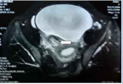
Fig. 3: Axial T2W MR image reveals a Focal lesion along the Right posterior lateral wall of Urinary bladder which is of Heterogenous signal intensity with hyperintense cystic areas in the centre (arrow). The lesion is seen to protrude into the bladder lumen. There is loss
of fat planes between the uterus (lower uterine segment) and the lesion.
Fig. 4: Sag T1 WI Reveals Focal lesion along the Right posterior lateral wall of Urinary bladder which is isointense with hyperintense areas(arrow) in the centre. The lesion is
J of Evolution of Med and Dent Sci/ eISSN- 2278-4802, pISSN- 2278-4748/ Vol. 4/ Issue 27/ Apr 02, 2015 Page 4753
DISCUSSION: Endometriosis is a benign condition affecting 15-20% of women with a child-bearing potential. Most commonly it affects organs such as the ovaries, uterine ligaments, Fallopian tubes, rectum and the cervico-vaginal region. Involvement of the urinary tract, however is rare and seen in about 4% of patients, the vesical location being the most frequent of these presentations (84%). Typically, the catamenial nature of bladder symptoms (frequency, urgency, dysuria and tenesmus) is pathognomonic. However, this may not be seen in all patients.1 Broadly two different etiologies appear to exist causing vesical endometriosis, one being spontaneous and the other post-Cesarean.2 In the former, the bladder lesion is a manifestation of the generalized pelvic disease, whereas after iatrogenic dissemination, growth of ectopic endometrium is usually limited to the bladder wall. On microscopy a solid endometrioma demonstrates abundant fibrosis, with small clusters of endometriotic glandular tissue.5
These lesions are typically iso to hypoechoic on USG. Transvaginal ultrasonography is more accurate and versatile than abdominal ultrasonography. On MRI most lesions show intermediate to hyperintense signal on T1W images and are hypointense on the T2W images. Punctate foci of high signal intensity (representing hemorrhage) may be seen on T1W images and enhancement is often seen after administration of contrast. We postulate that enhancement in an endometriosis on post-contrast studies is due to the presence of fibrosis. This may also cause a hypointense signal on the T2W images, which is otherwise, mainly due to the presence of hemorrhage. Endometriosis may also manifest as multiple homogeneously hyperintense cysts on T1W images.3
In conclusion, it is important to be acquainted with the various imaging characteristics of endometriosis and to have a high level of suspicion for detecting vesical endometriosis, especially following Cesarean section, since the clinical presentation is not always pathognomonic. MRI is preferred over CT scan, for diagnosis of pelvic endometriosis due to better lesional and anatomical characterization.
MRI is the best non-invasive method for evaluating endometriosis, with diagnostic accuracy close to laparoscopy.3,4 Its advantages over laparoscopy includes its ability to characterize endometriotic lesions and to evaluate extraperitoneal sites of involvement, contents of a pelvic mass, or lesions hidden by dense adhesions.
REFERENCES:
1. Shook TE, Nyberg LM. Endometriosis of the urinary tract. Urology 1988; 31:1-6.
2. Garcia Gonzalez JI, Entramiana Cameno J, Esteban Calvo JM, et al. Vesicle endometriosis after cesarean section : diagnostic and therapeutic aspects. Actas Urol Esp 1997; 21: 785-8.
3. Bis KG, Varchliotis TG, Agarwal R, et al. Pelvic endometriosis: MR imaging spectrum with laparoscopic correlation and diagnostic pitfalls. Radiographics 1997, 17: 639 – 55.
4. Fedele L. Bianchi S, Raffaelli R, et al. Pre-operative assessment of bladder endometriosis. Human Reprod. 1997; 12: 2519-22.
J of Evolution of Med and Dent Sci/ eISSN- 2278-4802, pISSN- 2278-4748/ Vol. 4/ Issue 27/ Apr 02, 2015 Page 4754
AUTHORS:
1. Shirish Paul Ganta 2. S. Venkateshwar Rao 3. N. Kiran Raju 4. V. N. Narvekar 5. Sivaleela 6. Ankum Rao 7. Devi Prathima 8. Srikanth
PARTICULARS OF CONTRIBUTORS:
1. Assistant Professor, Department of Radio-diagnosis, Asram Medical College, Eluru. 2. Associate Professor, Department of
Radio-diagnosis, Asram Medical College, Eluru.
3. Assistant Professor, Department of Radio-diagnosis, Asram Medical College, Eluru. 4. Professor & HOD, Department of
Radio-diagnosis, Asram Medical College, Eluru.
FINANCIAL OR OTHER COMPETING INTERESTS: None
5. Professor, Department of Radio-diagnosis, Asram Medical College, Eluru.
6. Post Graduate, Department of Radio-diagnosis, Asram Medical College, Eluru. 7. Post Graduate, Department of
Radio-diagnosis, Asram Medical College, Eluru. 8. Post Graduate, Department of
Radio-diagnosis, Asram Medical College, Eluru.
NAME ADDRESS EMAIL ID OF THE CORRESPONDING AUTHOR:
Dr. Shirish Paul Ganta, Assistant Professor,
Department of Radiodiagnosis, Asram Medical College, Eluru, West Godavari-534005, Andhra Pradesh.
E-mail: [email protected]