Insulin-Like Neuropeptide DAF-28 during Dauer
Formation in
Caenorhabditis elegans
Juan Carlos Fierro-Gonza´lez1, Astrid Cornils2, Joy Alcedo2, Antonio Miranda-Vizuete3,4*., Peter Swoboda1*.
1Department of Biosciences and Nutrition, Center for Biosciences at NOVUM, Karolinska Institute, Huddinge, Sweden,2Friedrich Miescher Institute for Biomedical Research, Basel, Switzerland,3Departamento de Fisiologı´a, Anatomı´a y Biologı´a Celular, Centro Andaluz de Biologı´a del Desarrollo - Consejo Superior de Investigaciones Cientı´ficas (CABD-CSIC), Universidad Pablo de Olavide, Sevilla, Spain,4Instituto de Biomedicina de Sevilla, Hospital Universitario Virgen del Rocı´o, Consejo Superior de Investigaciones Cientı´ficas, Universidad de Sevilla, Sevilla, Spain
Abstract
Thioredoxins comprise a conserved family of redox regulators involved in many biological processes, including stress resistance and aging. We report that the C. elegans thioredoxin TRX-1 acts in ASJ head sensory neurons as a novel modulator of the insulin-like neuropeptide DAF-28 during dauer formation. We show that increased formation of stress-resistant, long-lived dauer larvae in mutants for the gene encoding the insulin-like neuropeptide DAF-28 requires TRX-1 acting in ASJ neurons, upstream of the insulin-like receptor DAF-2. Genetic rescue experiments demonstrate that redox-independent functions of TRX-1 specifically in ASJ neurons are needed for the dauer formation constitutive (Daf-c) phenotype ofdaf-28mutants. GFP reporters oftrx-1anddaf-28show opposing expression patterns in dauers (i.e.trx-1is up-regulated anddaf-28is down-regulated), an effect that is not observed in growing L2/L3 larvae. In addition, functional TRX-1 is required for the down-regulation of a GFP reporter ofdaf-28during dauer formation, a process that is likely subject to DAF-28-mediated feedback regulation. Our findings demonstrate that TRX-1 modulates DAF-28 signaling by contributing to the down-regulation ofdaf-28expression during dauer formation. We propose that TRX-1 acts as a fluctuating neuronal signaling modulator within ASJ neurons to monitor the adjustment of neuropeptide expression, including insulin-like proteins, during dauer formation in response to adverse environmental conditions.
Citation:Fierro-Gonza´lez JC, Cornils A, Alcedo J, Miranda-Vizuete A, Swoboda P (2011) The Thioredoxin TRX-1 Modulates the Function of the Insulin-Like Neuropeptide DAF-28 during Dauer Formation inCaenorhabditis elegans. PLoS ONE 6(1): e16561. doi:10.1371/journal.pone.0016561
Editor:Ellen Nollen, University Medical Center Groningen, The Netherlands
ReceivedSeptember 24, 2010;AcceptedDecember 21, 2010;PublishedJanuary 27, 2011
Copyright:ß2011 Fierro-Gonza´lez et al. This is an open-access article distributed under the terms of the Creative Commons Attribution License, which permits
unrestricted use, distribution, and reproduction in any medium, provided the original author and source are credited.
Funding:This study was supported by grants from The Swedish Foundation for Strategic Research, The Swedish Research Council and The NordForsk Nordic C. elegans Network to PS; from Instituto de Salud Carlos III (Projects PI050065 and PI080557, co-financed by Fondo Social Europeo, FEDER) and Junta de Andalucı´a (Projects CVI-3629 and CVI-2697) to AMV; and from The Novartis Research Foundation to JA. The funders had no role in study design, data collection and analysis, decision to publish, or preparation of the manuscript.
Competing Interests:The Friedrich Miescher Institute is financially supported by the Novartis Research Foundation. The authors have declared that no competing interests exist and that they adhere to the journal’s policy on sharing of materials, methods and data.
* E-mail: [email protected] (PS); [email protected] (AMV)
.These authors contributed equally to this work.
Introduction
Thioredoxins comprise a conserved family of proteins charac-terized by the so-called thioredoxin fold and the highly conserved cysteine–glycine–proline–cysteine (CGPC) catalytic active site (reviewed in [1,2]). The two cysteines in the active site of thioredoxin are required for the reversible reduction of disulfide bonds in many target proteins. The functions of thioredoxins are extensive and mostly depend on their disulfide oxidoreductase attributes; in general, they can act as electron donors for metabolic enzymes, as antioxidants, or as redox regulators of signaling molecules and transcription factors (reviewed in [1]).
In some cases, thioredoxins have been reported to execute their specific functions in a redox-independent manner (reviewed in [2,3]). For instance, the regulation of phage T7 DNA polymerase activity by E. coli Trx1 [4,5], the cytokine function of human truncated thioredoxin (Trx80) [6], or the regulation of apoptosis signaling kinase 1 (ASK1) by human Trx1 [7], among others, are
all functions carried out independently of their oxidoreductase activity. More recently, thioredoxins have also been shown to promote folding of proteins independently of their redox activities (reviewed in [3]).
In the nematodeCaenorhabditis elegans, the gene trx-1encodes a thioredoxin found to be expressed in the ASJ sensory neuron pair [8,9]. These neurons are implicated in the regulation of aging, the response to stress conditions and the control of a state of developmental arrest called the dauer larva [10,11,12]. Previously, we and others have shown thattrx-1deletion shortens lifespan and increases sensitivity to oxidative stress, though it itself does not affect dauer formation [8,9]. These findings implicate TRX-1 in processes that regulate aging and stress resistance, which raises the question whether it is involved in mechanisms implicated in the regulation of dauer formation.
However, under circumstances in which genes involved in dauer development become inactivated due to mutations, animals arrest as dauers even under favorable conditions (dauer formation constitutive or Daf-c phenotype) [14]. Among the known daf-c
genes whose functions are linked to ASJ, four have previously been well characterized for their role in the dauer formation pathway:
daf-11 (abnormal dauer formation-11), daf-28, tax-2 (abnormal chemotaxis-2) andtax-4 (Figure S1; [12,15,16,17,18]). Moreover, mutations in these four daf-c genes have also been reported to extend lifespan [19,20,21], providing an additional link between dauer formation and aging. daf-11 encodes a transmembrane guanylyl cyclase, whiletax-2andtax-4encode subunits of a cyclic guanosine monophosphate (cGMP)-gated ion channel; these three genes are expressed in subsets of amphid sensory neurons, including ASI and ASJ [15,22]. DAF-11 regulates dauer formation by modulating the levels of cGMP in these neurons, to which TAX-2/TAX-4 respond [22], which then regulate the expression of transforming growth factor-beta (TGF-beta) [23] and insulin-like neuropeptides [18,24] (Figure S1). There are,40 insulin-like neuropeptides in C. elegans (www.wormbase.org), and several, including DAF-28, have been reported to be produced in ASJ, among other neurons [18,24]. To gain insight into whether TRX-1 plays a role in dauer formation inC. elegans, we analyzed at the cellular and genetic level its interaction with daf-c genes co-expressed in ASJ neurons.
We have identified theC. elegansthioredoxin TRX-1 as a novel modulator of the insulin-like neuropeptide DAF-28 during dauer formation. We found thattrx-1suppresses the Daf-c phenotype of alldaf-28insulin-like mutant alleles tested and that this suppression requires a functional DAF-2 insulin-like receptor. Genetic rescue experiments demonstrated that redox-independent functions of transgenic TRX-1 provided specifically in ASJ neurons can restore the suppression exerted bytrx-1deletion on the Daf-c phenotype ofdaf-28mutants. The suppression observed at the genetic level is also manifest at the cellular level specifically during dauer formation: GFP reporters of trx-1 and daf-28 display opposing expression patterns in dauers, which is in contrast to what is observed in growing L2/L3 larvae. Moreover, functional TRX-1 is required for the down-regulation of a GFP reporter ofdaf-28
during dauer formation, a mechanism that is likely mediated by DAF-28-dependent feedback regulation. Our data suggest that TRX-1 contributes to the regulation of insulin-like neuropeptide expression, in particular DAF-28, during dauer formation in response to a changing environment.
Results
trx-1has a novel synthetic dauer formation constitutive (Daf-c) phenotype
To investigate whethertrx-1plays a role in dauer formation, we first constructed double mutant combinations with different alleles ofdaf-11, since this gene is predicted to regulate dauer formation upstream of both the TGF-beta and insulin-like signaling (IS) pathways (Figure S1; [18,23]). Previously, it has been suggested that ASJ neurons are required for thedaf-11Daf-c phenotype, since laser ablation of ASJ suppresses the Daf-c phenotype of adaf-11(sa195)
mutant [12]. We tested whethertrx-1is also required for thedaf-11
Daf-c phenotype. Thetrx-1(ok1449)allele used in this study is a null mutation: a deletion in the coding region preventing translation of the protein (Table S1; [9]). trx-1(ok1449) enhanced the Daf-c phenotype of all daf-11 alleles tested at 15uC (Figure 1A and Table 1). Even at 25uC, two of the threedaf-11alleles tested showed a significant increase in dauer formation (Figure 1B and Table 2). Thus,trx-1has a novel synthetic Daf-c phenotype. In addition, the
slight suppression of thedaf-11(sa195)Daf-c phenotype exerted by
trx-1(ok1449)at 25uC (Figure 1B and Table 2), partially phenocopies the effect of killing ASJ in adaf-11(sa195)single mutant background [12]. Together, our findings indicate that most of the Daf-c phenotype ofdaf-11mutants is TRX-1-independent, while a small fraction requires TRX-1. This dual effect of thetrx-1mutation on
daf-11for dauer formation is reminiscent, although not identical, to that reported for mutations in the cGMP-gated ion channel genes
tax-2 and tax-4 [16]. tax-2 and tax-4 mutants show weak Daf-c phenotypes, which are epistatic to the strong daf-11 Daf-c phenotypes [16]. trx-1 deletion did not affect the weak Daf-c phenotype caused bytax-4(p678)(Figures 1A and 1B; Tables 1 and 2). Since mutations intax-4essentially remove the input fromdaf-11
signaling for dauer formation [16],trx-1deletion could not replicate the synthetic interaction observed withdaf-11mutants. These results further support the notion oftax-4acting downstream ofdaf-11for dauer formation [16], withtrx-1acting mostly independently of daf-11in the dauer formation pathway. We speculate that for the most part TRX-1 and DAF-11 are affecting a separate set of neurons for dauer formation (e.g. TRX-1 in ASJ and DAF-11 in ASI) (this work; [23]), while they share some modulatory functions in ASJ neurons.
trx-1deletion enhances the Daf-c phenotype of mutations in the TGF-beta signaling pathway
There is evidence that DAF-11 acts upstream of the IS and TGF-beta signaling pathways (Figure S1; reviewed in [25,26]). To determine iftrx-1interacts with any of these two genetic pathways for dauer formation, we first scored for dauers in double mutants containing trx-1(ok1449) and daf-c mutations in the TGF-beta signaling pathway. Previously, strong synergy was identified between daf-c mutations in several genes of the IS pathway, includingdaf-2anddaf-28, and mutations in genes of the parallel TGF-beta signaling pathway, represented here bydaf-7,daf-1and
daf-8[21,27]. We observed a marked increase in dauer formation in all threetrx-1;TGF-betadouble mutant combinations tested at 15uC (Figure 1A and Table 1). A straightforward interpretation of this synthetic enhancement observed is that the Daf-c phenotype of mutations in the TGF-beta signaling pathway is TRX-1-independent. SinceTGF-betasingle mutants already form nearly 100% dauers at 25uC ([28]; data not shown),trx-1;TGF-betadouble mutants did not display any further increase in dauer formation at that temperature (data not shown). We conclude that TRX-1 acts independently of the TGF-beta signaling pathway for dauer formation.
TRX-1 function is needed for the Daf-c phenotype of daf-28mutants
To investigate the interaction oftrx-1with the IS pathway for dauer formation, we first analyzed double mutants oftrx-1 with
daf-28. trx-1 suppressed the Daf-c phenotype of the two daf-28
alleles tested (Figures 1A and 1B; Tables 1 and 2), regardless of their nature (sa191 is a dominant-negative allele; tm2308 is predicted to be null) (Table S1). These results indicate that the suppression is not allele-specific and that the Daf-c phenotype of
daf-28mutants requires TRX-1 function for dauer formation. Our findings suggest that loss of TRX-1 function likely increases insulin-like signaling upon mutation ofdaf-28.
Suppression of thedaf-28Daf-c phenotype by trx-1(ok1449)depends on DAF-2 insulin-like receptor signaling
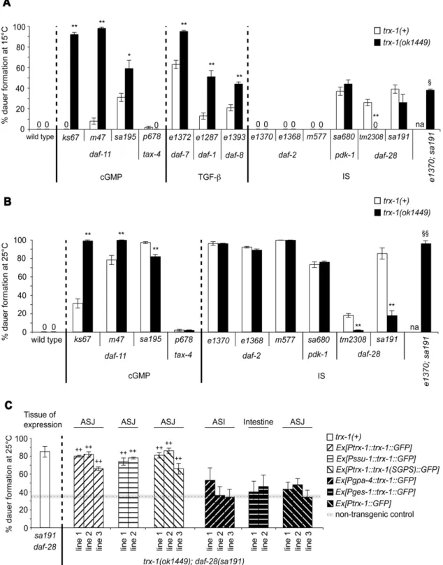
Figure 1. Redox-independent functions of TRX-1 in ASJ neurons modulate DAF-28 signaling during dauer formation. Dauer formation at 15uC (A) and 25uC (B) is shown for each of thedaf-cmutant alleles tested in a trx-1(+)wild-type background (white bars) or in combination with atrx-1(ok1449)deletion null mutant allele (black bars). (C) Rescue of the suppression of the Daf-c phenotype intrx-1(ok1449); daf-28(sa191)transgenic animals (patterned bars) expressing eitherPtrx-1::trx-1::GFP,Pssu-1::trx-1::GFP,Ptrx-1::trx-1(SGPS)::GFP, Pgpa-4::trx-1::GFP, Pges-1::trx-1::GFPorPtrx-1::GFP. The cellular site of expression for each transgenic extrachromosomal array is outlined above the graph in panel C. The non-transgenic control represents the average of all assays performed with non-non-transgenic progeny that segregated from the same non-transgenic parents (dotted line)6standard error of the mean (SEM, horizontal gray line). The white bar corresponding to thedaf-28(sa191)single mutant in panel C, has been taken from panel B for comparison. In all three panels, each bar represents the average of 2–3 independent assays6SEM, with more than 165 animals assayed in total per genotype. *p,0.05, **p,0.01,1
p,0.05 relative totrx-1(ok1449); daf-28(sa191),11
p,0.01 relative totrx-1(ok1449); daf-28(sa191), and++p
,0.001 relative to non-transgenic animals, by chi-squared test (see also Tables 1, 2 and 3). na: not assayed. The molecular identities of all mutant alleles presented here are shown in Table S1.
analyzed dauer formation in double mutants containing the trx-1(ok1449) deletion and hypomorphic (partial loss-of-function) mutant alleles ofdaf-2orpdk-1. The latter encodes a homologue of the mammalian Akt/PKB kinase PDK1, involved in transduc-ing DAF-2 signals on to the DAF-16 FOXO transcription factor to regulate dauer formation (Figure S1; [29]). Deletion mutations of
daf-16 and daf-18 (the latter encoding a homologue of the mammalian PTEN tumor suppressor), which act downstream of
daf-2for dauer formation, suppress the Daf-c phenotype ofdaf-2
mutants [27,30,31]. In addition, RNA interference of the gene
pptr-1(encoding a homologue of a B56 regulatory subunit of the mammalian PP2A holoenzyme), which acts downstream of both
daf-2 and pdk-1 for dauer formation, suppresses the Daf-c phenotypes of bothdaf-2andpdk-1mutants [32]. Thus, if TRX-1 acted downstream of DAF-2 and PDK-TRX-1, we would expect a suppressive effect of trx-1(ok1449) on the Daf-c phenotypes of either daf-2 or pdk-1 mutants. However, trx-1 deletion did not suppress the Daf-c phenotypes of three different hypomorphic daf-2mutants or of onepdk-1mutant at 15 and 25uC (Figures 1A and 1B; Tables 1 and 2). Moreover, at the intermediate temperature of 20uC,trx-1deletion did not suppress the Daf-c phenotype of daf-2(e1370)animals: the percent dauer formation6standard error of the mean was 2664 fordaf-2(e1370)and 2161 fortrx-1(ok1449); daf-2(e1370), with n.300 animals in total per genotype (p.0.1 by chi-squared test; 2 assays). Together, these data suggest that TRX-1 functions upstream of or in parallel to both DAF-2 and PDK-TRX-1 for dauer formation. To investigate whether the suppression of the
daf-28 Daf-c phenotype by trx-1(ok1449) depends on DAF-2 signaling, we constructed the triple mutant trx-1(ok1449); daf-2(e1370); daf-28(sa191). If the suppression ofdaf-28(sa191)by
trx-1(ok1449)was independent of DAF-2 signaling, we would expect the triple mutant to form dauers at levels comparable with those of
trx-1(ok1449); daf-28(sa191)double mutant animals, indicating that the suppression still persists even after mutation ofdaf-2. However,
trx-1(ok1449); daf-2(e1370); daf-28(sa191) triple mutant animals formed dauers at levels that were similar to those ofdaf-28(sa191)
single mutant animals, at both 15 and 25uC (Figure 1A and 1B; Table 1 and 2), suggesting that the suppression is no longer effective when daf-2 is mutated. Therefore, although genetic interactions using non-null mutants (cf. Table S1) need to be interpreted with some caution, these data demonstrate that the ability of trx-1(ok1449) to suppress the Daf-c phenotype of daf-28(sa191)depends on functional DAF-2.
Redox-independent functions of transgenic TRX-1 specifically in ASJ neurons restore the suppression of daf-28(sa191)bytrx-1(ok1449)
To investigate whether wild-type TRX-1 can restore the suppression of thedaf-28(sa191)Daf-c phenotype caused bytrx-1
deletion at 25uC, the transgene Ptrx-1::trx-1::GFP (see Materials and Methods) was used to transformtrx-1(ok1449); daf-28(sa191)
animals. Transgenic lines expressing this transgene restored the Daf-c phenotype back to daf-28(sa191) single mutant levels (Figure 1C and Table 3). Since the GFP-tagged transgene can be visualized in ASJ, this is in agreement with the hypothesis that wild-typetrx-1acts in ASJ neurons to modulate DAF-28 signaling during dauer formation. To confirm this notion, the expression of wild-type trx-1 genomic DNA fused to GFP was driven from another ASJ neuron-specific promoter,ssu-1[33]. Consistent with our hypothesis, this transgenePssu-1::trx-1::GFP also rescued the suppression of the Daf-c phenotype of daf-28(sa191) by trx-1(ok1449)(Figure 1C and Table 3).
C. elegans TRX-1 shares many conserved residues with its thioredoxin orthologues throughout evolution [9], including the active site, which has extensively been demonstrated to be essential for its oxidoreductase activity in bacteria [5], yeast [34], fruit fly
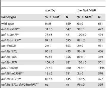
Table 1.Percent dauer formation at 15uC oftrx-1; daf-c double and triple mutants.
trx-1(+) trx-1(ok1449)
Genotype %±SEM{
N %±SEM{
N
wild type 060 478 060 442
daf-11(ks67)** 060 357 9262 300
daf-11(m47)** 863 586 9861 333 daf-11(sa195)* 3164 412 5968 172
tax-4(p678) 261 420 060 511
daf-7(e1372)** 6364 1257 9561 754 daf-1(e1287)** 1363 451 5166 485
daf-8(e1393)** 2163 1826 4462 1281
daf-2(e1370) 060 388 060 333
daf-2(e1368) 060 280 060 167
daf-2(m577) 060 250 060 187
pdk-1(sa680) 3764 863 4464 627
daf-28(tm2308)** 2663 617 060 578 daf-28(sa191) 3964 318 2668 238
daf-2(e1370); daf-28(sa191)1
na na 3861 365
{
Mean percentage of dauer larvae6standard error of the mean (SEM); 3 assays per genotype, except for wild type,tax-4(p678),daf-28(tm2308), daf-2(e1368), daf-2(m577)and the triple mutant: 2 assays. N: total (pooled) number of animals.
**p,0.01;
*p,0.05 by chi-squared test.
1
p,0.05 by chi-squared test with respect totrx-1(ok1449); daf-28(sa191). na: not assayed.
doi:10.1371/journal.pone.0016561.t001
Table 2.Percent dauer formation at 25uC oftrx-1; daf-c double and triple mutants.
trx-1(+) trx-1(ok1449)
Genotype %±SEM{
N %±SEM{
N
wild type 060 659 060 661
daf-11(ks67)** 3165 547 9961 422
daf-11(m47)** 7865 421 10060 474 daf-11(sa195)** 9761 595 8262 221
tax-4(p678) 261 853 260 931
daf-2(e1370) 9662 435 9660 466 daf-2(e1368) 9261 356 8961 349
daf-2(m577) 10060 621 10060 501
pdk-1(sa680) 7363 980 7661 1196
daf-28(tm2308)** 1862 781 260 570 daf-28(sa191)** 8566 445 1865 427
daf-2(e1370); daf-28(sa191)11
na na 9663 368
{
Mean percentage of dauer larvae6standard error of the mean (SEM); 3 assays per genotype, except for wild type,tax-4(p678),daf-2(e1368)and the triple mutant: 2 assays. N: total (pooled) number of animals.
**p,0.01 by chi-squared test.
11
p,0.01 by chi-squared test with respect totrx-1(ok1449); daf-28(sa191). na: not assayed.
[35], and in humans [36]. To investigate whether the redox activity of TRX-1 is necessary for rescue of trx-1(ok1449); daf-28(sa191)double mutants, we transformed these animals with Ptrx-1::trx-1(SGPS)::GFP,a transgene containing the mutated active site SGPS instead of the wild-type CGPC. The conversion of thioredoxin active-site cysteines to serines has been widely shown to eliminate its ability to function as oxidoreductase in classical reduction assays [5,36,37]. Interestingly, transgenic animals expressing the mutated transgenePtrx-1::trx-1(SGPS)::GFPrestored the Daf-c phenotype just asPtrx-1::trx-1::GFPdid (Figure 1C and Table 3), indicating that TRX-1 function in dauer formation does not require its oxidoreductase activity.
In contrast, the Daf-c restoration rescue could not be achieved whentrx-1(ok1449); daf-28(sa191)double mutants either expressed a transgene containing the ASI neuron-specificgpa-4promoter [38] with the complete coding region of wild-type trx-1fused to GFP (Pgpa-4::trx-1::GFP), a transgene that drives expression of wild-type
trx-1::GFP from the ges-1 promoter in the intestine ( Pges-1::trx-1::GFP) [39] or a transgene that only drives GFP expression from the trx-1promoter in ASJ neurons (Ptrx-1::GFP) (Figure 1C and Table 3). Together, these findings suggest that redox-independent functions of TRX-1 specifically in ASJ neurons are required for the Daf-c phenotype caused by defects indaf-28function.
TRX-1 contributes to the down-regulation ofdaf-28
expression during dauer formation, a process likely controlled by DAF-28-mediated feedback regulation
Previously, it was shown thatPdaf-28::GFPexpression in dauers is down-regulated by conditions of starvation and exposure to dauer pheromone [18]. To test whethertrx-1expression responds to these dauer-promoting signals, we analyzed Ptrx-1::GFP expression in both wild-type natural dauers, which are induced by a combination of both signals, and indaf-cmutant dauers, represented here by
daf-11anddaf-2. Sincetrx-1suppresses the Daf-c phenotype ofdaf-28
mutants (cf. above), one could expect that the response of Ptrx-1::GFPlevels to dauer-promoting signals might be opposite to that of
Pdaf-28::GFP. Consistent with our hypothesis, Pdaf-28::GFP was down-regulated and Ptrx-1::GFP was up-regulated in wild-type natural dauers, and indaf-11anddaf-2mutant dauers (Figures 2A and 2C). In contrast, these opposing expression levels observed in dauers were not seen in well-fed, growing L2/L3 larvae of the corresponding genotypes (Figure S2), suggesting a possible regula-tory effect of TRX-1 on DAF-28 (or vice versa) only during dauer formation. Therefore, to test whether TRX-1 function contributes to the down-regulation ofdaf-28expression during dauer formation, we analyzedPdaf-28::GFPexpression intrx-1mutant dauers. If the down-regulation ofdaf-28expression during dauer formation was independent of TRX-1, we would expecttrx-1mutant dauers to expressPdaf-28::GFPat levels comparable with those of wild-type natural dauers (cf. Figure 2A), indicating that the down-regulation still persists even after loss oftrx-1. Interestingly, we observed that
Pdaf-28::GFP expression in trx-1 mutant dauers is not down-regulated, but is in fact not significantly different from what is seen in well-fed, growing L2/L3 wild-type larvae (Figures 2B and 2D). These findings suggest that the up-regulation of TRX-1 specifically during dauer formation is, at least in part, causative of a reduction in DAF-28 signaling. Moreover, the up-regulation of Ptrx-1::GFP
expression indaf-28mutant dauers (Figures 2B and 2D), is further increased when compared with that seen in wild-type natural dauers (cf. Figures 2A and 2C). This result suggests thattrx-1up-regulation during dauer formation is likely subject to DAF-28-mediated feedback regulation. Together, these results are consistent with a model (Figure 3) in which TRX-1 modulates DAF-28 signaling by contributing to the down-regulation ofdaf-28expression exclusively during dauer formation, a modulatory process that is likely controlled by DAF-28-dependent feedback regulation.
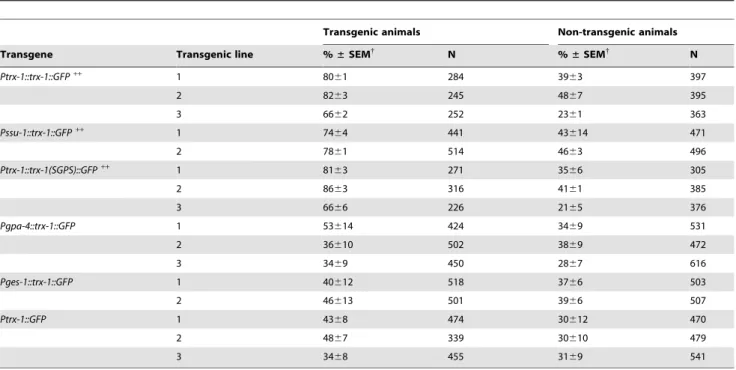
Table 3.Percent dauer formation at 25uC oftrx-1(ok1449); daf-28(sa191)double mutants expressing the indicated transgene.
Transgenic animals Non-transgenic animals
Transgene Transgenic line %±SEM{
N %±SEM{
N
Ptrx-1::trx-1::GFP++ 1 80
61 284 3963 397
2 8263 245 4867 395
3 6662 252 2361 363
Pssu-1::trx-1::GFP++ 1 74
64 441 43614 471
2 7861 514 4663 496
Ptrx-1::trx-1(SGPS)::GFP++
1 8163 271 3566 305
2 8663 316 4161 385
3 6666 226 2165 376
Pgpa-4::trx-1::GFP 1 53614 424 3469 531
2 36610 502 3869 472
3 3469 450 2867 616
Pges-1::trx-1::GFP 1 40612 518 3766 503
2 46613 501 3966 507
Ptrx-1::GFP 1 4368 474 30612 470
2 4867 339 30610 479
3 3468 455 3169 541
{
Mean percentage of dauer larvae6standard error of the mean (SEM); 2 assays per transgenic line, except forPtrx-1::GFPandPgpa-4::trx-1::GFP: 3 assays. N: total (pooled) number of animals.
++p
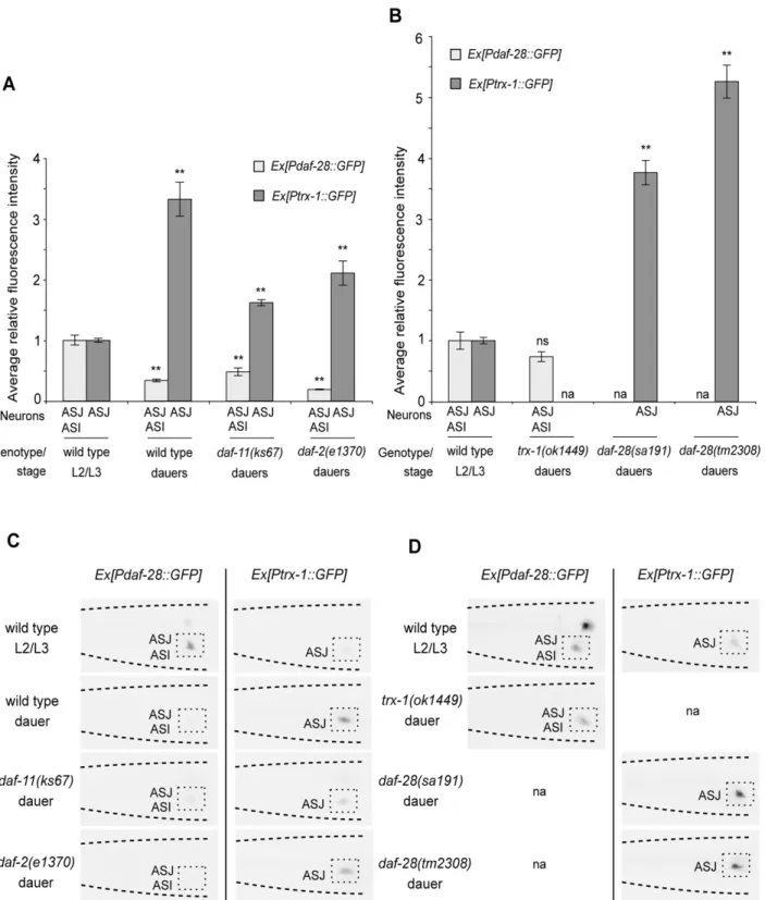
Figure 2. TRX-1 contributes to the down-regulation ofdaf-28insulin-like gene expression during dauer formation.Quantification of
Ptrx-1::GFPandPdaf-28::GFPexpression in dauers and well-fed, growing L2/L3 larvae in ASJ or ASI neurons. Average fluorescence intensity in ASJ or ASI neurons, normalized to that of well-fed, growing L2/L3 wild-type larvae, is shown for wild-type dauers and fordaf-2anddaf-11mutant dauers in (A), and fortrx-1anddaf-28mutant dauers in (B). Two independent transgenic lines were examined for each of the two transcriptionalPtrx-1::GFPand
Pdaf-28::GFPreporters, and the results were very similar; one transgenic line was tested fordaf-28(tm2308)dauers. The data derived from one transgenic line are presented. Each bar represents the average relative fluorescence intensity of 28–34 animals6standard error of the mean (SEM). **
p,0.01 relative to L2/L3 wild-type larvae, by Student’sttest (two-tailed, two-sample unequal variance); na: not assayed; ns: not significant. Shown in (C) and (D) are representative color-inverted images of animals assayed in (A) and (B), respectively, revealing the differences in GFP intensity in ASJ or ASI neurons (boxed). The tip of the head points to the left in all panels.
Discussion
Here, we have identified the thioredoxin TRX-1 as a novel modulator of the insulin-like neuropeptide DAF-28 during dauer formation inC. elegans. We found thattrx-1suppresses the Daf-c phenotypes of alldaf-28insulin-like mutant alleles tested, and that this suppression is dependent on a functional DAF-2 insulin-like receptor (Figures 1A and 1B; Tables 1 and 2). Genetic rescue experiments showed that redox-independent functions of TRX-1 specifically in ASJ neurons are needed for the Daf-c phenotype caused by defects indaf-28function (Figure 1C and Table 3). The suppression observed at the genetic level phenocopied that seen at the cellular level specifically during dauer formation:Ptrx-1::GFP
andPdaf-28::GFP showed opposing expression patterns in dauers (Figures 2A and 2C), which were not observed in growing L2/L3 larvae (Figure S2). In addition, Pdaf-28::GFP down-regulation during dauer formation required functional TRX-1, a mechanism that is likely controlled by DAF-28-dependent feedback regulation (Figures 2B and 2D). Taken together, our results suggest a model in which TRX-1 contributes to the adjustment ofdaf-28 insulin-like expression, and consequently its function, during dauer formation (Figure 3).
C. elegans neuropeptides are classified into three classes: the insulin-like proteins, the FMRF (Phe-Met-Arg-Phe)-amide pep-tides (referred to as FLPs) and the neuropeptide-like proteins or NLPs (reviewed in [40]). In addition to DAF-28, it might be possible that TRX-1 also modulates other neuropeptides co-expressed in ASJ neurons (Figure 3), such as the insulin-like proteins INS-9 and INS-1, or NLP-3 and FLP-21 ([24,41]; reviewed in [40]). If the effect of TRX-1 on dauer formation was solely dependent on DAF-28 signaling, we would expect that overexpression of trx-1 in the likely null mutant daf-28(tm2308)
results in no enhancement of the Daf-c phenotype. Interestingly, the observation that overexpression of wild-typetrx-1::GFPin ASJ was sufficient to enhance the weak Daf-c phenotype of the likely null mutant daf-28(tm2308) (Table S2), suggests that TRX-1 in part affects dauer formation independently of DAF-28, and potentially independently of DAF-2 (Figure 3). Moreover, the fact that trx-1 deletion, like daf-28(sa191) [21], enhanced the Daf-c phenotype of TGF-beta pathway mutations and of most daf-11
mutations, whereastrx-1deletion suppressed the Daf-c phenotype
of daf-28 mutations (Figures 1A and 1B; Tables 1 and 2), also supports the notion that TRX-1 to some extent modifies dauer formation independently of DAF-28, and possibly also of DAF-2 (Figure 3). Furthermore, because wild-type animals overexpressing wild-typetrx-1::GFP in ASJ were not induced to form dauers at 25uC (Table S2), other pathways and/or neurons might act in parallel to contribute, together with TRX-1, to the modulation of DAF-28 signaling. In fact, the cGMP pathway has previously been suggested to regulate DAF-28 signaling during dauer formation (Figure S1; [18]).
ASJ neurons have been shown to regulate dauer formation in concert with other sensory neurons [11,12], despite their discrete synaptic connectivity (ASK is the only sensory neuron in synaptic contact with ASJ; www.wormatlas.org). The localized function of TRX-1 in ASJ neurons for dauer formation (Figure 1C; Table 3) and the effect of TRX-1 ondaf-28expression (Figures 2B and 2D) support a model wherein TRX-1 modulates (the levels of) neurosecretory signals emanating from ASJ neurons to regulate dauer formation in a cell non-autonomous fashion. Although our results do not exclude that TRX-1 affects other neurons through direct synaptic connections, the control it exerts on neuropepti-dergic signaling would allow TRX-1 to function locally in ASJ neurons and affect dauer formation remotely. Such a model would explain the observation that deletion oftrx-1modifies the Daf-c phenotypes of mutations in genes not expressed in ASJ neurons (Figure 1A; Table 1) (e.g.daf-7is only expressed in ASI neurons) [42]. Similarly, TRX-1 could contribute to the down-regulation of
daf-28expression in ASI, either through ASJ-derived DAF-28 or other TRX-1-modified neuropeptides released from ASJ neurons. Previously, mammalian Trx1 has been proposed to participate in the redox regulation that mediates insulin secretion via NADPH as a signaling molecule [43]. However, we have shown in this report that TRX-1 function in dauer formation does not require its redox activity (Figure 1C and Table 3), suggesting that TRX-1 contributes to the down-regulation of daf-28 expression during dauer formation via mechanisms other than redox regulation of neuropeptide production and/or release.
It has recently been found in mammals that key neuronal lipid metabolites act as hypothalamic signaling mediators that monitor energy status and contribute to maintaining organism metabolic homeostasis (reviewed in [44]). We propose that the thioredoxin Figure 3. Model for TRX-1 function during dauer formation.TRX-1 is a novel modulator of the insulin-like neuropeptide DAF-28 during dauer formation. It modulates DAF-28 signaling, upstream of DAF-2, by specifically contributing to the down-regulation ofdaf-28expression during dauer formation; this modulatory process is likely subject to DAF-28-mediated feedback regulation (black dashed line). In addition, TRX-1 possibly affects dauer formation independently of both DAF-28 and DAF-2 by modulating other neuropeptides co-expressed in ASJ neurons (see Results and Discussion for details).
TRX-1 might similarly act as a neuronal modulator in ASJ neurons to monitor metabolic status and thus adjust neuropeptide expression, including the insulin-like neuropeptide DAF-28, during dauer formation in response to adverse conditions (Figure 3). In this context, ASJ neurons would monitor the choice between reproductive development and dauer formation by responding to intra-neuronal fluctuations of TRX-1 (Figure 3). In turn, these TRX-1 fluctuations would be triggered in response to signals from the environment and peripheral tissues that reflect the animal’s global energy status. Interestingly, theC. elegansASI neurons have been suggested to mediate responses to nutrient availability through neuroendocrine signals that promote adult longevity by sensing similar energy-monitoring molecules within them [45]. Moreover, ASJ neurons have been proposed to influence dauer formation through sulfated neuroendocrine signals [33]. Thus, together with previous observations by others, our findings anticipate a neuroendocrine network in the nematode involved in adjusting the animal’s global energy status in response to a changing environment, both during development and adulthood. Future work is needed to identify and dissect the components of such a neuroendocrine network that, including TRX-1, are involved in regulating mechanisms designed to monitor energy homeostasis in varying environmental conditions in the nematode, and most likely also in higher organisms.
Materials and Methods
Nematode strains and culture conditions
The standard methods used for culturing C. elegans were described previously ([46]; reviewed in [47]). Strains and transgenes used in this work are summarized in Table S3. All strains were grown at 20uC, except fordaf-cmutant strains, which were grown at 15uC.
Transgene injection constructs and germline transformation
The Ptrx-1::trx-1::GFP translational fusion construct (cf. Figure 1C; Tables 3 and S2) and thePdaf-28::GFPtranscriptional fusion construct (cf. Figures 2 and S2) were previously reported [9,48]. For the Ptrx-1::GFP transcriptional fusion construct (cf. Figures 1C, 2 and S2; Table 3),,1 kb of sequence upstream of thetrx-1bsplice variant, plus the first 65 nucleotides of the coding sequence were amplified by PCR and cloned in-frame with GFP into the pPD95.77 vector. The Ptrx-1::trx-1(SGPS)::GFP transla-tional fusion construct (cf. Figure 1C and Table 3) was obtained by site-directed mutagenesis of thetrx-1active site, as specified in the QuikChange II Site-Directed Mutagenesis Kit (Stratagene), using thePtrx-1::trx-1::GFPtranslational fusion construct as a template. The Pssu-1::trx-1::GFP, Pgpa-4::trx-1::GFP and Pges-1::trx-1::GFP
translational fusion constructs (cf. Figure 1C and Table 3) included ,0.5 kb (ssu-1)or,2.5 kb (gpa-4andges-1) of promoter sequence upstream of the respective start codon fused to the full-lengthtrx-1
genomic DNA fragment, in-frame with GFP, into the pPD95.77 vector. For the experiments shown in Figures 2 and S2, 80 ng/ml of Ptrx-1::GFPor 50 ng/ml ofPdaf-28::GFP were coinjected with
20 ng/ml of the injection markerPelt-2::mCherry(a gift from Gert Jansen, Rotterdam), into trx-1(ok1449); daf-28(sa191) or trx-1(ok1449) animals. These extrachromosomal arrays were then crossed into wild-type,daf-11(ks67),daf-2(e1370),daf-28(sa191)or
daf-28(tm2308) animals. For the rescue experiments shown in Figure 1C and Table 3, 40 ng/ml ofPtrx-1::trx-1::GFP, Pssu-1::trx-1::GFP, Ptrx-1::trx-1(SGPS)::GFP, Pgpa-4::trx-1::GFP, Pges-1::trx-1::GFP or Ptrx-1::GFP were coinjected with 30 ng/ml of the
injection marker Punc-122::DsRed [49], into trx-1(ok1449);
daf-28(sa191)animals. For the overexpression experiments shown in Table S2, 100 ng/ml of Ptrx-1::trx-1::GFP were coinjected with 30 ng/ml of the injection markerPunc-122::DsRedinto wild-type or daf-28(tm2308)animals. Germline transformation was performed as described elsewhere [50].
Construction of double and triple mutants
To construct trx-1; daf-c double mutants, trx-1 homozygous males were crossed to the appropriatedaf-cmutant, and F1 cross progeny hermaphrodites were grown singly at 25uC. We singled F2 dauers and allowed them to recover at 15uC. Recovered dauers were considered to be homozygous for thedaf-cmutation. To construct the trx-1; tax-4 double mutant, trx-1; daf-2
homozygous hermaphrodites were crossed to tax-4homozygous males. F2 progeny were grown singly at 25uC from F1 cross progeny.tax-4homozygous candidates were selected further from F2s that did not segregate dauers at 25uC. To confirm the presence of the tax-4 mutation, we performed a soluble compound chemotaxis assay, as described elsewhere [51]. To construct the triple mutant trx-1(ok1449); 2(e1370); daf-28(sa191), we took advantage of the fact that daf-28 dauers recover well at 25uC, whereasdaf-2dauers do not recover or only recover with difficulties at this temperature [21,52]. In brief,trx-1
homozygous males where crossed totrx-1; daf-2 double mutant hermaphrodites. The resulting male progeny were crossed to trx-1; daf-28hermaphrodites, and F1 cross progeny hermaphrodites were grown individually at 25uC. We then picked many F2 dauers from several F1s onto new, seeded plates and kept them at 25uC. Those dauers that recovered after ,14–24 h, were considered to be daf-28homozygotes. F3 progeny of these daf-28homozygous candidates that remained arrested as dauers after ,17–24 h at 25uC were potentialdaf-2homozygotes. We singled these dauers and transferred them to 15uC for recovery. We tested all candidates for the presence of the daf-28 and daf-2
mutations by sequencing. In all cases, the presence of thetrx-1
deletion was demonstrated by performing PCR on single-worm lysates, based on methods previously described ([53]; reviewed in [54]).
Analysis of dauer formation
For dauer formation assays, 8–20 gravid hermaphrodites were allowed to lay eggs for a given time at the test temperature (Tables S4 and S5), and then removed. Dauers and non-dauers (L3 and L4 larvae and adult animals) on the agar and side of the plate were then counted at specific time points after egg-laying ended (Table S4). The scoring time points were selected for each genotype so that all animals had passed the pre-dauer or L2 stages at the time of scoring. Corresponding single and double mutant strains were always assayed in parallel. In all cases, dauers were discriminated from non-dauers based on the absence of pharyngeal pumping, intestinal re-organization and radial shrinkage of the body [13]. More than 165 animals were counted in a total of 2–3 independent assays per genotype. Dauers of selected key genotypes used in this study were subjected to morphological analysis using differential interference contrast (DIC) microscopy at an optical magnification of x800 or x1000, and also tested for resistance to 1% sodium dodecylsulfate (SDS) [13]. All dauers analyzed were identical to wild-type dauers in their morphological features (i.e. pharynx and alae) and SDS-resistance.
Microscopy and fluorescence imaging
The average relative intensity of a transcriptionalPtrx-1::GFPor
specificdaf-cgene as compared to growing L2/L3 wild-type larvae. Animals were visualized on a Zeiss Axioplan fluorescence microscope at an optical magnification of x640. Worms were put into M9 buffer on a very thin 2% agarose pad containing an anesthetic (15 mM NaN3). Previously, it had been shown that
exposure to 20 mM NaN3or less during 60 min [55],,30 mM NaN3for 20 min [56] or 50 mM NaN3for 5 min [57] can induce
evident physiological changes in the worm. All animals assayed in our study have been exposed to only 15 mM NaN3 for a
maximum of 10–15 min prior to image acquisition, thereby avoiding the induction of evident physiological changes. A Hamamatsu CCD camera and Openlab software (Improvision) were used for image acquisition at the brightest focal plane and a fixed exposure time. Pixel intensity in the entire ASJ cell body was determined from captured images in the form of maximum gray values by using NIH ImageJ software. An exception was made for
Pdaf28::GFPexpression intensity in dauers, which was measured from either ASJ or ASI, due to the fact that overall very low expression levels hindered correct neuron identification. Since the brightest expressing cell was always measured for every dauer, the data forPdaf-28::GFPexpression intensity in dauers, therefore, is necessarily a conservative (over-) estimate. In all cases, fold differences with respect to growing L2/L3 wild-type larvae were calculated to show the average relative intensity among the assayed animals. Wild-type or mutant dauers and growing L2/L3
daf-cmutant larvae were always assayed in parallel with growing L2/L3 wild-type larvae at the test temperature. In brief, wild-type,
trx-1(ok1449), daf-28(sa191) and daf-28(tm2308) dauers were collected from crowded starved plates maintained at 20uC, while
daf-11(ks67)anddaf-2(e1370)dauers were picked from uncrowded well-fed plates grown at 25uC; growing L2/L3daf-cmutant larvae were collected from uncrowded well-fed plates maintained at 20uC. Growing L2/L3 wild-type larvae were always picked from uncrowded well-fed plates. Twenty-eight to thirty-six animals were analyzed per genotype and condition. Two independent extra-chromosomal transgenic lines were examined for each of the two transcriptional Ptrx-1::GFP and Pdaf-28::GFP reporters, and the results were very similar; one transgenic line was tested for daf-28(tm2308)dauers. The data derived from one transgenic line are presented in Figures 2 and S2. To clarify whether the expression of
Pdaf-28::GFPin ASJ and ASI changed in a similar fashion among dauers of the four genotypes assayed, we focused our attention on dauers showing expression in at least three of the four possible neuronal cell bodies in the head (ASJL/R, ASIL/R). This approach seeks to exclude expression changes that are due to transgenic extrachromosomal array variability or to transgene mosaicism. The expression of Pdaf-28::GFPchanged in a similar manner in all cell bodies examined (n = 80 dauers; data not shown), suggesting that similar changes ofPdaf-28::GFPexpression occur in ASJ and ASI.
Supporting Information
Figure S1 A speculative model describing the genetic pathways that regulate dauer formation. Not all genes
known to act in these pathways are shown. Solid lines represent active regulation, and dashed lines represent inactive regulation. Arrows indicate positive regulation and crossbars indicate repressive regulation. See text for details and references. (TIF)
Figure S2 Ptrx-1::GFPlevels are similar toPdaf-28::GFP
levels in growing L2/L3 larvae. The opposing expression levels observed in dauers (cf. Figures 2A and 2C) were not seen in growing L2/L3 larvae. Average fluorescence intensity in ASJ or ASI neurons, normalized to that of growing L2/L3 wild-type larvae, is shown for growing L2/L3 larvae mutant for the indicated daf-c genes. Two independent transgenic lines were examined for each of the two transcriptionalPtrx-1::GFPand Pdaf-28::GFP reporters, and the results were very similar. The data derived from one transgenic line are presented. Each bar represents the average relative fluorescence intensity of 28–36 animals6standard error of the mean (SEM).
(TIF)
Table S1 Molecular identities of all mutant alleles used in this study.
(DOC)
Table S2 Percent dauer formation at 256C of animals overexpressing the extrachromosomal array transgene
Ptrx-1::trx-1::GFPat 100 ng/ml.
(DOC)
Table S3 Strains and extrachromosomal arrays used in this study.
(DOC)
Table S4 Egg-laying periods and post-egglay scoring time points for the analysis of dauer formation.
(DOC)
Table S5 Percent dauer recovery at 156C of the egg-laying defective (egl) mutants used in this study.
(DOC)
Acknowledgments
We thank Gautam Kao, Simon Tuck and Gert Jansen for plasmids; the CaenorhabditisGenetics Center, theC. elegansGene Knockout Consortium, the National Bioresource Project for the nematode C. elegans, Makoto Koga, Silvia Ferna´ndez-Martı´nez and Jaime Santo-Domingo for strains; the Swoboda and Bu¨rglin labs for excellent discussions; and Manuel Mun˜oz and Peter Askjaer for their insightful comments on the manuscript.
Author Contributions
Conceived and designed the experiments: JCFG AC JA AMV PS. Performed the experiments: JCFG. Analyzed the data: JCFG JA AMV PS. Contributed reagents/materials/analysis tools: AC AMV. Wrote the paper: JCFG JA AMV PS.
References
1. Lillig CH, Holmgren A (2007) Thioredoxin and related molecules—from biology to health and disease. Antioxid Redox Signal 9: 25–47.
2. Meyer Y, Buchanan BB, Vignols F, Reichheld JP (2009) Thioredoxins and Glutaredoxins: Unifying Elements in Redox Biology. Annu Rev Genet 43: 335–367.
3. Berndt C, Lillig CH, Holmgren A (2008) Thioredoxins and glutaredoxins as facilitators of protein folding. Biochim Biophys Acta 1783: 641–650. 4. Huber HE, Russel M, Model P, Richardson CC (1986) Interaction of mutant
thioredoxins ofEscherichia coliwith the gene 5 protein of phage T7. The redox
capacity of thioredoxin is not required for stimulation of DNA polymerase activity. J Biol Chem 261: 15006–15012.
5. Russel M, Model P (1986) The role of thioredoxin in filamentous phage assembly. Construction, isolation, and characterization of mutant thioredoxins. J Biol Chem 261: 14997–15005.
7. Liu Y, Min W (2002) Thioredoxin promotes ASK1 ubiquitination and degradation to inhibit ASK1-mediated apoptosis in a redox activity-independent manner. Circ Res 90: 1259–1266.
8. Jee C, Vanoaica L, Lee J, Park BJ, Ahnn J (2005) Thioredoxin is related to life span regulation and oxidative stress response inCaenorhabditis elegans. Genes Cells 10: 1203–1210.
9. Miranda-Vizuete A, Fierro Gonza´lez JC, Gahmon G, Burghoorn J, Navas P, et al. (2006) Lifespan decrease in aCaenorhabditis elegansmutant lacking TRX-1, a thioredoxin expressed in ASJ sensory neurons. FEBS Lett 580: 484–490. 10. Alcedo J, Kenyon C (2004) Regulation of C. elegans longevity by specific
gustatory and olfactory neurons. Neuron 41: 45–55.
11. Bargmann CI, Horvitz HR (1991) Control of larval development by chemosensory neurons inCaenorhabditis elegans. Science 251: 1243–1246. 12. Schackwitz WS, Inoue T, Thomas JH (1996) Chemosensory neurons function in
parallel to mediate a pheromone response inC. elegans. Neuron 17: 719–728. 13. Cassada RC, Russell RL (1975) The dauerlarva, a post-embryonic
develop-mental variant of the nematodeCaenorhabditis elegans. Dev Biol 46: 326–342. 14. Swanson MM, Riddle DL (1981) Critical periods in the development of the
Caenorhabditis elegansdauer larva. Dev Biol 84: 27–40.
15. Coburn CM, Bargmann CI (1996) A putative cyclic nucleotide-gated channel is required for sensory development and function in C. elegans. Neuron 17: 695–706.
16. Coburn CM, Mori I, Ohshima Y, Bargmann CI (1998) A cyclic nucleotide-gated channel inhibits sensory axon outgrowth in larval and adultCaenorhabditis elegans: a distinct pathway for maintenance of sensory axon structure. Development 125: 249–258.
17. Komatsu H, Mori I, Rhee JS, Akaike N, Ohshima Y (1996) Mutations in a cyclic nucleotide-gated channel lead to abnormal thermosensation and chemosensa-tion inC. elegans. Neuron 17: 707–718.
18. Li W, Kennedy SG, Ruvkun G (2003) daf-28 encodes a C. elegans insulin superfamily member that is regulated by environmental cues and acts in the DAF-2 signaling pathway. Genes Dev 17: 844–858.
19. Apfeld J, Kenyon C (1999) Regulation of lifespan by sensory perception in Caenorhabditis elegans. Nature 402: 804–809.
20. Hahm JH, Kim S, Paik YK (2009) Endogenous cGMP regulates adult longevity via the insulin signaling pathway inCaenorhabditis elegans. Aging Cell 8: 473–483. 21. Malone EA, Inoue T, Thomas JH (1996) Genetic analysis of the roles ofdaf-28 andage-1 in regulatingCaenorhabditis elegans dauer formation. Genetics 143: 1193–1205.
22. Birnby DA, Link EM, Vowels JJ, Tian H, Colacurcio PL, et al. (2000) A transmembrane guanylyl cyclase (DAF-11) and Hsp90 (DAF-21) regulate a common set of chemosensory behaviors inCaenorhabditis elegans. Genetics 155: 85–104.
23. Murakami M, Koga M, Ohshima Y (2001) DAF-7/TGF-beta expression required for the normal larval development inC. elegansis controlled by a presumed guanylyl cyclase DAF-11. Mech Dev 109: 27–35.
24. Pierce SB, Costa M, Wisotzkey R, Devadhar S, Homburger SA, et al. (2001) Regulation of DAF-2 receptor signaling by human insulin andins-1, a member of the unusually large and diverseC. elegansinsulin gene family. Genes Dev 15: 672–686.
25. Fielenbach N, Antebi A (2008)C. elegansdauer formation and the molecular basis of plasticity. Genes Dev 22: 2149–2165.
26. Hu PJ (2007) Dauer. In:TheC. elegansResearch Community, ed. WormBook. doi/10.1895/wormbook.1.144.1, http://www.wormbook.org.
27. Ogg S, Paradis S, Gottlieb S, Patterson GI, Lee L, et al. (1997) The Fork head transcription factor DAF-16 transduces insulin-like metabolic and longevity signals inC. elegans. Nature 389: 994–999.
28. Vowels JJ, Thomas JH (1992) Genetic analysis of chemosensory control of dauer formation inCaenorhabditis elegans. Genetics 130: 105–123.
29. Paradis S, Ailion M, Toker A, Thomas JH, Ruvkun G (1999) A PDK1 homolog is necessary and sufficient to transduce AGE-1 PI3 kinase signals that regulate diapause inCaenorhabditis elegans. Genes Dev 13: 1438–1452.
30. Mihaylova VT, Borland CZ, Manjarrez L, Stern MJ, Sun H (1999) The PTEN tumor suppressor homolog inCaenorhabditis elegansregulates longevity and dauer formation in an insulin receptor-like signaling pathway. Proc Natl Acad Sci U S A 96: 7427–7432.
31. Ogg S, Ruvkun G (1998) TheC. elegansPTEN homolog, DAF-18, acts in the insulin receptor-like metabolic signaling pathway. Mol Cell 2: 887–893. 32. Padmanabhan S, Mukhopadhyay A, Narasimhan SD, Tesz G, Czech MP, et al.
(2009) A PP2A regulatory subunit regulatesC. elegansinsulin/IGF-1 signaling by modulating AKT-1 phosphorylation. Cell 136: 939–951.
33. Carroll BT, Dubyak GR, Sedensky MM, Morgan PG (2006) Sulfated signal from ASJ sensory neurons modulates stomatin-dependent coordination in Caenorhabditis elegans. J Biol Chem 281: 35989–35996.
34. Muller EG (1995) A redox-dependent function of thioredoxin is necessary to sustain a rapid rate of DNA synthesis in yeast. Arch Biochem Biophys 318: 356–361.
35. Pellicena-Palle´ A, Stitzinger SM, Salz HK (1997) The function of theDrosophila thioredoxin homologue encoded by the deadhead gene is redox-dependent and blocks the initiation of development but not DNA synthesis. Mech Dev 62: 61–65.
36. Tonissen K, Wells J, Cock I, Perkins A, Orozco C, et al. (1993) Site-directed mutagenesis of human thioredoxin. Identification of cysteine 74 as critical to its function in the "early pregnancy factor" system. J Biol Chem 268: 22485–22489. 37. Oblong JE, Berggren M, Gasdaska PY, Powis G (1994) Site-directed mutagenesis of active site cysteines in human thioredoxin produces competitive inhibitors of human thioredoxin reductase and elimination of mitogenic properties of thioredoxin. J Biol Chem 269: 11714–11720.
38. Jansen G, Thijssen KL, Werner P, van der Horst M, Hazendonk E, et al. (1999) The complete family of genes encoding G proteins ofCaenorhabditis elegans. Nat Genet 21: 414–419.
39. Aamodt EJ, Chung MA, McGhee JD (1991) Spatial control of gut-specific gene expression duringCaenorhabditis elegansdevelopment. Science 252: 579–582. 40. Li C, Kim K (2008) Neuropeptides. In:TheC. elegansResearch Community, ed.
WormBook. doi/10.1895/wormbook.1.142.1, http://www.wormbook.org. 41. Nathoo AN, Moeller RA, Westlund BA, Hart AC (2001) Identification of
neuropeptide-like protein gene families inCaenorhabditis elegansand other species. Proc Natl Acad Sci U S A 98: 14000–14005.
42. Ren P, Lim CS, Johnsen R, Albert PS, Pilgrim D, et al. (1996) Control ofC. eleganslarval development by neuronal expression of a TGF-beta homolog. Science 274: 1389–1391.
43. Ivarsson R, Quintens R, Dejonghe S, Tsukamoto K, in ’t Veld P, et al. (2005) Redox control of exocytosis: regulatory role of NADPH, thioredoxin, and glutaredoxin. Diabetes 54: 2132–2142.
44. Wolfgang MJ, Lane MD (2006) Control of energy homeostasis: role of enzymes and intermediates of fatty acid metabolism in the central nervous system. Annu Rev Nutr 26: 23–44.
45. Bishop NA, Guarente L (2007) Two neurons mediate diet-restriction-induced longevity inC. elegans. Nature 447: 545–549.
46. Brenner S (1974) The genetics ofCaenorhabditis elegans. Genetics 77: 71–94. 47. Stiernagle T (2006) Maintenance of C. elegans. In:The C. elegansResearch
Community, ed. WormBook. doi/10.1895/wormbook.1.101.1, http://www. wormbook.org.
48. Kao G, Nordenson C, Still M, Ronnlund A, Tuck S, et al. (2007) ASNA-1 positively regulates insulin secretion inC. elegansand mammalian cells. Cell 128: 577–587.
49. Loria PM, Hodgkin J, Hobert O (2004) A conserved postsynaptic transmem-brane protein affecting neuromuscular signaling in Caenorhabditis elegans. J Neurosci 24: 2191–2201.
50. Mello CC, Kramer JM, Stinchcomb D, Ambros V (1991) Efficient gene transfer inC. elegans: extrachromosomal maintenance and integration of transforming sequences. EMBO J 10: 3959–3970.
51. Wicks SR, de Vries CJ, van Luenen HG, Plasterk RH (2000) CHE-3, a cytosolic dynein heavy chain, is required for sensory cilia structure and function in Caenorhabditis elegans. Dev Biol 221: 295–307.
52. Malone EA, Thomas JH (1994) A screen for nonconditional dauer-constitutive mutations inCaenorhabditis elegans. Genetics 136: 879–886.
53. Edgley M, D’Souza A, Moulder G, McKay S, Shen B, et al. (2002) Improved detection of small deletions in complex pools of DNA. Nucleic Acids Res 30: e52.
54. Ahringer J (2006) Reverse genetics. In:TheC. elegansResearch Community, ed. Wormbook. doi/10.1895/wormbook.1.47.1, http://www.wormbook.org. 55. Massie MR, Lapoczka EM, Boggs KD, Stine KE, White GE (2003) Exposure to
the metabolic inhibitor sodium azide induces stress protein expression and thermotolerance in the nematodeCaenorhabditis elegans. Cell Stress Chaperones 8: 1–7.
56. Kell A, Ventura N, Kahn N, Johnson TE (2007) Activation of SKN-1 by novel kinases inCaenorhabditis elegans. Free Radic Biol Med 43: 1560–1566. 57. An JH, Blackwell TK (2003) SKN-1 linksC. elegansmesendodermal specification