• Nenhum resultado encontrado

Rev. bras. ortop. vol.46 número6 en a03v46n6

N/A
N/A
Protected

Academic year: 2018

Share "Rev. bras. ortop. vol.46 número6 en a03v46n6"

Copied!
9
0
0

Texto

(1)

2 artigo 476

UPDATING ARTICLE

1 – Assistant Professor with Doctoral Degree; Assistant of the Hip Group of the School of Medical Sciences, Santa Casa de São Paulo – São Paulo, SP, Brazil. 2 – Orthopedist; Former Intern of the Hip Group of the School of Medical Sciences, Santa Casa de São Paulo – São Paulo, SP, Brazil.

3 – Orthopedist; Assistant of the Hip Group of the School of Medical Sciences, Santa Casa de São Paulo – São Paulo, SP, Brazil.

4 – Teaching Instructor with Master’s Degree; Assistant of the Hip Group of the School of Medical Sciences, Santa Casa de São Paulo – São Paulo, SP, Brazil. 5 – Teaching Instructor with Doctoral Degree; Senior Member of the Hip Group of the School of Medical Sciences, Santa Casa de São Paulo – São Paulo, SP, Brazil. 6 – Assistant Professor with Doctoral Degree; Head of the Hip Group of the School of Medical Sciences, Santa Casa de São Paulo – São Paulo, SP, Brazil.

Study conducted at the Department of Orthopedics and Traumatology of the School of Medical Sciences, Santa Casa de São Paulo – Director: Prof. Dr. Osmar Avanzi.

Mailing address: Rua Dr. Cesário Motta Júnior, 112, Prédio Ortopedia, 2º andar, Sala Quadril – 01221-020 – São Paulo, SP, Brazil. Email: dot.quadril@hotmail.com

Study received for publication: 1/11/2011, accepted for publication: 3/25/2011.

PROPOSAL FOR STANDARDIZATION OF RADIOGRAPHIC

STUDIES ON THE HIP AND PELVIS

Giancarlo Cavalli Polesello1, Tarsila Sato Nakao2, Marcelo Cavalheiro de Queiroz3, Daniel Daniachi3, Walter Ricioli Junior3,

Rodrigo Pereira Guimarães4, Emerson Kiyoshi Honda5, Nelson keiske Ono6

INTRODUCTION

The diagnosis of hip and pelvic disorders is based on detailed clinical history, physical examination and laboratory tests, as appropriate for each condition.

Plain radiography is still the initial examination of choice, although computed tomography and nuclear mag-netic resonance are useful for diagnostic confirmation(1,2). In view of the importance of radiography, there is a need to standardized radiographic studies, both in relation to execution and in radiographic series, according to the different pathological conditions. The aim of this article is to propose standardization for the main radiographic views of the hip and pelvis, and with regard to performing specific series for different pathological conditions, provi-ding technical guidance for achieving these aims.

RADIOGRAPHIC VIEWS

A) Non-traumatic series

1) Anteroposterior (AP) pelvic radiograph:

ABSTRACT

Diagnoses of hip and pelvis disorders are based on the de-tailed medical history, physical examination and laboratory tests, as appropriate for each condition. Plain radiography is still the initial examination of choice and, because of its importance, there is a need to standardize radiographic stu-dies, both in relation to execution and in radiographic series,

- Patient in supine or orthostatic position;

- Beam incident on median line just above the pubic symphysis, feet rotated internally from 15 to 20° (for correction of the neck anteversion angle), so that the greater trochanter does not overlap the femoral neck (Figure 1);

- It should be possible to visualize the coccyx aligned with the pubic symphysis, 2.5 cm cranial in the fe-male sex and 1.5 cm in the fe-male sex. The obturator foramens should be symmetrical(3) (Figure 2);

- The iliopectineal line, ilioischial or Köehler’s line(4), teardrop (lower limit of the acetabulum), acetabular roof and edges of the anterior and posterior walls can be observed (Figure 3); and

- The AP pelvic radiograph is the main view in the radiographic series of the hip and of the pelvis; how-ever, their performance with stress is controversial in literature. Conrozier et al(5) and Vanni et al(6)

demonstrated that there is only a decrease of the articular space in patients with coxarthrosis, in a

according to the different pathological conditions. The aim of this paper was to propose standardization for the main radio-graphic views of the hip and pelvis, and with regard to per-forming specific series for different pathological conditions, and to provide technical guidance for achieving these aims.

Keywords - Hip/pathogy; Hip/radiography; Pelvis/pathogy; Pelvis/radiography

7KHDXWKRUVGHFODUHWKDWWKHUHZDVQRFRQIOLFWRILQWHUHVWLQFRQGXFWLQJWKLVZRUN

(2)

comparison with unstressed radiography. However, in patients with normal hips or in cases of initial arthrosis, the use of stressed radiography is not necessary(6-8).

2) Lequesne’s false profile(9):

- It is a false profile, as it corresponds to the profile of the head and of the proximal femur, and not of the acetabulum (Figure 4);

- Patient in orthostatic position, with the back tilted 65° anteriorly in relation to the film chassis, both lower limbs in external rotation, with the affected limb (limb furthest from the chassis) perpendicular to the chassis and the contralateral limb parallel to the chassis (Figure 5);

- When properly executed, observe between the femo-ral heads the distance corresponding to the diameter of a femoral head (Figure 6); and

- It is a useful view for the visibility of the medial and anterosuperior impingement of the coxofemoral joint. Therefore, it is important for the evaluation of coxarthrosis and acetabular dysplasia(2,10,11).

3) Ducroquet’s profile:

- Patient positioned supine, affected hip with flexion of 90° and abduction of 45° (this radiograph therefore requires good hip mobility) (Figure 7);

- Beam centered vertically on the coxofemoral joint; - We can observe the profile of the femoral neck, with good visibility of the anterosuperior region of the femoral head-neck transition, the most frequent site of CAME type femoroacetabular impingement. Besides the neck, we can visualize the acetabular roof

Figure 1 – $QWHURSRVWHULRU SHOYLF UDGLRJUDSK SRVLWLRQLQJ RI

SDWLHQWVXSLQHZLWKWKHORZHUOLPEVURWDWHGLQWHUQDOO\IURPWR GHJUHHVEHDPLQFLGHQWRQWKHPHGLDQOLQHMXVWDERYHWKH SXELFV\PSK\VLV

Figure 2 – $QWHURSRVWHULRU SHOYLF UDGLRJUDSK H[HFXWHG ZLWK

FRUUHFW WHFKQLTXH 2EVHUYH WKH DOLJQPHQW RI WKH FRFF\[ ZLWK WKHSXELFV\PSK\VLV7KHFRFF\[VKRXOGEHORFDWHGFUDQLDOO\QR IXUWKHUWKDQFPIURPWKHSXELFV\PSK\VLV

Figure 3 – $QWHURSRVWHULRU SHOYLF UDGLRJUDSK DQG WKH PDLQ

(3)

Figure 4 – /HTXHVQHjVIDOVHSURILOHUDGLRJUDSKFDOOHGIDOVHSURILOH

DVLWFRUUHVSRQGVWRWKHSURILOHRIWKHKHDGDQGRIWKHSUR[LPDO IHPXUDQGQRWRIWKHDFHWDEXOXP

Figure 5 – /HTXHVQHjV IDOVH SURILOH UDGLRJUDSK 2EVHUYH

SDWLHQWjVSRVLWLRQLQJZLWKWKHDIIHFWHGOHIWORZHUOLPEIXUWKHUIURP WKHFKDVVLV3DWLHQWLQRUWKRVWDWLFSRVLWLRQZLWKWKHEDFNWLOWHG £DQWHULRUO\LQUHODWLRQWRWKHILOPFKDVVLVERWKORZHUOLPEV LQH[WHUQDOURWDWLRQZLWKWKHDIIHFWHGOLPESHUSHQGLFXODUWRWKH FKDVVLVDQGWKHFRQWUDODWHUDOOLPESDUDOOHOWRWKHFKDVVLV

Figure 6 – /HTXHVQHjVIDOVHSURILOHUDGLRJUDSKH[HFXWHGZLWK

FRUUHFWWHFKQLTXH2EVHUYHWKHGLVWDQFHEHWZHHQWKHWZRKHDGV FRUUHVSRQGLQJWRWKHGLDPHWHURIRQHRIWKHP

Figure 7 – 'XFURTXHWSURILOHYLHZ3DWLHQWLQVXSLQHSRVLWLRQ

(4)

i.e., loses sphericity. Its normal value should not exceed 55°)(3,12,13) (Figure 9).

4) Arcelin’s surgical profile or cross table view: - Patient in supine position with flexion of 90 degrees of the contralateral hip;

- The X-ray tube should be angled 45° cranially in the horizontal plan, towards the thigh root (does not require mobilization of the affected hip, and is ideal for traumatized patients) (Figure 10); and

- Observe the femoral neck in profile and the head-neck transition.

5) Lauenstein pelvic radiograph (frog position): - Patient in supine position with double abduction of the lower limbs; beam incident on median line, just above the pubic symphysis, oriented vertically (Figure 11).

Given the superimposition of images on the femo-ral side and on the acetabular side, it is the same as a frontal pelvic radiograph, and its usefulness in adults is questionable.

Figure 8 – 5DGLRJUDSKLQ'XFURTXHWSURILOHYLHZ

Figure 9 – 0HDVXUHPHQWRIWKHDOSKDDQJOHLQWKH'XFURTXHW

SURILOHYLHZDQJOHIRUPHGE\WKHORQJLWXGLQDOD[LVRIWKHIHPRUDO QHFNDQGOLQH$&$qSRLQWRIORVVRIVSKHULFLW\RIWKHKHDGQHFN MXQFWLRQ&qFHQWHURIWKHKHDGUqUDGLXVRIWKHIHPRUDOKHDG

B) Traumatic series 1) Alar(14):

- Patient in supine position with rotation of 45° over the affected side; beam centered vertically on the thigh root (Figure 12);

- It evidences the iliac wing, sacroiliac joint, posterior column and anterior acetabular wall (Figure 13); - Indicated mainly for physical trauma, especially acetabular fractures(15,16).

2) Protrusive or foraminal view(14):

- Patient in supine position rotated 45° over the unaf-fected side; beam centered vertically on the thigh root (Figure 14);

- It evidences anterior column and posterior acetabu-lar wall (Figure 15); and

(5)

Figure 11 – $/DXHQVWHLQSHOYLFUDGLRJUDSK3DWLHQWLQVXSLQH

SRVLWLRQZLWKGRXEOHDEGXFWLRQRIWKHORZHUOLPEVEHDPLQFLGHQW RQ WKH PHGLDQ OLQH MXVW EHORZ WKH SXELF V\PSK\VLV RULHQWHG YHUWLFDOO\%/DXHQVWHLQSHOYLFUDGLRJUDSK

Figure 12 – $ODUREOLTXHYLHZRISHOYLV3DWLHQWLQVXSLQHSRVLWLRQ

ZLWK URWDWLRQ RI £ RYHU WKH DIIHFWHG VLGH EHDP FHQWHUHG YHUWLFDOO\RQWKHWKLJKURRW

A

B

Figure 10 – $6XUJLFDOSURILOHYLHZ3DWLHQWVXSLQHWKH;UD\WXEHVKRXOGEHDQJOHG£FUDQLDOO\LQWKHKRUL]RQWDOSODQHWRZDUGV

WKHWKLJKURRW%5DGLRJUDSKLQVXUJLFDOSURILOHYLHZ

(6)

3) Pelvic inlet(17);

- Patient in horizontal supine position, with beam in-cident in the craniocaudal direction with angulation of 60° (Figure 16);

- When properly executed, observe the promontory over-lapping the anterior cortex of the S1 vertebral body(17); - Indicated mainly for physical trauma (pelvic frac-ture); and

- It allows us to evaluate the integrity of the pelvic ring, as well as anteroposterior and rotational deviations.

4) Pelvic outlet(17):

- Patient in horizontal supine position, with beam in-cident in the caudocranial direction with angulation of 45° (Figure 17);

- Technique properly executed when the upper part of the pubic symphysis is at the same level as the second sacral body;

- Indicated mainly for physical trauma (pelvic fracture); - It allows us to evaluate sacral fractures (to observe the wedge format when intact and to evaluate the outline of the foramens), as well as fractures of the posterior portion of the iliac wing and of the pubic ra-mus, sacroiliac disjunction and vertical deviations(17).

C) Suggestions of views by condition

1) Coxarthrosis:

The AP pelvic radiograph is still the main exami-nation, where it is possible to classify the degree of arthrosis.

Figure 13 – $ODUSHOYLFUDGLRJUDSKRIWKHOHIWKLS

Figure 14 – )RUDPLQDO REOLTXH YLHZ RI WKH SHOYLV 3DWLHQW LQ

VXSLQH SRVLWLRQ ZLWK URWDWLRQ RI £ RYHU WKH XQDIIHFWHG VLGH EHDPFHQWHUHGYHUWLFDOO\RQWKHWKLJKURRW

(7)

Another very useful view, particularly for initial cases of arthrosis, is Lequesne’s false profile, as it evidences anterosuperior and medial impingement, often not clearly observed in the AP view (Figure 18), which can lead to inappropriate indications and surgeries.

2) Alterations in the acetabular morphology and depth: The AP pelvic radiograph allows us to visualize alterations in the acetabular version, dysplasia, ace-tabular protrusion and coxa profunda.

Alterations in the acetabular depth should be based on the ilioischial line, and are called coxa profunda when the floor of the acetabulum touches the line and acetabular protrusion when the femoral head surpas-ses such line (Figure 19).

Lequesne’s view is also useful in the evaluation of acetabular dysplasia, measuring the angle of anterior coverage of the femoral head, whose normal value is 25° or higher(2) (Figure 20).

3) Femoroacetabular impingement:

With the AP radiograph we can evaluate the presen-ce of deformity in the proximal portion of the femur, alterations in the acetabular version and dysplasia.

The Lequesne, Ducroquet and Dunn views are used to evaluate the sphericity of the cervicocepha-lic junction, mainly in the anterolateral portion, as well as the acetabular coverage of the femoral head. Through Lequesne’s false profile view we can visu-alize potential excessive acetabular coverage (Pincer impingement)(18).

Figure 17 – 2XWOHWYLHZRIWKHSHOYLV$3DWLHQWLQKRUL]RQWDO

VXSLQHSRVLWLRQZLWKEHDPLQFLGHQWLQWKHFDXGRFUDQLDOGLUHFWLRQ ZLWKDQJXODWLRQRI£%2XWOHWSHOYLFUDGLRJUDSK

A

B

Figure 16 – ,QOHWYLHZRISHOYLV$3DWLHQWLQKRUL]RQWDOVXSLQH

SRVLWLRQZLWKEHDPLQFLGHQWLQWKHFUDQLRFDXGDOGLUHFWLRQZLWK DQJXODWLRQRI£%,QOHWSHOYLFUDGLRJUDSKH[HFXWHGZLWKFRUUHFW WHFKQLTXH 2EVHUYH WKH SURPRQWRU\ RYHUODSSLQJ WKH DQWHULRU FRUWH[RIWKH6YHUWHEUDOERG\

A

(8)

Figure 19 – $3SHOYLFUDGLRJUDSKZLWKFR[DSURIXQGDRIWKHULJKW

KLSDQGSURWUXVLRQRIWKHOHIWKLS

Figure 20 – /HTXHVQHjVIDOVHSURILOHUDGLRJUDSK9LVLELOLW\RI

WKHDQJOHRIDQWHULRUFRYHUDJHRIWKHIHPRUDOKHDG7KHOLQHV DUHGUDZQSDVVLQJWKURXJKWKHFHQWHURIURWDWLRQRIWKHIHPRUDO KHDGZLWKRQHYHUWLFDODQGWKHRWKHUSDVVLQJDFURVVWKHPRUH RVVLILHGHGJHRIWKHDFHWDEXODUSRUWLRQ

Figure 18 – $3YLHZRIOHIWKLSZLWKVOLJKWDQWHURVXSHULRULPSLQJHPHQW/HTXHVQHjVIDOVHSURILOHYLHZRIWKHVDPHSDWLHQWZLWKPRUH

HYLGHQWDQWHURVXSHULRULPSLQJHPHQW

As described previously, the Dunn and Ducroquet views are useful to measure the alpha angle, important in the study of CAME impingement.

4) Fractures:

a) Pelvis – AP, inlet and outlet;

b) Acetabulum – AP, alar and foraminal; and

(9)

REFERENCES

1. Clohisy JC, Carlisle JC, Trousdale R, Kim YJ, Beaule PE, Morgan P, Steger- May K, Schoenecker PL, Millis M. Radiographic evaluation of the hip has limited reliability. Clin Orthop Relat Res. 2009;467(3):666-75.

2. Godefroy D, Chevrot A, Morvan G, Rousselin B, Sarazin L. [Plain films of pelvis]. J Radiol. 2008;89(5 Pt 2):679-90.

3. Tannast M, Siebenrock KA, Anderson SE. Femoroacetabular impingement: ra-diographic diagnosis--what the radiologist should know. AJR Am J Roentgenol. 2007;188(6):1540-52.

4. Armbuster TG, Guerra J Jr, Resnick D, Goergen TG, Feingold ML, Niwayama G, Danzig LA. The adult hip: an anatomic study. Part I: the bony landmarks. Radiology. 1978;128(1):1-10.

5. Conrozier T, Lequesne MG, Tron AM, Mathieu P, Berdah L, Vignon E. The effects of position on the radiographic joint space in osteoarthritis of the hip. Osteoarthritis Cartilage. 1997;5(1):17-22.

6. Vanni GF, Stucky JM, Schwarstmann CR. Avaliação radiológica do espaço articular na artrose do quadril: estudo comparativo em decúbito e ortostatismo. Rev Bras Ortop. 2008;43(10):460-4.

7. Auleley GR, Duche A, Drape JL, Dougados M, Ravaud P. Measurement of joint space width in hip osteoarthritis: influence of joint positioning and radiographic procedure. Rheumatology (Oxford). 2001;40(4):414-9

8. Vignon E, Conrozier T, Piperno M, Richard S, Carrillon Y, Fantino O. Radio-graphic assessment of hip and knee osteoarthritis. Recommendations: recom-mended guidelines. Osteoarthritis Cartilage. 1999;7(4):434-6.

9. Lequesne MG, Laredo JD. The faux profil (oblique view) of the hip in the stand-ing position. Contribution to the evaluation of osteoarthritis of the adult hip. Ann Rheum Dis. 1998;57(11):676-81

10. Lequesne M, Laredo JD. The faux profil view of the hip may detect joint space narrowing when lacking on the anteroposterior radiograph in incipient osteoar-thritis. Arthritis Rheum. 1998;41(Suppl):145.

11. Conrozier T, Bochu M, Gratacos J, Piperno M, Mathieu P, Vignon E. Evaluation of the ‘Lequesne’s false profile’ of the hip in patients with hip osteoarthritis. Osteoarthritis Cartilage. 1999;7(3):295-300.

12. Meyer DC, Beck M, Ellis T, Ganz R, Leunig M. Comparison of six radiographic projections to assess femoral head/neck asphericity. Clin Orthop Relat Res. 2006;445:181-5.

13. Polesello GC, Queiroz MC, Ono NK, Honda EK, Guimarães RP, Ricioli W. Tratamento artroscópico do impacto femoroacetabular. Rev Bras Ortop. 2009;44(3):230-38.

14. Reilly MC. Fractures of the acetabulum. In: Bucholz RW, Heckman JD, Court-Brown CM, editors. Rockwood & Green’s fractures in adults. 6th ed. Philadel-phia: Lippincott Williams & Wilkins; 2006. p.1666- 714.

15. Judet R, Judet J, Letournel E. Fractures of the acetabulum: classification and surgical approaches for open reduction. Preliminary report. J Bone Joint Surg Am. 1964;46:1615-46.

16. Letournel E. Acetabulum fractures: classification and management. Clin Orthop Relat Res. 1980;(151):81-106.

17. Starr AJ, Malekzadeh AS. Fractures of the pelvic ring. Bucholz RW, Heckman JD, Court-Brown CM, editors. Rockwood & Green’s fractures in adults. 6th ed. Philadelphia: Lippincott Williams & Wilkins; 2006. p. 1585-664.

Imagem

Figure  1  –  $QWHURSRVWHULRU SHOYLF UDGLRJUDSK SRVLWLRQLQJ RI SDWLHQWVXSLQHZLWKWKHORZHUOLPEVURWDWHGLQWHUQDOO\IURPWR GHJUHHVEHDPLQFLGHQWRQWKHPHGLDQOLQHMXVWDERYHWKH SXELFV\PSK\VLV
Figure 4 –  /HTXHVQHjVIDOVHSURILOHUDGLRJUDSKFDOOHGIDOVHSURILOH DVLWFRUUHVSRQGVWRWKHSURILOHRIWKHKHDGDQGRIWKHSUR[LPDO IHPXUDQGQRWRIWKHDFHWDEXOXP
Figure 11 –  $/DXHQVWHLQSHOYLFUDGLRJUDSK3DWLHQWLQVXSLQH SRVLWLRQZLWKGRXEOHDEGXFWLRQRIWKHORZHUOLPEVEHDPLQFLGHQW RQ WKH PHGLDQ OLQH MXVW EHORZ WKH SXELF V\PSK\VLV RULHQWHG YHUWLFDOO\%/DXHQVWHLQSHOYLFUDGLRJUDSK
Figure  14  –  )RUDPLQDO REOLTXH YLHZ RI WKH SHOYLV 3DWLHQW LQ VXSLQH SRVLWLRQ ZLWK URWDWLRQ RI £ RYHU WKH XQDIIHFWHG VLGH EHDPFHQWHUHGYHUWLFDOO\RQWKHWKLJKURRW
+2

Referências

Documentos relacionados

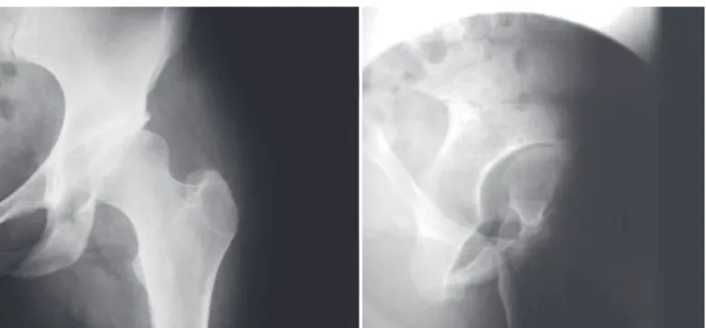
In our study, we evaluated the percentage of loss of the humeral head diameter, the depth of the HS lesion and the residual articular arc through the axial section with the largest

No statistically valid correlation was observed be- tween the subscapularis tendon lesion and its corres- ponding repair, with the occurrence of residual Popeye deformity (p =

We consider this an initial study on the adjuvant therapeutic approach using low-level laser therapy in the postoperative period of carpal tunnel syndrome. Other

WeitereBeitrage zu tramatish-mechanischen Enstenhung der “spontanen” Knorpelalblosungen (sogen. Osteochondritis dissecans). Transchondral fractures of the talus. Ferkel

The second group comprised patients treated using the posterior route associated with Smith-Petersen osteotomy, with a mean age of 27.3 years, preoperative kyphosis of 72.9°,

The results obtained in this study demonstrate that the bone marrow centrifugate was not able to induce a better quality of muscle healing, increasing the number of

Study conducted at the Knee Surgery Group of the Orthopedics and Traumatology Clinic of Hospital São Lucas da Pontifícia Universidade Católica do Rio Grande do Sul..

Miller et al (7) presented a report of fracture of the medial and lateral extremity of the clavicle with the use of a reconstruction plate and another with locking T-plate,