10 artigo 475
ORIGINAL ARTICLE
1 – Head Surgeon of the Shoulder Group of the Lifecenter, Belo Horizonte and Clínica Ortopédico-BH hospitals – Belo Horizonte, MG, Brazil. 2 – Third-year Resident of Hospital Belo Horizonte – Belo Horizonte, MG, Brazil.
3 – Surgeon of the Shoulder Group, Hospital Lifecenter and Clínica Ortopédico-BH – Belo Horizonte, MG, Brazil. Study conducted at the Lifecenter, Belo Horizonte and Clínica Ortopédico-BH hospitals– Belo Horizonte, MG.
Mailing address: Rua Vicente Guimarães, 35/1.002, Belvedere – 30320-640 – Belo Horizonte, MG. Email: [email protected] Study received for publication: 2/13/2011, accepted for publication: 5/25/2011..
“ROCAMBOLE-LIKE” BICEPS TENODESIS:
TECHNIQUE AND RESULTS
Glaydson Gomes Godinho1, Fabrício Augusto Silva Mesquita2, Flávio de Oliveira França3, José Márcio Alves Freitas3
7KHDXWKRUVGHFODUHWKDWWKHUHZDVQRFRQIOLFWRILQWHUHVWLQFRQGXFWLQJWKLVZRUN
This article is available online in Portuguese and English at the websites: www.rbo.org.br and www.scielo.br/rbort ABSTRACT
Objective: To present a new technique for bicipital teno-desis and its results: accomplished partially via arthroscopy and grounded in concepts of the normal and pathological anatomy of the tendon of the biceps long head. It is based on the predisposition of this tendon towards becoming atta-ched to the intertubercular sulcus after rupture or tenotomy (auto-tenodesis). Methods: Evaluations were conducted on 63 patients (63 shoulders), aged from 32 to 77 years (average 55), consisting of 32 females (51%) and 31 males (49%). Thirty-five of the patients (55.6%) were over 60 years of age and 28 patients (44.4%) were under 60 years of age. Eighteen were sports participants (28.6%). Fourteen had in-juries associated with the subscapularis (22.2%). The average follow up was 43 months (ranging from 12 to 74 months). The right shoulder accounted for 48 cases (76.2%), of which one was a left-handed individual and 47 were right-handed. The left shoulder accounted for 15 (23%) of the patients, of whom two were left-handed and 13 were right-handed. There were no bilateral occurrences. The statistical analysis were
done using SPSS version 18. Pearson’s chi-square test and continuity corrections were used to investigate the statistical significance of associations between variables. Associations were taken to be statistically significant when p was less than 0.05. Results: Residual Popeye deformity was perceived by seven patients (11.1%); it was only observed by the examiner in 15 cases (23.8%); and neither the patient nor the examiner observed it in 41 cases (65%). There were no statistically valid influences from age, participation in contact or thro-wing sports, subscapularis tendon-associated injury or Popeye deformity. Fifty-eight patients (92.06%) were satisfied, two patients were dissatisfied (3.17%) and three patients were indifferent (4.76%). Conclusion: The technique presented high patient satisfaction rates (92.06%) and residual defor-mity was perceived by 11.1% of the patients. The appearance did not have any statistically valid correlation with ages over or under 60 years (p = 0.883), sports practice (p = 0.195) or subscapularis-associated injury (p = 0.958).
Keywords – Arthroscopy; Shoulder/injuries; Shoulder/surgery
INTRODUCTION
The origin of the biceps tendon is located in the supraglenoid tubercle in 30% of cases; in the labrum, in 45%; and mixed in the labrum and tu-bercle, in 25%. Its course is oblique in the joint, emerges under the transverse humeral ligament in-side the intertubercular groove, is intra-articular and extrasynovial. The average length is 102 mm (89-146 mm). The proximal area of the glenoid (ar-ticular) averages 8.4 x 3.4 mm. At the exit point of the intertubercular groove (extra-articular) it mea-sures 4.5 x 2.1 mm(1).
The long head of biceps tendon is a frequent cause of pain in the shoulder.
The pathology of the long head of biceps tendon can be a result of trauma or micro-instability, or a consequence of a chronic inflammatory process due to excessive or degenerative use.
Figure 1 – 7UDQVIL[DWLRQRIWKHELFHSVWHQGRQZLWKGRXEOHURZ UHSDLUVXWXUHDSSUR[LPDWHO\FPIURPLWVRULJLQ
Lesions vary in degree from tendinitis, delami-nation and subluxation on the medial border of the intertubercular groove, to its full dislocation, which can also cause a glenohumeral articular block(3).
It was demonstrated that the diameter of unhealthy tendons is significantly larger than in normal tendons, and that the force required to cause the distal migra-tion of the biceps is also significantly greater than that necessary in normal tendons(1,2).
Multiple surgical techniques were described for the treatment of bicipital degeneration with the per-formance of tenodesis(4,5).
Tenodesis was proposed by several authors as an alternative to avoid the cosmetic alteration caused by residual deformity (“Popeye”) and possible muscle discomfort after tenotomy(4).
Some techniques use the fixation of the tendon in the intertubercular groove, while others use fixation in soft parts, partially or totally by arthroscopic route. In-terference anchors are generally used in these cases(5). The aim of this study is the presentation of a tenode-sis technique developed by the institution’s group of shoulder specialists, based on the normal and patho-logical anatomy of the long biceps tendon. The shape formed by suturing the stump on itself suggests the figure of a “Swiss roll”, exacerbating the diameter of the articular extremity of the tendon, which is normally larger than the extra-articular diameter. Consequently, there is an even more pronounced block of its distal migration, performed in a simple, low-cost manner.
METHODS
A retrospective study was conducted with 81 pa-tients (81 shoulders), operated in the period between 4/23/03 and 7/15/09, on whom we performed biceps tenodesis, according to the technique to be presented. The study was authorized by the medical ethics committees of the institutions involved.
The criteria of indication of the bicipital tenodesis were: 1) patients submitted to the treatment of dege-nerative lesions involving approximately 50% of the diameter of the long head of biceps tendon, associated with rotator cuff lesions or separate; 2) subluxations and dislocations of this tendon(6).
Surgical technique
Patient positioned in lateral decubitus with vertical and longitudinal traction mechanisms on the arm to be operated. General anesthesia associated with brachial plexus block.
Classic arthroscopic posterior, lateral, anterior su-perior and anterior inferior portals.
Joint inspection and identification of the bicipital lesion and its extension, besides that of the associated lesions(7).
The biceps tendon is initially transfixed with dou-ble-row repair sutures, approximately 1.0 cm from its origin in the supraglenoid tubercle and upper labrum. Number 1, monofilament suture thread is used for this purpose, replaced after the passage of number 2 nonabsorbable thread (Figure 1).
The tendon is sectioned close to the upper labrum, taking care to preserve its length as much as possible (Figure 2).
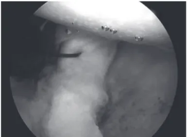
After the tenotomy, the arm is removed from the longitudinal traction mechanism, the elbow is flexed to relax the biceps and the tendon is pulled outside through the anterior superior portal and maintained with maximum exposure while being fixed with a pair of forceps. At this time, the extremity is rolled around itself and transfixed with number 2 nonabsorbable thread, producing a configuration that resembles a “Swiss roll”, after which the technique was named (Figures 3A and 3B).
Figure 2 – %LFHSVWHQRWRP\
Figure 3A – ([WHUQDOL]DWLRQDQGVXWXULQJRIWKHWHQGRQVWXPS RQLWVHOI
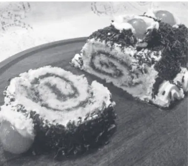
Figure 3B – 6ZLVVUROOWKHGLVKZKRVHDSSHDUDQFHLQVSLUHGWKH QDPHRIWKHWHFKQLTXH
the tendon stump migrates to the entrance of the intertubercular groove, where it is blocked. This is followed by the performance of the rotator cuff repair procedure, when necessary.
In the case of deinsertion of the subscapularis ten-don, this tendon is reinserted prior to the bicipital tenodesis.
The immobilization time, when dealing with an isolated procedure of the biceps, is three weeks with the use of a velpeau sling. Flexion/extension exercises are allowed once a day, with restriction of the last 30 degrees of extension to avoid forcing the distal migration of the biceps.
When isolated, tenodesis allows patients to resume contact and throwing sports 90 days after surgery. When associated with rotator cuff repair procedures, this resumption will depend on the extension of the cuff lesion, whose protocol is resumption of sports six months after surgery.
The exclusion criteria were: patients with follow-up time of less than 12 months after surgery, those who did not attend the clinical revision and did not present complete medical records.
A total of 63 patients (77.8%), with 31 (49%) ma-les and 32 (51%) femama-les, were evaluated. Age ranged from 32 to 77 years, averaging 55 years. Thirty-five patients (55.0%) were aged 60 years or over.
The minimum follow-up was 12 months, avera-ging 43 months, with a maximum of 74. The right shoulder corresponded to 48 cases (76.2%), with one left-handed patient and 47 right-handed patients. The left shoulder represented 15 (23.8%), with two left--handed patients and 13 rightleft--handed patients. There was no bilateral occurrence.
Eighteen patients (28.6%) played sports with direct involvement of the arms.
In 14 patients (22.2%) there was concomitant dein-sertion of the subscapularis tendon.
Table 1 – $QDO\VLV RI WKH FRUUHODWLRQ EHWZHHQ WKH REMHFWLYH HYDOXDWLRQRIWKHSDWLHQWDQGRIWKHH[DPLQHUZLWKUHJDUGVWRWKH SUHVHQFHRI3RSH\HGHIRUPLW\
Objective evaluation of the examiner Without Popeye
Objective evaluation of
the patient Total P-value Without
Popeye Popeye
:LWKRXW
3RSH\H &DVHV
41 0 41
3RSH\H &DVHV 7 22
7RWDO &DVHV 7
Pearson’s chi-square test, with continuity correction, of the association between the objective evaluation of the examiner and the objective evaluation of the patients from the sample.
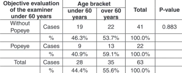
Table 2 q 2FFXUUHQFH RI 3RSH\H GHIRUPLW\ DFFRUGLQJ WR WKH H[DPLQHUUHODWHGWRWKHYDULDEOHDJHRYHURUXQGHU\HDUV
Objective evaluation of the examiner
under 60 years
Age bracket
Total P-value under 60
years
over 60 years
:LWKRXW
3RSH\H &DVHV 22 41
3RSH\H &DVHV 22
7RWDO &DVHV
3HDUVRQVFKLVTXDUHWHVWZLWKFRQWLQXLW\FRUUHFWLRQRIWKHDVVRFLDWLRQEHWZHHQWKHREMHFWLYH HYDOXDWLRQRIWKHH[DPLQHUDQGWKHDJHEUDFNHWRIWKHSDWLHQWVIURPWKHVDPSOH
fatigue and influence of the associated subscapularis tendon lesion and of playing sports involving direct action of the arms on the results were also evaluated. The patients were asked the following question: “Do you notice any modification in the contour of your operated arm in comparison to the non-operated arm? Are you satisfied, dissatisfied or indifferent in relation to your arm’s appearance?”
The examiner was expected to verify the presence or absence of residual deformity in the arm, as well as pain and/or muscle fatigue, and to perform the clini-cal evaluation of elbow and forearm flexion strength.
STATISTICAL ANALYSIS
The statistical analysis was carried out according to the resources of version 18 of the SPSS statistical program. Descriptive measures (mean and standard deviation, minimum and maximum) were used for the quantitative variables with frequency distributions for the qualitative variables(8,9).
Contingency tables were used to associate the observer’s objective evaluation with the following variables: age bracket, physical activity, presence of lesions of the subscapularis tendon in the operated patients and objective evaluation of the patients. Pear-son’s chi-square test, with continuity correction, was adopted to test the statistical significance of the as-sociation between and among such variables.
A significance level of 5% was considered in all the statistical tests used. Therefore associations whose p value was below 0.05 are considered statistically significant(8,9).
RESULTS
The results indicated that there is correlation between the evaluation of Popeye deformity car-ried out by the observer (examiner) and the patient. However, the observer’s evaluation to identify the presence of Popeye is more concise than that of the patient (p = 0.001) (Table 1).
We were able to observe that the variable age of patients did not have statistical significance in the cross-evaluation with the occurrence of Popeye de-formity (p = 0.883) (Table 2 and Figure 4).
The statistical analysis showed absence of valid cor-relation between the practice of sports and the occurrence of Popeye deformity (p = 0.195) (Table 3 and Figure 5).
No statistically valid correlation was observed between the subscapularis tendon lesion and its cor-responding repair with the occurrence of residual Pop-eye deformity (p = 0.958) (Table 4).
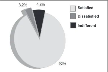
Fifty-eight patients (92%) declared themselves satisfied; two (3.2%) dissatisfied; and three (4.8%) indifferent.
No difference of strength of elbow flexion or forearm supination was observed during the physical evaluation in any patient.
Weak residual pain was reported by two patients, which correspond to the unsatisfactory results (3.2%) (Figure 6).
Table 3 – $QDO\VLVRIWKHFRUUHODWLRQEHWZHHQSK\VLFDODFWLYLW\ DQG3RSH\HGHIRUPLW\DFFRUGLQJWRWKHH[DPLQHU
Objective evaluation of the observer
Physical activity
Total P-value
No Yes
:LWKRXW
3RSH\H &DVHV 41
3RSH\H &DVHV 22
7RWDO &DVHV
Figure 5 – $QDO\VLVRIWKHFRUUHODWLRQEHWZHHQSK\VLFDODFWLYLW\ DQG3RSH\HGHIRUPLW\DFFRUGLQJWRWKHH[DPLQHUDQGWKHSDWLHQW S
Figure 6 – 'HJUHHRIVDWLVIDFWLRQRIWKHSDWLHQWVDFFRUGLQJWRWKH HVWKHWLFDQGIXQFWLRQDOUHVXOW
DISCUSSION
The long head of biceps tendon is a frequent cause of pain in the shoulder. Gilcreest(10) was the first au-thor to describe tenodesis of the long head of the bi-ceps, fixing it in the coracoid process.
Since then, multiple techniques have been de-scribed, some with fixation of the tendon in the inter-tubercular groove(11,12) and others with fixation in soft parts, partially or totally by arthroscopic approach(13). Almeida et al(14), in analyzing a group of 77 pa-tients submitted to arthroscopic tenotomy of the long head of biceps tendon, noted that there were no com-plaints of esthetic deformity in 50 (64.9%) patients, while 27 (35.1%) expressed some complaint.
In our sample, we observed that the patient per-ceived residual Popeye deformity after tenodesis in 11.1% of the cases (seven patients). The complete absence of residual deformity was observed in 65% (41 patients). The results indicated that there is a re-lationship between the evaluation of the observer and that of the patient; however, the observer’s evaluation to identify the presence of Popeye is more concise than that of the patient (p = 0.001) (Table 1).
Szabó et al(3) used the criterion of age to indicate tenotomy or tenodesis, preferring tenodesis for more active patients under 60 years of age. The same authors noted that patients aged between 45 and 55 years appear more concerned about the cosmetic aspect of the residual deformity after spontaneous rupture of the biceps long head, which is not the case in patients over 65 years of age, partly due to the atrophy and loss of muscle tone related to age(3).
Table 4 – $QDO\VLVRIWKHFRUUHODWLRQEHWZHHQ3RSH\HGHIRUPLW\ DQGOHVLRQRIWKHVXEVFDSXODULVWHQGRQ
Objective evaluation of the observer
Subscapularis lesion
Total P-value
No Yes
:LWKRXW3RSH\H &DVHV 11 41
3RSH\H &DVHV 17 22
7RWDO &DVHV 47 16
3HDUVRQVFKLVTXDUHWHVWZLWKFRQWLQXLW\FRUUHFWLRQRIWKHDVVRFLDWLRQEHWZHHQWKH REMHFWLYHHYDOXDWLRQRIWKHREVHUYHUDQGWKHVXEVFDSXODULVOHVLRQLQWKHSDWLHQWV IURPWKHVDPSOH
Figure 4 – 2FFXUUHQFH RI 3RSH\H GHIRUPLW\ DFFRUGLQJ WR WKH HYDOXDWLRQRIWKHSDWLHQWDQGRIWKHH[DPLQHUUHODWLQJWRWKHYDULDEOH DJHRYHURUXQGHU\HDUVS
Popeye deformity observed by the
patient
Popeye deformity observed only by
the observer
Absence of deformity observed by the patient and
observer Age 60 years
or over
Age under 60 years
Popeye deformity observed by the patient
Deformity observed only by
the observer
Absence of deformity observed
by the patient and observe Practicing sports
Not practicing sports
Satisfied
Dissatisfied
Besides the concern with the cosmetic aspect, the possible deficit of strength at the level of the elbow and forearm is a recommendation for tenodesis instead of tenotomy, in patients under 55 years of age(3).
Almeida et al(14) did not find statistical significance when they performed the cross-evaluation between the patients’ ages and the esthetic complaint of residual deformity.
The mean age of the patients in our sample was 55 years, ranging between 32 and 77 years. When we di-vided the study group into two subgroups, over and un-der 60 years of age, respectively, we were able to note that the patients’ age did not have statistical significance in the cross-evaluation with the occurrence of Popeye deformity (p = 0.883) (Table 2 and Figure 4).
Tenodesis is recommended in physiologically young, active patients under 50 years of age, either in associa-tion with or without arthroscopic subacromial decom-pression(15).
Eighteen patients played contact or throwing sports in our study group, corresponding to 28.6%. The sta-tistical analysis showed absence of valid correlation between these sports and the occurrence of Popeye deformity (p = 0.195) (Table 3 and Figure 5).
A question that is always raised in relation to the type of technique described concerns its safety when there is deinsertion of the subscapularis tendon, reinserting this tendon in the lesser tubercle in the same surgery.
This condition typically occurs in throwing athletes,
requiring the correction of both deformities(15).
In our sample, we observed the association of lesion caused by complete deinsertion of the subscapularis tendon and bicipital lesion, with performance of teno-desis in 14 patients (22.2%).
No statistically valid correlation was observed be-tween the subscapularis tendon lesion and its corres-ponding repair, with the occurrence of residual Popeye deformity (p = 0.958) (Table 4).
Walch et al(16) observed the presence of Popeye
de-formity in 50% of the patients submitted to arthroscopic tenotomy. However, it was observed that the cosmetic deformity of the arm does not usually cause the patient discomfort. Furthermore, no patient characterized its result as fair or poor with a basis on the cosmesis.
The degree of satisfaction of the patients showed 58 satisfied (92.06%), two dissatisfied (3.17%) and three patients (4.76%) who declared themselves indif-ferent with regards to the residual deformity (Popeye) observed (Figure 6).
CONCLUSION
“Swiss roll” biceps tenodesis presents high per-centages of satisfaction (92.06%); residual Popeye deformity is perceived by 11.1% of the patients. Its appearance has no statistically valid correlation with the age bracket over or under 60 years (p = 0.883), with sports (p = 0.195) or with the associated lesion of the subscapularis tendon and its repair (p = 0.958).
REFERENCES
1. Yamaguchi K, Bindra R. Disorders of the bíceps tendon. In: Ianotti JP, Willians GR Jr, editors. Disorders of the shoulder: diagnosis and management. Phila-delphia: Lippincott Williams and Wilkins; 1999. p. 159-90.
2. Ahmad SC, DiSipio C, Lester J, Gardner RT, Levine NW, Bigliani L. Factors affecting dropped biceps deformity after tenotomy of the long head of the biceps tendon. Arthroscopy. 2007;23(5):537-41.
3. Szabó I, Boileau P, Walch G. The proximal biceps as a pain generator and results of tenotomy. Sports Med Arthrosc Rev. 2008;16(3):180-6.
4. Boileau P, Krishnan SG, Coste JS, Walch G. Arthroscopic biceps tenodesis:a new technique using bioabsorbable interference screw fixation. Arthroscopy. 2002;18(9):1002-12.
5. Mazzoca DA, Bicos J, Santangelo S, Romeo AA, Arciero AR. The biome-chanical evaluation of four fixation techniques for proximal bíceps tenodesis. Arthroscopy. 2005;21(11):1296-306.
6. Crenshaw AH, Kilgore WE. Surgical treatment of bicipital tenosynovites. J Bone Joint Surg Am.1966;48(8):1496-502.
7. Godinho GG, Souza JMG, Bicalho LA. Reparo das rupturas do manguito rotador do ombro pela videoartroscopia cirúrgica: técnica. Rev Bras Ortop. 1996;31(4):284-8.
8. Cohen J. Statistical power analysis for the behavioral sciences. 2nd. ed. New Jersey: Editora Lawrence Erlbaum Associates; 1988.
9. Triola MF. Introdução à estatística. 7a. ed. Rio de Janeiro: LTC Editora; 1998.
10. Gilcreest EL. Two cases of spontaneous rupture of the long head of the bíceps flexor cubiti. Surg Clin North Am. 1926;6:539-54.
11. Lippman RK. Frozen shoulder, periarthritis, bicipital tenosynovitis. Arch Surg. 1943;47:283-96.
12. Hitchcock HH, Bechtol CO. Painful shoulder; observations on the role of the tendon of the long head of the biceps brachii in its causation. J Bone Joint Surg Am. 1948;30(2):263-73.
13. Mazzoca DA, Bicos J, Santangelo S, Romeo AA, Arciero AR. The biome-chanical evaluation of four fixation techniques for proximal biceps tenodesis. Arthroscopy. 2005;21(11):1296-306.
14. Almeida A, Roveda G, Scheifler C. Avaliação da deformidade estética após a tenotomia da cabeça longa do bíceps na artroscopia do ombro. Rev Bras Ortop. 2008;43(7):271-8.
15. Busconi BB, DeAngelis N, Guerrero EP. The proximal bíceps tendon:Tricks and pearls. Sports Med Arthrosc Rev. 2008;16(3):187-94.