12 artigo 371
ORIGINAL ARTICLE
1 – Orthopedist and Traumatologist; Head of the Foot and Ankle Surgery Clinic of the Instituto de Ortopedia e Traumatologia de Passo Fundo, RS; Member of the Sociedade Brasileira de Ortopedia e Traumatologia (Brazilian Society of Orthopedics and Traumatology) and of the Sociedade Brasileira de Cirurgia do Pé e Tornozelo (Brazilian Society of Foot and Ankle Surgery).
2 – Orthopedist and Traumatologist of the Vitória Apart Hospital – Vitória, ES; Specialist in Foot and Ankle Surgery; Member of the Sociedade Brasileira de Ortopedia e Traumatologia.
3 – Orthopedist and Traumatologist; Preceptor of the Instituto de Ortopedia e Traumatologia de Passo Fundo, RS; Member of the Sociedade Brasileira de Ortopedia e Trau-matologia and of the Sociedade Brasileira de Cirurgia do Joelho (Brazilian Society of Knee Surgery).
4 – Orthopedist and Traumatologist, Instituto de Ortopedia e Traumatologia de Passo Fundo, RS; Master’s and Doctoral Degree from the Universidade Federal do Rio Grande do Sul. Study conducted at the Instituto de Ortopedia e Traumatologia de Passo Fundo, RS.
Mailing address: Instituto de Ortopedia e Traumatologia de Passo Fundo, Rua Uruguai 2.050 – 99010-220 – Passo Fundo, RS, Brazil. Email: [email protected] / [email protected]
Study received for publication: 8/18/2010, accepted for publication: 5/25/2011.
TREATMENT OF OSTEOCHONDRAL LESIONS OF THE TALUS BY
MEANS OF THEARTHROSCOPY-ASSISTED
MICROPERFORATION TECHNIQUE
Everton de Lima1, Felipe de Queiroz2, Osmar Valadão Lopes Júnior3, Leandro de Freitas Spinelli4
7KHDXWKRUVGHFODUHWKDWWKHUHZDVQRFRQIOLFWRILQWHUHVWLQFRQGXFWLQJWKLVZRUN
This article is available online in Portuguese and English at the websites: www.rbo.org.br and www.scielo.br/rbort
ABSTRACT
Objective: To evaluate patients affected by osteochon-dral fractures of the talus who were treated surgically by means of arthroscopy-assisted microperforation. Methods: A retrospective study was carried out on 24 patients with osteochondral lesions of the talus who underwent micro-perforation assisted by videoarthroscopy of the ankle. They were evaluated using the American Orthopaedic Foot and Ankle Society (AOFAS) score system before and after the operation. Results: There were 19 men and 5 women, with a
mean age of 35.3 years (minimum of 17 years and maximum of 54 years). The minimum
follow-up was two years (maximum of 39 months). All the patients showed an improvement in AOFAS score after surgery, with an average improvement of around 22.5 points. Conclusion: Videoarthroscopy-assisted microperforation is a good option for treating osteochondral lesions of the talus and provides good functional results.
Keywords - Osteochondral; Talus/injuries; Talus/surgery; Arthroscopy; Ankle
INTRODUCTION
The evolution of orthopedics produced the develo-pment of minimally invasive surgical techniques for the diagnosis and treatment of orthopedic pathologies. Arthroscopic surgery of the ankle allows the approach to intra-articular structures without extensive inci-sions, increasing the diagnostic capacity and allowing the execution of less aggressive surgical correction techniques.
Munro(1), in 1856, was the first author to describe the existence of free bodies in the ankle joint. Barth(2), in 1898, considered the osteochondral lesion of the ta-lus as being an intra-articular fracture. Kappis(3) used the term “osteochondritis dissecans of the talus” for
703 MICROPERFORATION TECHNIQUE
that the most severely affected zones would be zone 4 (medial and central) and, in second place, zone 6 (mid-lateral). Medial lesions, besides being more fre-quent, are also larger and deeper than lateral lesions. Fractures of the lateral portion of the talar dome occur when inversion force affects the foot in dorsiflexion, while fractures of the medial portion are produced upon inversion on the foot in equinus(4).
Clinically, patients with osteochondral lesions of the talus refer to nonspecific pains of low intensity involving the ankle joint. They also report edema, clicking, blocks and a buckling sensation in the affec-ted ankle. A previous history of traumatism involving the ankle joint is common in most cases. Regarding the physical examination, patients generally present medial or lateral hypersensitivity in the ankle accom-panied by limitation of range of motion and edema, and may present signs of ankle instability(8).
The diagnosis of osteochondral lesions of the talus requires a high suspicion rate. The period between the onset of symptoms and the definitive diagnosis can range from four months to up to two years(9,10). In many cases, the radiological alterations are slight and only appear some months after the onset of the symptoms. Nowadays, computed tomography and particularly nuclear magnetic resonance of the ankle are essential in the early diagnosis of osteochondral lesions of the talus(8,11,12). The osteochondral lesion is classified radiologically, according to Berndt and Harty(4), in four stages: I – small area of compres-sion of subchondral bone; II – partially detached osteochondral fragment; III – completely detached osteochondral fragment; IV – displaced osteochon-dral fragment(4).
The treatment of osteochondral lesion of the ta-lus can be a major challenge due to the low intrinsic reparability of the articular damage(13). Most authors defend surgical treatment as the most appropriate means of treatment of osteochondral lesions of the talus(9,14-16), while some still believe that the conserva-tive treatment is the best conduct to be employed(17). In this context, videoarthroscopy of the ankle offers an adequate treatment with lower morbidity and an accelerated return to sports and daily activities(8), be-sides offering the chance of a careful articular inspec-tion and lavage after the procedure for removal of loose debris(18).
During the videoarthroscopy procedure, Van Ber-gen et al(18) suggest that the treatment of
osteochon-dral lesion defects smaller than 15 mm be executed by debridement and stimulation of the spongy bone. For cystic lesions, retrograde drilling associated with bone graft is a good alternative. Osteochondral autograft or autologous chondrocyte implantation are recommen-ded for secondary cases, as well as for larger lesions. Takao et al(19) propose the same procedure. Giza et al(13) also use the autologous chondrocyte graft, but in the treatment of patients who do not respond to cu-rettage of the cyst and subsequent microperforations. Nery and Carneiro(15) carry out the same procedure as an alternative method in cases in which the patient continues with complaints after the conservative or surgical treatment, also with good results.
More recently, Nery et al(20) analyzed the results of autologous chondrocyte implantation in patients submitted to previous surgical treatment without the obtainment of satisfactory results in terms of healing of the lesion and the remission of symptoms. The authors noted that autologous chondrocyte implanta-tion is a safe and effective method for the treatment of osteochondral lesions of the talus. Cohen et al(21)
also present autologous chondrocyte implantation as a promising technique for chondral lesions of the knee and of the talus. Other authors still advocate the use of mosaicplasty(22) or fresh talus allograft(23). Gras et al(24) propose the use of navigation in association with arthroscopy aiming to improve the accuracy of the retrograde perforation of cystic osteochondral lesions.
The objective of this study is to evaluate patients with osteochondral lesion of the talus treated with videoarthroscopy-guided microperforations.
MATERIALS AND METHODS
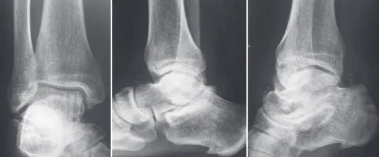
Figure 1 – $QWHURSRVWHULRUODWHUDODQGREOLTXHUDGLRJUDSKLHVVKRZLQJPLQLPXPDOWHUDWLRQRIWKHPHGLDOSRUWLRQRIWKHWDOXV
Figure 2 – 0DJQHWLFUHVRQDQFHLPDJLQJLQFRURQDODQGVDJLWWDOWRD[LDOFURVVVHFWLRQVVKRZLQJWKHRVWHRFKRQGUDOOHVLRQLQWKH
PLGPHGLDODVSHFWRIWKHWDOXV
All the patients were evaluated through anteropos-terior, lateral and oblique radiographies (Figure 1) and by nuclear magnetic resonance (NMR) (Figure 2). The scoring system of the American Orthopaedic Foot & Ankle Society (AOFAS)(25)was used for the functional evaluation of results.
SURGICAL TECHNIQUE
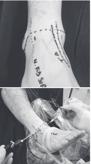
As regards anesthesia, the patients were submitted to epidural block or general anesthesia. A tourniquet was applied in the proximal region of the thigh and the lower limb was positioned in a leg holder. The ankle arthroscopy was executed through the antero-medial and anterolateral portals (Figure 3)(14). The surgeon used a scope with a diameter of 2.9 mm and
30° of angulation, accompanied by a compatible can-nula for visualization of the articular compartment. The evaluation of the cartilage condition was per-formed by direct visualization and through the use of a probe for palpation and evaluation of its integrity. A grasper is used to remove fragments and articular debris. The procedure was executed with cutaneous traction, without a distractor, performed by manual traction.
705
Figure 3 – 3RVLWLRQLQJ RI WKH DQWHURPHGLDO DQG DQWHURODWHUDO
SRUWDOV
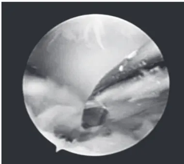
Figure 4 – 2VWHRFKRQGUDOOHVLRQ
Figure 5 – 2VWHRFKRQGUDOOHVLRQDIWHUGHEULGHPHQW
MICROPERFORATION TECHNIQUE
1.5 mm drill at low rotation (Figure 6). The tourni-quet was released after the complete performance of perforations to visualize whether the bleeding at the lesion site was satisfactory (Figure 7). After closing the portals, an occlusive dressing was applied in all the cases. The patients were kept without load for 45 days, but with early mobility through passive and active ex-ercises since the immediate postoperative period.
RESULTS
The study group consisted of 19 men and five women, with mean age of 35.3 years (minimum of
17 and maximum of 54), with minimum follow-up of two years (maximum of 39 months). Nineteen pa-tients (79.2%) reported ankle sprain, but were unable to define the sprain mechanism. The right side was the most affected (70.8%) (Table 1). None of the patients presented type I lesion according to the classification of Berndt and Harty(4), while 50.0% presented type II, 41.7% type III and 8.3% type IV.
Figure 6 – 3HUIRUDWLRQVRIWKHOHVLRQ
Figure 7 – )LQDODSSHDUDQFHDIWHUSHUIRUDWLRQV
and two patients (8.3%) in zone 7 (posteromedial). The affected surface was never larger than 1.2 mm.
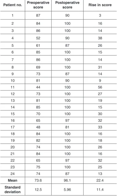
Table 2 contains the data referring to the results through an evaluation of the AOFAS score, obtained in the pre- and postoperative period of the osteochon-dral lesion of the talus. All 24 patients evaluated in the study presented a rise in the AOFAS score, averaging 22.4 points (± 11.4 standard deviation), changing the mean preoperative score from 73.6 (± 12.5) (mini-mum: 44/maxi(mini-mum: 87 points) to 96.1 points (± 5.96) (minimum: 81/maximum: 100). In the patients free
from complications (22 patients), there was a rise in the AOFAS score of 23.9 (± 10.6) points. Two patients exhibited superficial infection in one of the surgical portals. The diagnosis was made two weeks after sur-gery and resolved with the used of oral antibiotics for seven days. No patient required an additional surgical procedure.
DISCUSSION
Osteochondral lesions of the talus are hard to diag-nose and treat(6). The symptoms are usually nonspe-cific with late radiological findings. The use of com-puted tomography (CT) and of nuclear magnetic resonance (NMR) for the investigation of cases of pain in the ankle without a defined cause is essential for the diagnosis and early treatment of lesions. In the Table 1 – 'LVWULEXWLRQRIWKHDQDO\]HGSDWLHQWV
Nº Sex Age Side Lesion mechanism
Lesion site (zone)
Classification Follow-up (months)
1 M 5LJKW 6SUDLQ 4 II 2 M /HIW 6SUDLQ 4 III 24
) 5LJKW 6SUDLQ II 4 M 26 5LJKW 6SUDLQ 7 III 24
M 47 /HIW 6SUDLQ 4 III 6 M /HIW 6SUDLQ 6 IV 7 M 5LJKW 6SUDLQ 4 II
M 5LJKW 6SUDLQ 6 III 24
M 42 5LJKW )UDFWXUH 6 II 24 10 M 47 5LJKW 6SUDLQ 4 II 24 11 M 24 5LJKW 6SUDLQ 4 III 24 12 M 5LJKW 6SUDLQ 7 II 20
M /HIW 6SUDLQ 4 II 24 14 ) 20 5LJKW 6SUDLQ 4 II 24
) 5LJKW 6SUDLQ 4 III 24 16 M 5LJKW 6SUDLQ 4 II 17 ) /HIW 6SUDLQ III
) 20 /HIW 6SUDLQ 4 III 24
M 5LJKW 6SUDLQ 4 IV 26 20 M 17 5LJKW )UDFWXUH 6 III 24 21 M /HIW 6SUDLQ 4 III 24 22 M 5LJKW )UDFWXUH 6 II
M 40 5LJKW 6SUDLQ 6 II 24 24 M 5LJKW 6SUDLQ 4 II 24
0HDQIROORZXSqPRQWKV0HDQDJHq\HDUV:RPHQq0HQq 5LJKWVLGHq/HIWVLGHq
707 MICROPERFORATION TECHNIQUE
usual radiological views it is often difficult to locate a lateral or medial lesion, and to determine whether it is anterior, medial or posterior. NMR allows multiplanar evaluation and offers the advantage of visualizing the articular cartilage and the subchondral bone of the ta-lus, besides evaluating the edema and the surrounding soft tissues(8).
The analysis of age bracket, distribution between left and right sides, sex and the greater incidence in the medial talar dome in our sample coincide with the observations of other authors(14,17,26,27). A clinical his-tory involving ankle inversion followed by persistent chronic pain in the tibiotalar joint is the classical pre-sentation of osteochondral lesions of the talus. Ferkel
et al(8) found a previous history of trauma involving the ankle joint in 37 of the 50 patients reported in
their study. Anderson et al(26) reported osteochondral fractures of the talus in 57% of the patients with an-kle sprain. In our study, all the patients referred to a history of trauma in the origin of the osteochondral fracture of the talus.
Intra-articular lesions of the talus of any origin present a reserved prognosis(14,17). Invariably they cause pain, functional limitation and deterioration in the quality of life of patients. The option for ar-throscopic treatment for osteochondral lesions of the talus is due to the relative ease and low morbidity of this procedure. Although having imaging resources, at present there are not criteria able to predict the de-velopment of each case. It is known that there is pro-gression of fractures from the less severe to the most severe stages(28,29) and strong correlation between the size of the lesion and its prognosis(30,31). In this survey there was homogeneity in relation to the size of the lesions, which ranged between 0.8 and 1.2 mm. Most patients presented an improvement of symptoms, seen on the AOFAS scale.
Parisien(32) obtained 88% of good results after ar-throscopic treatment in 18 patients with osteochondral lesions of the talus. The treatment consisted of partial synovectomy, debridement of the osteochondral lesion and microperforations. Even after the short follow-up period, which varied from three months to three years, Parisien recommends arthroscopic excision due to the reduced morbidity, short hospitalization period and faster recovery. In another study, Ogilvie-Harris and Sarrosa(27) described significant improvement of pain, edema and claudication after arthroscopic treat-ment in 33 patients, consisting of the removal of the cartilaginous fragments, debridement and abrasion of the base until bleeding occurs in the subchondral bone. Nevertheless, the study reports persistence of pain in 24% of the patients due to loose chondral and osteochondral residual dendrites at the lesion site(27).
Various studies demonstrate good and excellent results after arthroscopic treatment of osteochondral lesions of the talus. However, it was observed that most studies present a short postoperative follow-up period(10,29,33-37). The importance of the follow-up pe-riod after the arthroscopic treatment of osteochondral lesions of the talus is emphasized in the study by Hunt and Sherman(35), in which 54% of the patients presen-ted unsatisfactory results after 66 months of follow-up. Table 2 – 7DEOHIRUHYDOXDWLRQRISDWLHQWVE\WKH$2)$6V\VWHP
Patient no. Preoperative score
Postoperative
score Rise in score
1
2 100 16
100 14
4
61 26
6 100
7 100 14
100
14
10
11 44 100
12 100 27
100
14 100
70 100
16
17
100 16
100
20 74 100 26
21 100 16
22
100
24 74
Mean
Standard
The increase of the mean AOFAS score obtained in this study is equivalent to most of the results encoun-tered in literature. The choice of the AOFAS table for evaluation allows us to evaluate the evolution of pain and of functions (distance walked, gait abnormalities, limitation of activities and need for support etc.)(25).
As regards the postoperative period, there is no consensus concerning the turnaround time required for the complete reestablishment of the osteochondral lesion. The information obtained through the simple radiological study during the clinical reviews is ques-tionable, since full repair of the bone defect is very slow and often fails to occur(17,32). Hyer et al(38)
men-tion that rehabilitamen-tion should be individualized by patient according to the physiotherapist, and can start after healing, which occurs between six and seven weeks for microperforation or internal fixation pro-cedures, and the patient should remain without sup-port in this period. The physiotherapy includes active and passive exercises for gain of mobility, control of edema, stretching and proprioceptive training.
CONCLUSION
The videoarthroscopy-assisted microperforation tech-nique consists of a good option for the treatment of osteo-chondral lesions of the talus and provides good functional results with an increase in the mean AOFAS score.
REFERENCES
1. Munro A. Microgeologie. Berlin, Germany: The Billroth; 1856.
2. Barth A. Die Enstenhung und das Wachsthum der freien Gelenkkorper. Arch Klin Chir. 1898;56:507-73.
3. Kappis M. WeitereBeitrage zu tramatish-mechanischen Enstenhung der “spontanen” Knorpelalblosungen (sogen. Osteochondritis dissecans). Dtsch Z Chir. 1922;171:13-29.
4. Berndt AL, Harty M. Transchondral fractures of the talus. J Bone Joint Surg Am. 1959;41:988-1020.
5. Ferkel RD, Sgaglione NA, Del Pizzo W. Arthroscopic treatment of osteochon-dral lesions of the talus: techinque and results. Orthop Trans. 1990; 14:172-3.
6. Roach R, Frost A. Osteochondral injuries of the foot and ankle. Sports Med Arthrosc. 2009;17(2):87-93.
7. Elias I, Zoga AC, Morrison WB, Besser MP, Schweitzer ME, Raikin SM. Osteo-chondral lesions of the talus: localization and morphologic data from 424 pa-tients using a novel anatomical grid scheme. Foot Ankle Int. 2007;28(2):154-61.
8. Ferkel DR, Zanotti MR, Komenda AG. Arthoscopic Treatment of chron-ic osteochondral lesions of the talus: long-term results. Am J Sports Med.
2008;36(9):1750-62.
9. Loomer R, Fischer C, Loyd-Smith R, Sisler J, Cooney T. Osteochondral Lesions of the talus. Am J Sports Med 1993;21(1):13-9.
10. Pritsch M, Horoshovski H, Farine I. Arthroscopic treatment of osteochondral lesions of the talus. J Bone Joint Surg Am. 1986;68(6):862-5.
11. Van Bergen CJA, De Leeuw PAJ, Van Dijk CN. Treatment of osteochondral defects of the talus. Rev Chir Orthop Reparatrice Appar Mot. 2008;94(8 Suppl):398-408.
12. Amendola A, Panarella L. Osteochondral lesions: medial versus lateral, persis-tent pain, cartilage restoration options and indications. Foot & Ankle Clin North Am. 2009;14(2):215-27.
13. Giza E, Sullivan M, Ocel D, Lundeen G, Mitchell ME, Veris L, Walton J. Matrix-induced autologous chondrocyte implantation of talus articular defects. Foot Ankle Int. 2010;31(9):747-53.
14. Ferkel RD. Artroscopy of the ankle and foot. In: Mann RA, Coughlim MJ. Sugery of the foot and ankle. 8th ed. Philadelphia: Mosby/Elsevier; 2007. p.1643-83.
15. Nery CAS, Carneiro MF. Tratamento artroscópico das fraturas osteocondrais do talo. Rev Bras Ortop. 1995;30(8):567-74.
16. White KS, Sands AK. Osteochondral lesions of the talus. Cur Orthop Pract. 2009;20(2):123-9.
17. Mukherjee SK, Young AB. Dome fractures of the talus a report of ten cases. J Bone Joint Surg Br. 1973;55(2):319-26.
18. Van Bergen CJA, Leeuw PAJ, Van Dijk CN. Potential pitfall in the microfracturing technique during the arthroscopic treatment of an osteochondral lesion. Knee Surg. Sports Traumat Arthrosc. 2009;17(2):184-7.
19. Takao M, Innami K, Komatsu F, Matsushita T. Retrograde cancellous bone plug transplantation for the treatment of advanced osteochondral lesions with large subchondral lesions of the ankle. Am J Sports Med. 2010;38(8):1653-60.
20. Nery C, Lambello C, Réssio C, Asaumi I. Implante autólogo de condrócitos no tratamento das lesões osteocondrais do talo. Rev ABTPe. 2010;4(2):113-23.
21. Cohen M, Nery C, Pecin MS, Réssio CR, Asaumi ID, Lombello CB. Implante autólogo de condrócitos para o tratamento de lesão do côndilo femoral e talo. Einstein. 2008;6(1):37-41.
22. .LOLo$ .DEXNoXRJOX< *O 0 2]ND\D 8 6|NF 6 (DUO\ UHVXOWV RI RSHQ mosaicoplasty in osteochondral lesions of the talus. Acta Orthop. Traumatol. Turc. 2009;43(3):235-42.
23. Hahn DB, Aanstoos ME, Wilkins RM. Osteochondral lesions of the talus treated with fresh talar allografts. Foot Ankle Int. 2010;31(4):277-82.
24. *UDV)0DULQWVFKHY,0OOHU0.ORV./LQGQHU50FNOH\7+RIPDQQ*2
Arthroscopic-controlled navigation for the retrograde drilling of osteochondral lesions of the talus. Foot Ankle Int. 2010;31(10):897-904.
25. Kitaoka HB, Alexander IJ, Adelar RS. AOFAS Clinical Rating Systems for the ankle-hindfoot, hallux and lesser toes. Foot Ankle Int. 1994;15(7):135-49.
26. Anderson LF, Crichton MB, Grattan-Smith MB, Cooper RA, Brazie D. Os-teochondral Fractures of the dome of the talus. J Bone Joint Surg Am. 1989;71(8):1143-52.
27. Ogilvie-Harris DJ, Sarrosa EA. Arthroscopic Treatment of Osteochondritis dis-secans of the talus. Arthroscopy 1999;15(8):805-8.
28. O`Farrell TA, Costello BG. Osteochondritis dissecans of the talus. J Bone Joint Surg Br. 1982;64(4):494-7.
29. Robinson DE, Wilson IG, Harris WJ, Kelly AJ. Arthroscopic treatment of osteo-chondral lesions of the talus. J Bone Joint Surg Br. 2003;85(7):989-93.
30. Chuckpaiwong B, Berkson EM, Theodore GH. Microfracture for osteochondral lesions of the ankle: outcome analysis and outcome predictors of 105 cases. Arthroscopy 2008;24(1):106-12.
31. Choi WJ, Park KK, Kim BS, Lee JW. Osteochondral lesion of the talus. Is there
a critical defect size for poor outcome? Am J Sports Med. 2009;37(10):1974-80. 32. Parisien JS. Arthroscopic treatment of osteochondral lesions of the talus. J
Sports Med Am. 1986;14(3):211-7.
33. Baker CL, Andrews JR, Ryan JB. Arthroscopic treatment of transchondral talar dome fractures. Arthroscopy. 1986;2(2):82-7.
34. Frank A, Cohen P, Beaufils P, Lamare J. Atrhroscopic treatment of osteochon-dral talar dome. Arthroscopy. 1989;5(1):57-61.
35. Hunt SA, Sherman O. Arthroscopic treatment of osteochondral lesions of the ta-lus with corelation of outcome scoring systems. Arthroscopy. 2003;19(4):360-7.
36. Kumai T, Takakura Y, Higashiyama I, Tami S. Arthroscopic drilling for the treatment of osteochondral lesions of the talus. J Bone Joint Surg Am. 1999;81(9):1229-35.
37. Van Buecken K, Barrack RL, Alexander AH, Ertl JP. Arthroscopic treatment of transchondral talar dome fratctures. Am J Sports Med. 1989;17(3):350-6.