1
Arquivos Brasileiros de Cardiologia - Volume 85, Nº 2, Agosto 2005
Clinical-Radiographic Correlation
Case 2/2005 - Forty Days Old Infant with Transposition of the Great Arteries and
Ventricular Septal Defect
Edmar Atik
Instituto do Coração do Hospital das Clínicas - FMUSP - São Paulo, SP - Brazil
Editor: Edmar Atik
Mailing address: Edmar Atik InCor Av. Dr. Eneas C. Aguiar, 44 -05403-000 - São Paulo, SP - Brazil - E-mail: [email protected] English version by Bridge Inglês Personalizado
Clinical data - A 40-day Caucasian female infant showing tiredness and cyanosis at crying since 3 days of life. Cardiac murmur was auscultated on third day of life. A low ponderal gain (440 g) was displayed in the first month. She was showing mild tachypnea, acyanotic, and normal pulse at physical examination. Her weight was 3,440 g, 156bpm heart rate, 60 bpm respiratory rate, blood pressure of 90/60 mmHg, and 88% oxygen saturation. The aorta was not palpated. Mild systolic impulsions at left sternal edge were detected in precordium and
ictus cordis was located in the 4th left intercostal space,
mus-cular +/++, limited by two digital pulps. Hyperphonetic sounds with the first more intense in mitral area and the second in tricuspid, in relation to pulmonary one. Full, rough, systolic murmur was found in the 3rd and 4th intercostal spaces, spreading
to the mitral and especially to the aortic area. Liver was palpated at 4cm from right costal fold. EKG displayed biventricular overload signs with RS complexes in V2, V3, and V4. SAP: +50o, SAQRS:
+120o, SAT: +70o.
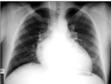
Radiographic Imaging - Showing cardiac area increase at right atrium and left ventricle’s expense, forming oval-shaped ima-ging. Narrow vascular peduncle and clearly increased pulmonary vascular network were detected (fig.1).
Diagnostic Impression - Imaging implying transposition of the great arteries associated to large interventricular communication-and/or artery canal-type intracavitary communication.
Differential Diagnosis - Tricuspid failure congenital cyanogenic cardiopathies, such as in Ebstein anomaly, distinguish from those followed by right atrial increase and with enlarged pulmonary vascular network. However, Ebstein anomaly is interventricular communication rarely associated. Total anomalous pulmonary venous drainage comes usually with right ventricular increase and cardiopathies restraining blood from right atrium, such as pulmonary stenosis-free tricuspid atresia.
Diagnostic Acknowledgement - Transposition of the great ar-teries with interventricular communication is proposed by clinical elements due to systolic murmur radiated to aortic area, with second hyperphonetic sound in heart failure neonate and with mild arterial unsaturation. Radiographic imaging confirms such deficiency association connected to 5.8 mm interatrial communi-cation, after Rashkind technique. A 9 mm long VSD was shown in outlet muscle position and pulmonary artery sprang mildly in ventricular septum. Catheterization showed the same aspects and there was also a left sinus-originated coronary ostium. Pressures found were as follows: RA: 10, LA: 5, RV: 60/10, LV: 60/5, PT: 60/20-33, Aorta: 60/40-47 mmHg. Both mean atrial pressures started to be 10 mmHg, after interatrial communication opening.
Procedure - Jatene-type anatomic correction was successfully indicated and carried out. Single coronary ostium originated at Valsalva sinus on the right, with left components behind dilated pulmonary arteries. Besides, two discontinued infundibula were between atrioventricular and semilunar valves.