1
Original Article
Comparative Study of 18 F-Fluorodeoxyglucose
Imaging with a Dual-Head Coincidence Gamma
Camera with Dobutamine Stress Echocardiography for
the Assessment of Myocardial Viability
Pedro Silvio Farsky, Leopoldo Soares Piegas, Vera Marcia Gimenes, Ana Claudia G. Petisco,
Paulo S. Duarte, Luiz Roberto Martins, Mario Issa, Paulo Paredes Paulista,
José Eduardo Moraes Rego Sousa
Instituto Dante Pazzanese de Cardiologia e Fleury - Centro de Medicina Diagnóstica - São Paulo, SP - Brazil
Mailing address: Pedro Silvio Farsky - Rua Caravelas, 423
- 04012-090 - São Paulo, SP - Brazil - E-mail: [email protected] Sent for publishing on: 06/23/2004
Accepted on: 02/23/2005
The benefit of myocardial revascularization (RM) surgery was first demonstrated in patients with severe left ventricular dysfunction with CASS registration1. A longer survival, in five years, was verified
in patients with left ventricular fraction of ejection (LVFE) lower than 0.26 and submitted to MR. However, those patients showed high surgical risk and higher operative complication rate.
At that time, akinetic myocardial segments were characte-rized as fibrotic, and therefore, insusceptible to improvement. However, Rahimtoola2 verified that some myocardial segments
recover contractility when properly reperfused. Those were called hibernating myocardia.
Identification of viable myocardium held important prognostic implications. Correction of arterial flow may lead to recovery of segmental, and even global, myocardial contractility, which pro-motes improvement of symptomatology and even survival3,4.
Myocardial viability can be detected by four different methods: metabolic function, cell membrane integrity, perfusion and con-tractile reserve. Metabolic function can be assessed through ca-pitation of 18 F-fluorodeoxyglucose (FDG). Cell membrane integrity can be assessed through tracer capitation, especially 201 thallium. Myocardial perfusion depends on microvascular integrity and can be assessed through contrast echocardiography. Contractile re-serve analysis was first assessed by postextrasystolic potentiation, during contrast ventriculography. However, the currently used me-thod is dobutamine stress echocardiography (DSE).
Among the methods mentioned, DSE and 201 thallium myo-cardial scintigraphy are the most commonly used in our milieu. DSE is widely employed due to its low cost in relation to the others and its availability in many cardiology centers. However, it does depend on operator’s experience and provides a lower sensi-tivity, especially in akinetic segments5. 201 thallium myocardial
scintigraphy is a strong scientific-based method, but when compared with positron emission tomography it shows lower sensitivity. The greatest part of fixed defects, from light to moderate level through 201 thallium myocardial scintigraphy, are regarded as viable by positron emission tomography (PET)6.
PET was regarded as gold standard for myocardial viability detection for long time. Moreover, its clinical application has been limited by short half-life and low availability of FDG, associated to equipment high cost. FDG capitation, through conventional gamma
Objective
To compare Dual-Head Coincidence Gamma Camera (DCD-AC) with Dobutamine Stress Echocardiography (DSE) in viability assessment, using functional recovery as the gold standard.
Methods
Twenty-one patients were prospectively studied, with coronary artery disease and severe left ventricular dysfunction undergoing DSE and DCD-AC at baseline and DSE three months after myocardial revascularization (MR).
Results
From 290 segments analyzed, 83% were akinetic, 15% hypokinetic and 2% dyskinetic at rest. DSE identified 68% of these segments as non-viable. DCD-AC identified 56% of these segments as normal (dysfunctional segments with preserved metabolism and perfusion), 30% as viable (preserved metabolism and reduced perfusion) and 14% as non-viable (absent perfusion and metabolism). Among DSE non-viable segments, DCD-AC classified 80% as normal or viable and 19.9% as non-viable (p<0.001). In hypokinetic segments viability and normal segments were detected in a higher proportion by both methods (p<0.001). DSE sensitivity and specificity were 48.3% and 78.1% respectively. DCD-AC sensitivity and specificity was 92.2% and 20.0%. DCD-AC identifies a higher incidence of function improvement in normal segments than in viable and non-viable.
Conclusion
DCD-AC classified as normal or viable most of the non-viable DSE segments. In assessment of functional recovery, three months after MR, DCD-AC showed a high sensitivity but low specificity.
Key words
2
cameras and use of ultra-high energy collimators, was initially used, but with limited image quality. With the event of scintillation cameras with coincidence principle and attenuation correction pro-grams (AC), a significant improvement in image quality was achieved, but such method is still with no validation in clinical practice.
Definition of myocardial viability involves recovery of segmental myocardial contractility with proper restoring revascularization of compromised segment. Therefore, different identification methods of myocardial viability must be compared with contractility recovery after surgical or percutaneous myocardial revascularization.
So, such work compared myocardial viability detection through capitation of FDG, through AC and DSE, in patients with severe left ventricular dysfunction. Functional recovery three months after MR was used as gold standard.
Methods
Twenty-one patients (20 men, mean of age of 58.6±10 years old) with multi-arterial coronary disease and severe left ventricular dysfunction, defined by LVFE <0.40, echocardiography at rest (Sim-pson method), submitted to MR, were prospectively studied. The study was carried out in patients with indication of MR and results from AC and DSE were not used for procedure definition. All patients were under sinus rhythm and were ill-taken from at least one myo-cardial infarction, with more than 15 days from the beginning of the study. Thirteen patients showed angina due to exercise and four did not show angina. Eight showed stable heart failure (class
≥ III New York Heart Association). Nine were diabetic and 15 hypertensive. Beta-blockers administration was interrupted on the day of DSE. The mean of LVFE was 0.32±5.9. There were no patients with significant valve disease or left ventricular aneurysm. The study was previously approved by the Ethics Committee in Research and information on free and clear consent was provided by all patients.
Myocardial viability was assessed by DSE, as by AC, before surgery. Regional and global contractility were assessed through echocardiography, at rest and under stress by dobutamine.
Myocardial contractility recovery was analyzed three months after myocardial revascularization, through transthoracic echocardiography at rest and through dobutamine stress echocardiography.
For assessment purposes, left ventricle was divided into 17 segments, according to standards by American Heart Association7.
With fasting patients, 50 g of glucose were administrated 50 minutes prior to FDG endovenous administration (2.22 MBq/Kg) and insulin administration. Images were acquired by using dual-head coinci-dence gamma camera operated through coincicoinci-dence mode, with correction attenuation (Vértex Plus MCD/AC ADAC Laboratories).
For perfusion study, Setamibi-[Tc-99m] (MIBI) (555 MBq) was utilized. Images were acquired with dual-head coincidence gamma camera, equipped with low energy and high resolution collimator (Vértex Plus MCD/AC).
Images were obtained in 48 projections with 180° 64x64 matrix. Transaxial tomography images were reconstructed through filtered back projection by using a Butterworth convolution filter with Autoespect plus software (Philips Medical System). Images with transmission attenuation correction were divided into short axle, long axle, long vertical axle and long horizontal axle.
Segments were qualitatively classified in accordance to MIBI or FDG uptake: normal = 0, discreet reduction = 1, moderate reduction = 2, important reduction = 3 and absent = 4.
Every myocardial was classified as normal, if perfusion was shown normal or discreetly reduced; as viable, if FDG uptake was relatively increased in a perfusion defect (perfusion-metabolism disagreement); and as non-viable, if there was reduced perfusion and FDG uptake (absence of perfusion-metabolism).
Segment contraction was assessed through transthoracic e-chocardiography (HP Sonos 5500 and ATL HDI 5000, Philips Me-dical System), according to recommendations from American E-chocardiography Society8. Myocardial contraction was assessed as
normal = 1, hypokinetic = 2, akinetic = 3 and dyskinetic = 4. Dobutamine infusion started from 5 µg/kg/min, then increasing to 10 and 20 µg/kg/min, every 3 minutes.
In pre-surgery DSE, segments were classified as: 1) normal, in the presence of preserved contraction at rest, with hyperdynamic response to dobutamine infusion; 2) viable, hypokinetic or akinetic segments with improvement of at least one point in contraction score due to dobutamine infusion; e 3) non-viable, segments with reduced contraction at rest and absence of improvement due to dobutamine infusion8. Segments classified as normal were excluded
from the analysis. Only segments with contraction changes at rest were considered.
In DSE, carried out three months after MR, segments were classified according to echocardiographic assessment compared to pre-operative period contractility, in: 1) functional recovery segments, which means segments that showed contractility improvement in relation to pre-operative period; 2) contractile reserve, segments without contraction improvement at rest, but with improvement due to dobutamine infusion; 3) absence of improvement; and, finally, 4) worsening of contraction in relation to pre-operative period. Thickening assessment, instead of contractility, was carried out at cipoal region, due to anomalous movement at post-operative period.
Quantitative data were described as mean ±1 standard devia-tion and qualitative data as propordevia-tions.
Chi-square test was used for comparisons between proportions, and the t-test of Student, was used for the analysis of quantitative variables. In the presence of two or more categories in at least one of the variable, residue analysis was applied for identification of more relevant proportions9. The level described of p<0.05 was
regarded as significant.
Results
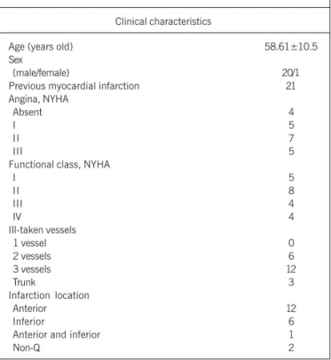
Clinical characteristics of patients are shown in table I. All patie-nts were ill-taken by previous myocardial infarction. Time interval between the infarction and their inclusion in the study was longer than 15 days. All patients had stable coronary artery disease. All patients showed significant obstruction in anterior descending artery and, at least, another artery with lesion higher than 75%.
3
Twenty patients had improvement in their functional level and none of them showed angina.
From a total of 357 segments, 51 were excluded for showing normal contractility at rest, 12 because they were not submitted to revascularization and four due to improper echocardiography analysis. From 290 assessed segments, 83% were akinetic, 15%, hypokinetic and 2% dyskinetic.
DSE classified 94 (32%) segments as viable and 196 (68%), as non-viable. On the other hand, AC classified 162 (56%) seg-ments as normal, 87 (30%), as viable and 41 (14%), as non-viable segments (fig. 1).
Among segments classified as viable through DSE, there was a greater incidence of normal and viable segments through AC (p<0.05) and, among segments classified as non-viable thr-ough DSE, there was a greater incidence of non-viable segmen-ts through AC (p<0.05).
According to contractility at rest, hypokinetic segments showed a greater incidence of viable segments through DSE and normal and viable segments through AC (p<0.001). A larger number of non-viable segments occurred among akinetic segments through both methods (p<0.006) (fig. 2).
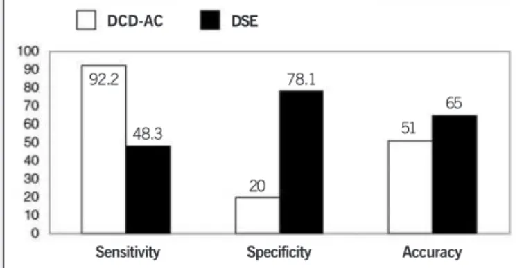
Regional contraction recovery, either at rest or under dobuta-mine, was detected in 116 (43%) segments. DSE identified 90 segments as viable, from which 56 recovered their contraction. AC identified 231 segments as normal or viable, from which only 107 recovered their contractility (fig. 3).
Discussion
Contractile reserve detection through DSE needs a preserved contractile system and, therefore, a greater quantity of func-tioning myocytes, whereas AC assesses the perfusion and meta-bolism of glucose.
In this work, myocardial viability detection through DSE and
DCD-AC was compared. Data obtained showed differences between the two methods, regarding sensitivity and specificity. DSE showed lower sensitivity, but its specificity was satisfactory, whereas AC showed high sensitivity, but reduced specificity. Results from DSE can be explained by to many factors. Studied population showed at least one previous myocardial infarction, multilateral coronary disease and a high proportion of akinetic segments (83% from dysfunction segments). Akinetic segments showed a larger quantity of fibrosis, dedifferentiation and loss of contractile elements7. Those
segments require a longer time to recover from post-MR myocardial contraction, and the three-month assessment may be underesti-mated the total number of those segments. Pervious studies8,9 in
patients with severe left ventricular dysfunction showed a lower prevalence of akinetic segments, which reflected the important ventricular dysfunction level among the assessed population.
Hypokinetic segments were mostly classified as viable, through DSE, and normal or viable, through DCD-AC. Obviously, those segments have preserved myocardial cells and sufficient contractile apparatus to allow for myocardial contraction. Some of those regions were considered as non-viable due to a discreet improve-ment of segimprove-ment contraction, which was not sufficient to change the contractility score during dobutamine infusion.
A significant number of segments show a normal or viable pattern through DCD-AC, without showing post-MR contraction
Table I - Data expressed through mean ± SD or number of patients. NYHA- New York Heart Association
Clinical characteristics
Age (years old) 58.61±10.5
Sex
(male/female) 20/1
Previous myocardial infarction 21 Angina, NYHA
Absent 4
I 5
II 7
III 5
Functional class, NYHA
I 5
II 8
III 4
IV 4
Ill-taken vessels
1 vessel 0
2 vessels 6
3 vessels 12
Trunk 3
Infarction location
Anterior 12
Inferior 6
Anterior and inferior 1
Non-Q 2
Fig. 1 - Number of normal, viable, and non-viable segments through DCD-AC. divided according to DSE viability (p<0.001). DSE = dobutamine stress echocar-diography. DCD-AC = dual-head coincidence gamma camera and attenuation correction programs.
Normal Viable Non-viable
Viable Non-viable
Fig. 2 - Number of normal, viable, and non-viable segments through DCD-AC, divided according to DSE viability and contractility detection (p<0.001). DSE = dobutamine stress echocardiography. DCD-AC = dual-head coincidence gamma camera and attenuation correction programs.
Viable Non-viable Viable Non-viable
Hypokinetic
Normal Viable Non-viable
4
improvement. Those findings can be explained by the following reasons: 1) hypokinetic segments were considered as non-viable due to discreet or absence of myocardial contraction improvement, during dobutamine infusion, which was insufficient to allow for a change of the echocardiographic score; 2) presence of viable tissues islands, intertwined by a significant quantity of fibrotic tissue, which did not allow for post-MR contractility improve-ment10, and 3) hibernating myocardium, with preserved FDG
up-take and that does not show response to inotropic stimulation, can have other changes of cell functions involved11.
One hundred and sixteen segments recovered their contractile function. DCD-AC showed good sensitivity, but reduced specificity, and DSE showed lower sensitivity and specificity than the literature. Those differences can be attributed to the great quantity of akinetic segments and to the short time interval for functional recovery. A significant number of truly viable segments was classified as non-viable through DSE and showed FDG uptake. That fact can be explained by the severity of changes in cardiomyocytes (damage and loss of myofibrils and increase of interstitial fibrosis), ill-taken level of coronary flow reserve and by the down regulation of β -receptors in patients with heart failure5,10. Additionally, the DSE
negative predictive value decreases with the increase in the number of akinetic segments11. Those results confirm data published by
Shimoni et al.12, with similar sensitivity and specificity.
Contractility assessment, three months after MR, may have underestimated the number of segments with functional recovery. Bax et al.13 studied 26 patients, before and after MR. Contractile
function was early (three months) and tardively (14 months) reas-sessed, after MR. Only 32% of hibernating segments showed improvement of contractile function at early assessment, with an additional improvement of 61%, tardively. Pagano et al.14 carried
out the control study after six months of MR, mentioning that a longer period would be necessary.
Accuracy of both techniques was compared with functional re-covery after MR. Despite such gold standard being imperfect, when placing excessive importance on sub-endocardium, it is the most suitable for clinical assessment15. That gold standard does not assess
remodeling, arrhythmias and prevention from new infarctions. Myocardial contraction assessment, under dobutamine infusion,
allows for identification of segments that show contraction recovery only under inotropic stimulus.
This study made use of assessment per segment, which is suitable for validation and comparison between different techniques. However, some studies use the improvement of fraction of ejection, measured at rest, as an indication of myocardial viability. The measurement of global left ventricular fraction of ejection has the influence of hypercontractile myocardial segments, which com-pensate for viable segments with contraction reduction. Improve-ment in viable segImprove-ment contraction with revascularization allows for hypercontractile segments retake over normal contraction, wi-thout modifying global fraction of ejection.
Due to the remarkable attenuation effect through positron annihilation energy transmission, attenuation correction program, through transmission method, is necessary to reach validation of positron emission tomography with dedicated equipment16.
This is the first study assessing Ac use in MR submitted patients, by using regional contractility recovery analysis as myocardila via-bility gold standard. This study allows for the analysis of results from this new technology in clinical practice.
Left ventricle was divided into 17 segments7. Such model
provides the best harmony with anatomical data and has the best adjustment with usually employed methods in echocardiography and in nuclear cardiology.
DSE interpretation is visual, therefore a dependent operator. Contractility division in four categories gathers, in the same cate-gory, segments with small contractility differences. As the con-tractility assessment is subjective, the division into a larger number of categories could lead to misinterpretation. The 17-segment model used may have caused some discrepancy in left ventricular division between different techniques.
Only lower doses of dobutamine were used. The utilization of high doses allows for identifying biphasic response, which has grea-ter sensitivity17. In post-operative assessment, the use of high doses
would allow for the identification of ischemic segments, occasioned by failure in revascularization. Even in lower doses of dobutamine, a high incidence of ventricular arrhythmias and hypotension took place. Lower doses area widely used for viability assessment18,19.
Functional recovery was assessed three months after surgical procedure. Akinetic segments may require a longer time for con-tractility recovery, but the necessary time is still unknown. A serial mode analysis would allow for a better information on the necessary time for myocardial functional recovery.
Both methods use din this study identified a greater pro-portion of viable and normal segments among hypokinetic seg-ments. Most DSE non-viable segments were classified as nor-mal or viable through AC.
In post-three-moth functional recovery, AC showed high sen-sitivity, but reduced specificity, whereas DSE showed low sensitivity and good specificity.
Acknowledgements
This study was funded by FAPESP through assistance-research (process 01/09471-9), and technical support from Fleury - Centro de Medicina Diagnóstica.
Fig. 3 - DSE and DCD-AC sensitivity, specificity and accuracy. DSE = dobutamine stress echocardiography. DCD-AC = dual-head coincidence gamma camera and attenuation correction programs.Fig. 3 - DSE and DCD-AC sensitivity, specificity and accuracy. DSE = dobutamine stress echocardiography. DCD-AC = dual-head coincidence gamma camera and attenuation correction programs.
Sensitivity
DCD-AC DSE
Specificity Accuracy
92.2
48.3
20 78.1
5
1. Alderman EL, Fisher LD, Litwin P et al. Results of coronary artery surgery inpa-tients with poor left ventricular function (CASS). Circulation 1983; 68: 785-95. 2. Rahimtoola SH. The hibernating myocardium. Am Heart J 1989; 117: 211-21.
3.Bounous EP, Mark DB, Pollock BG et al. Surgical survival benefits for coro-nary disease patients with left ventricular dysfunction. Circulation 1988; 78(3 Pt 2): I151-I157.
4. Lee KS, Marwick TH, Cook SA et al. Prognosis of patients with left ventricular dys-function, with and without viable myocardium after myocardial infarction. Relative efficacy of medical therapy and revascularization. Circulation 1994; 90: 2687-94. 5. Ausma J, Cleutjens J, Thone F, Flameng W, Ramaekers F, Borgers M. Chronic hi-bernating myocardium: interstitial changes. Mol Cell Biochem 1995; 147: 35-42. 6. Bonow RO, Dilsizian V, Cuocolo A, Bacharach SL. Identification of viable myocar-dium in patients with chronic coronary artery disease and left ventricular dysfunc-tion. Comparison of thallium scintigraphy with reinjection and PET imaging with 18F-fluorodeoxyglucose. Circulation 1991; 83: 26-37.
7. Cerqueira MD, Weissman NJ, Dilsizian V et al. Standardized myocardial segmenta-tion and nomenclature for tomographic imaging of the heart: a statement for healthcare professionals from the Cardiac Imaging Committee of the Council on Clini-cal Cardiology of the American Heart Association. Circulation 2002; 105: 539-42. 8. Schiller NB, Shah PM, Crawford M et al. Recommendations for quantitation of the left ventricle by two-dimensional echocardiography. American Society of Echocardiography Committee on Standards, Subcommittee on Quantitation of Two-Dimensional Echocardiograms. J Am Soc Echocardiogr 1989; 2: 358-67. 9. Pereira JCR. Análise de dados quantitativos. Estratégias metodológicas para as
ciências de Saúde, Humanas e Sociais. 1999.
10. Bristow MR, Ginsburg R, Minobe W et al. Decreased catecholamine sensitivity and beta-adrenergic-receptor density in failing human hearts. N Engl J Med 1982; 307: 205-11.
11. Panza JA, Dilsizian V, Laurienzo JM, Curiel RV, Katsiyiannis PT. Relation between
thallium uptake and contractile response to dobutamine. Implications regarding myocardial viability in patients with chronic coronary artery disease and left ven-tricular dysfunction. Circulation 1995; 91: 990-8.
12. Shimoni S, Frangogiannis NG, Aggeli CJ et al. Identification of hibernating myocar-dium with quantitative intravenous myocardial contrast echocardiography: com-parison with dobutamine echocardiography and thallium-201 scintigraphy. Circu-lation 2003; 107: 538-44.
13. Bax JJ, Visser FC, Poldermans D et al. Time course of functional recovery of stunned and hibernating segments after surgical revascularization. Circulation 2001; 104(12 Suppl 1): I314-I318.
14. Pagano D, Bonser RS, Townend JN, Ordoubadi F, Lorenzoni R, Camici PG. Pre-dictive value of dobutamine echocardiography and positron emission tomography in identifying hibernating myocardium in patients with postischaemic heart failure. Heart 1998; 79: 281-8.
15. Kaul S. There may be more to myocardial viability than meets the eye. Circulation 1995; 92: 2790-3.
16. Hasegawa S, Uehara T, Yamaguchi H et al. Validity of 18F-fluorodeoxyglucose imaging with a dual-head coincidence gamma camera for detection of myocardial viability. J Nucl Med 1999; 40: 1884-92.
17. Afridi I, Qureshi U, Kopelen HA, Winters WL, Zoghbi WA. Serial changes in re-sponse of hibernating myocardium to inotropic stimulation after revascularization: a dobutamine echocardiographic study. J Am Coll Cardiol 1997; 30: 1233-40.
18. Perrone-Filardi P, Pace L, Prastaro M et al. Assessment of myocardial viability in patients with chronic coronary artery disease. Rest-4-hour-24-hour 201Tl tomog-raphy versus dobutamine echocardiogtomog-raphy. Circulation 1996; 94: 2712-19. 19. Bax JJ, Cornel JH, Visser FC et al. Prediction of recovery of myocardial dysfunction
after revascularization. Comparison of fluorine-18 fluorodeoxyglucose/thallium-201 SPECT, thallium-fluorodeoxyglucose/thallium-201 stress-reinjection SPECT and dobutamine echocardiography. J Am Coll Cardiol 1996; 28: 558-64.