1
Arquivos Brasileiros de Cardiologia - Volume 85, Nº 2, Agosto 2005
Case Report
Takotsubo Cardiomiopathy. A Rare Cause of
Cardiogenic Shock Simulating Acute Myocardial
Infarction
Jayro Thadeu Paiva de Vasconcelos, Sebastião Martins, João Francisco de Sousa,
Antenor Portela
Hospital São Marcos - Teresina, PI - Brazil
Mailing address: Antenor Portela - Rua Aviador Irapuan Rocha, 2101/1002 - 64048-230 - Teresina, PI, Brazil
E-mail: [email protected] Received for publishing on 08/11/2004 Accepted on 01/21/2005
Takotsubo Cardiomiopathy is a rare cause of acute left ventricular aneurysm, in the absence of coronariopathy, only recently described in world literature. Symptoms may be similar to those from acute myocardial infarction with typical thoracic pain. The image of dumbbell or Takotsubo (a device used in Japan to capture octopus) suggestive ventricular ballooning is characteristic of that new syndrome and there is usually the disappearing of dyskinetic movement up to the 18th day from the beginning of the symptoms, in average.
We report a case of cardiogenic shock caused by an acute left ventricular aneurysm, similar to Takotsubo or Dumbbell, in a patient without obstructive coronary lesion. The case fulfills all criteria for Takotsubo cardiomiopathy, a pathology most frequent in Japan and that can simulate acute myocardial infarction.
Case Report
A 70-year-old, female patient, with precordial discomfort under constriction for 6 hours, without irradiation, followed by difficulty to breathe, with stressed worsening in the last 3 hours. In the morning before the beginning of the symptoms, the family informed and intense emotion motivated by family discussion. There was no report of morbid history or use of medications.
A patient showing stressed respiratory discomfort, pale, with abounding sudoresis. Tachycardic rhythmic sounds without other noises, bullous rales of medium and thin bubbles up to pulmonary apexes. Blood pressure was 90x60 mmHg, heart rate was 135 b.p.m, respiratory rate was 35 i.p.m., axillary temperature was 37°C. During the exam in the emergency room, the patient showed stressed worsening of respiratory discomfort, needing an urgent oro-tracheal intubation and mechanical ventilation. Dopamine IV was started. Electrocardiogram (ECG) of 12 derivations showed sinus tachycardia with non-specific changes of ventricular repolarization. Dosage of CKMB - mass collected at the admission was 22 u.
The patient was transferred to this service with the diagnostic hypothesis of non-Q infarction and cardiogenic shock.
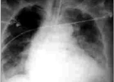
She was under mechanical ventilation, tachycardic with rhyth-mic sounds, heart rate of 145 b.p.m, bullous rales up to the upper third of both pulmonary fields, blood pressure was 80x50 mmHg. The thoracic radiography showed right pneumothorax, of moderate size and signs of pulmonary congestion (fig. 1). The ECG showed changes in ventricular repolarization and sinus tachycardia (fig. 2). The CKMB-mass was 29 u, creatinine of 1,2 mg% and glycemia 140 mg%. The pneumothorax was immediately drained. An echocardiogram performed by the bed, showed left ventricular aneurysm of anterior wall, compromising the middle and apical regions (fig. 3). After a fast hemodynamic stabilization with careful infusion of fluids, guided by the echocardiogram, ins-titution of dobutamine at 12 mcg/kg/min and noradrenaline at 8 mcg/min, the patient was sent to hemodynamics laboratory, where the coronary angiography showed coronary arteries wi-thout obstructive lesions (fig. 4) and the left ventriculography showed anterior wall aneurysm in a shape similar to Takotsubo or Dumbbell (fig. 5). The patient was kept under mechanical ventilation, with vasoactive drugs. Successive measurements of CKMB revealed a peak of 45 u in approximately 40 hours of evolution. After 48 hours there was an improvement of the features, with possibility of removal of mechanical ventilation and progressive discontinuity of vasoactive drugs. A new echocardiogram, by the bed, performed 72 hours after admission, did not show abnormalities of segmental contraction (fig. 6). The patient was discharged from ICU four days after her admission and was discharged on the tenth day, under use of nitrates, antagonist of calcium canals and acetylsalicylic acid.
Changes in segmental contractions without significant coronary lesions were already described, which may result from myocarditis, coronary spasm, pheochromocytoma and subarachnoidal hemor-rhage, more frequently1-4.
The presence of transient dyskinetic movement of left ventricular (LV) anterior wall, with stress of kinetics of ventricular base, as-sociated to thoracic pain, electrocardiographic changes that can vary from supraunlevelling of segment ST to discreet changes of ventricular repolarization and absence of obstructive coronariopathy, takes characteristics of syndrome and was first described by Satoh et al5. The symptoms can be similar to those of acute myocardial
infarction with typical thoracic pain6. The LV morphology at
2
Arquivos Brasileiros de Cardiologia - Volume 85, Nº 2, Agosto 2005
Takotsubo Cardiomiopathy. A Rare Cause of Cardiogenic Shock Simulating Acute Myocardial Infarction
(a device used in Japan to capture octopus), justifies its denomi-nation5,6. The reversibility of contractile change of LV and the
absence of significant obstructive coronariopathy are the impor-tant aspects for the diagnosis, and, on average, up to the 18th
day from the beginning of the symptoms the total reestablishing of the ventricular function is observed, with a variation from 3 to 50 days6,7. It is more usual in women after the 5th decade of
age, usually unleashed by emotional factors, surgery or acute disease8. Many cases have been described in Japan, United
Sta-tes and Europe8. In Brazil, there are few reports9. The
submis-sion with cardiogenic shock is particularly less frequent, which only occurs in 4% of the cases6,8.
The differential diagnosis must be done with pheochromocyto-ma, changes in segmental contraction secondary to encephalic vascu-lar accident and subarachnoidal hemorrhage, besides acute coronary
syndromes6,7,8. Upon the submission with supraunlevelling of segment
ST, differentiating such situation from acute myocardial infarction is practically impossible without the findings of coronary angiography and many of those patients have thrombolytic inadvertently9.
Here, the emotional stress was the unleashing event. Despite the association with spontaneous pneumothorax has already been found10, there is a strong evidence in that report suggesting
mothorax due to accident during central vain puncture, as pneu-mothorax was not seen in the first radiography. The patient was sent with thoracic draining, optimization of volemia and
vaso-Fig. 1 - Thoracic radiography showing pulmonary congestion and right pneumothorax.
Fig. 2 - ECG showing changes in ventricular repolarization on anterior wall.
Fig. 3 - Echocardiogram, at the bed, showing LV middle apical aneurysm.
Fig. 4 - Coronary angiographies showing arteries without obstructive lesions.
Fig. 5 - Left ventriculographies in systole and diastole showing LV aneurysm.
3
Arquivos Brasileiros de Cardiologia - Volume 85, Nº 2, Agosto 2005
Takotsubo Cardiomiopathy. A Rare Cause of Cardiogenic Shock Simulating Acute Myocardial Infarction
active drugs, for the stabilization of vital signs and fast sent to hemodynamics laboratory for a possible intervention. With the findings from coronary angiography and left ventriculography; ab-sence of neurological changes at the clinical exam, previous pa-thological history incompatible with pheochromocytoma, sudden start of symptoms, making improbable the diagnosis of myocarditis, the possibility of Takotsubo syndrome was raised. As the patient had her vital signs kept and having in view the reversibility of ventricular condition, the option was not instituting circulatory assistance with intra-aortic balloon. The clinical and echocardio-graphic evolution with complete resolution of the segmental con-traction change confirmed the diagnosis. The subsequent treatment is not totally defined yet, as there is who considers the non-utilization of other measures in addition to those of support, having in view the reversibility of the features, and who advocates the use of inhibitors of the angiotensin converter enzyme, beta-blockers and antagonists of calcium canals, whenever possible5,9,11.
The mechanism that leads to the bad acute ventricular perfor-mance in the syndrome of Takotsubo is unknown. Despite the spasm of the anterior interventricular artery has been initially conjectured6,7,
it was not confirmed in more judicious analyses performed afterwards, as the presence of coronary spasm in those cases was more sporadic than uniform7,12,13. The decrease of flow reserve and the increase
in time of coronary flow are always present. However, as they stay
after the normalization of the ventricular function, they do not totally explain the acute changes12. Specimens from posterior and apical
wall biopsy have shown a discreet interstitial fibrosis and cellular infiltrated7. Sorology for many viral agents, usually involved in
myo-cardial aggressions, were extensively investigated. However, it was not possible to establish a correlation with that nosologic entity7.
Scintigraphic analyses with 123IMIBG revealed a reduction in the retention of the radiotracer and an increase in its elimination in the apical region of the LV in acute stage, which suggested a disorder of adrenergic neurotransmission14.
Studies with technecium-99m showed a defect in the capture at LV apex, with normalization between 25 and 90 days, suggesting a mitochondrial transitory defect7,15.
Recently, concomitant analyses with thallium 201 and with a positron emission tomography (SPECT), using pentadecaenoic acid marked with iodine 123 (I-BMIPP), showed a defect of fatty acid metabolism in an area greater than that associated to the defect of perfusion, which suggested a more extensive metabolic disorder13.
Cardiomyopathy of Takotsubo or ventricular ballooning is a less frequent cause of left ventricular aneurysm, in the absence of obstructive coronariopathy, which can simulate acute myocardial infarction and that, in this report, had a greater manifestation cardiogenic shock. Its conduction is essentially done with hemo-dynamic support measures.
1. Miklozek CL, Crumpacker CS, Royal HD, Come PC, Sullivan JL, Abelmann WH. Myo-carditis presenting as acute myocardial infarction. Am Heart J 1988; 115: 768-76. 2. Shaw TRD, Bafferty P, Tait GW. Transient shock and myocardial impairment
cau-sed by pheochromocytoma crisis. Br Heart J 1987; 57: 194-8.
3. Kono T, Morita H, Kuroiwa T, Onaka H, Takatsuka H, Fujiwara A. Left ventricular wall motion abnormalities in patients with subarachnoid hemorrhage: neurogenic stunned myocardium. J Am Coll Cardiol 1994; 24: 636-40.
4. Braunwald E, Kloner RA. The stunned myocardium; prolonged, postischemic ven-tricular dysfunction. Circulation 1982; 66: 1146-9.
5. Satoh H, Tateishi H, Uchida T. Takotsubo-type cardiomyopathy due to multivessel spasm. In: Kodama K, Haze K, Hon M. (eds). Clinical aspects of Myocardial Injury: From Ischemia to Heart Failure. Tokyo: Kagakuhyouronsya Co. 1990: 56-64.
6. Tsuchihashi K, Ueshima K, Uchida T. Transient left ventricular apical ballooning wi-thout coronary artery stenosis: a novel heart syndrome mimicking acute myocar dial infarction. J Am Coll Cardiol 2001; 38: 11-8.
7. Abe Y, Kondo M, Matsuoka R, Araki M, Dohyama K, Tanio H. Assessment of cli-nical features in transient left ventricular apical ballooning. J Am Coll Cardiol 2003, 41: 737-42.
References
8. Seth PS, Aurigemma GP, Krasnow JM, Tighe DA, Untereker WJ, Meyer TE. A syndro-me of transient left ventricular apical wall motion abnormality in the absence of co-ronary disease; a perspective from United States. Cardilogy 2003; 100: 61-6. 9. Desmet WJR, Adriaenssens BFM, Dens JAY. Apical balooning of the left ventricle:
first series in white patients. Heart 2003; 89: 1027-31.
10. Akashi YJ, Sakakibara M, Miyake F. Reversible left ventricular dysfunction “takotsubo” cardiomyopathy associated with pneumotorax. Heart 2002; 87: e1. 11. Akashi YJ, Nakazawa, Sakakibara M, Miyake F, Koike H, Sasaka K. The clinical
features of takotsubo cardiomyopathy. Q J Med 2003; 96: 563-73.
12. Mesquita ET (http://esquina.cardiol.br/sbc.udt/colunas sbc/anteriores/09/003.asp-9kb) - SBC/RJ.
13. Kurisu S, Inoue I, Kawagoe T et al. Myocardial perfuison and fatty acid metabo-lism in patients with tako-tsubo like left ventricular dysfunction. J Am Coll Cardiol 2003, 41: 743-8.
14. Mesquita CT, Pessoa MCP, Felix RCM et al. Avaliação da neurotransmissão adre-nérgica cardíaca em pacientes com síndrome de takotsubo. Rev SOCERJ 2003; 16(supl. A): 97.