217
METABOLIC INVESTIGATION OF UROLITHIASIS Clinical Urology
International Braz J Urol
Official Journal of the Brazilian Society of Urology
Vol. 29 (3): 217-220, May - June, 2003
METABOLIC INVESTIGATION OF PATIENTS WITH UROLITHIASIS IN A
SPECIFIC REGION
LUIS A. B. PERES, ANDRÉ S. MOLINA, MARCOS H.L. GALLES
State University of West Paraná, UNIOESTE, Cascavel, Paraná, Brazil
ABSTRACT
Objective: To assess the prevalence of the main metabolic alterations found in patients with recent diagnosis of urolithiasis in the West region of Paraná state, Brazil.
Materials and Methods: We made a retrospective study on 425 patients with evidence of recent formation of renal stones. Laboratory assessment consisted in 3 samples of 24-hour urine with dosing of calcium, uric acid, citrate, oxalate, sodium and creatinine. A urine culture was also made and qualitative cystinuria and urinary pH following 12-hour fasting and water restriction were evalu-ated.
Results: In 96.5% of patients a cause was detected for the urolithiasis. Metabolic alterations most frequently found were: hypercalciuria (38.3%), hypocitraturia (29.6%) and hyperexcretion of uric acid (21.6%). Low urinary volume (17.9%), urinary tract infection (12.9%), hyperparathyroid-ism (3.3%), renal tubular acidosis (1.2%), cystinuria (0.9%) and anatomical alterations (12.7%) were also observed.
Conclusions: Hypercalciuria, hypocitraturia and hyperuricuria are the most frequent meta-bolic disorders in the population under study and these data are in accordance to the literature.
Key words: urolithiasis; metabolism; metabolic disease; risk factors; calcium oxalate; calcium Int Braz J Urol. 2003; 29: 217-220
INTRODUCTION
Renal lithiasis affects 12% of the United States of America population and its recurrence can reach 50%. Significant advancements in surgical ap-proach have occurred, but they did not change the natural history of urolithiasis (1,2). In Brazil there is a report that 5% of the population has urolithiasis, which corresponds to more than 7 million of patients with lithiasis. It is a pathology that affects young people, with its peak incidence during the third de-cade of life and a very high recurrence, reducing pro-ductivity of such individuals (3). The dissemination of preventive metabolic investigation programs and nephrologic approach is necessary, however the high costs of laboratory tests and the poor adhesion of
pa-tients to treatment and to dietary orientation limit the success of the medical approach.
The objective of the present work is to show the prevalence of major metabolic alterations found in patients with renal lithiasis coming from urban and rural zones of West region of Paraná state, Brazil.
MATERIALS AND METHODS
con-218
METABOLIC INVESTIGATION OF UROLITHIASIS
firmation of presence of stones in urinary tract in the past 6 months.
Laboratory investigation consisted in 3 blood and 24-hour urine samples, with a 30-day interval between them. Calcium, uric acid, citrate, sodium, creatinine and oxalate were evaluated in 24-hour urine, and calcium, uric acid, creatinine and parathor-mone in blood. Qualitative cystinuria, urinary pH following a 12-hour fasting and water restriction and urine culture were also performed.
General nutritional orientations were offered to all patients following the second samples collec-tion, consisting in: 6 grams of salt/day, 1 gram/Kg of weight of proteins, restriction of purines to 150 mg, adjustment of calcium intake to 1000 mg and increase in fluid intake to maintain a urinary volume above 20 mL/kg/day.
Laboratory methods employed and reference values adopted for 24-hour urine samples were: cal-cium – atomic absorption spectrophotometry method (< 4.0 mg/kg), uric acid – uricase enzymatic method (up to 750 mg for females and 800 mg for males), citrate – citrate-lyase enzymatic method (> 320 mg), sodium – selective ion method (< 150 mEq), creati-nine – alkaline picrate method (> 1000 mg) and uri-nary volume – volumetric measurement in Becker by visual analysis. For plasma dosing the methods employed were: calcium – colorimetric method (8.5 – 10.5 mg/dL), uric acid – uricase colorimetric method (2.0 to 7.0 mg/dL), creatinine – alkaline pi-crate method (0.7 a 1.4 mg/dL) e parathormone – intact molecule assay. For assays in single urine sample the methods were: qualitative cystinuria – sodium nitroprusside test, and urinary pH – mea-surement by reactive strips with methyl red and bromthymol blue indicator system. Urinary volume was considered to be decreased, when at least one of the samples presented a 24-hour urinary volume lower than 15 mL/kg.
RESULTS
1,023 patients with lithiasis were assessed out of a total of 5,207 attended patients, among whom only 425 concluded the metabolic study. Patients mean age was 32.2 years (ranging from 2 months to 67
years), 61.2% were male and 38.8% were female. As for race, 85% were Caucasian.
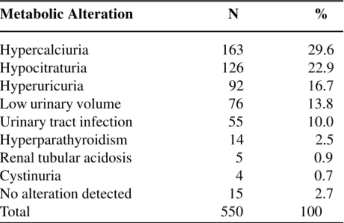
Among the 425 patients with lithiasis under study, at least one alteration was found in 410 (96.5%). 604 diagnoses were made, showing that some patients present more than one alteration. Metabolic alterations found were: hypercalciuria in 38.3%, hypocitraturia in 29.6%, hyperexcretion of uric acid in 21.6%, low urinary volume in 17.9%, urinary tract infection in 12.9%, hyperparathyroid-ism in 3.3%, renal tubular acidosis in 1.2% and cysti-nuria in 0.9% (Table-1). Anatomical alterations were found in 12.7%. In 3.5% of patients no alterations were found.
Among the anatomical alterations found (Table-2), we observed 22 renal cysts (40.7%), pyelocaliceal duplications (20.4%), 5 atrophic kidneys (9.3%), 5 stenoses of ureteropelvic junction -UPJ (9.3%), 5 single kidneys (9.3%), 3 neurogenic bladders (5.5%), 1 pelvic kidney (1.8%), 1 horse-shoe kidney (1.8%) and 1 medullary sponge kidney (1.8%).
DISCUSSION
Nephrolithiasis is a disease with high preva-lence and recurrence, being one of the most common diseases of the urinary tract (4). We do not have avail-able data about prevalence of renal lithiasis in the general population in the West region of Paraná state, Brazil. In this study we observed that approximately
Table 1 – Metabolic alterations found.
Metabolic Alteration N %
219
METABOLIC INVESTIGATION OF UROLITHIASIS
20% of attended patients in a general nephrologic out-patient service, which receives out-patients coming from urban and rural zones, have the diagnosis of urolithi-asis. The risk of terminal chronic renal insufficiency (CRI) is small in theses patients, but some conditions, if left untreated, present a high risk of evolving to renal failure (cystinuria, hyperparathyroidism, ox-aluria, etc.). The prompt recognition of such condi-tions is important to prevent CRI (5).
Urolithiasis affects preferably young males (6). In our study there was a predominance of male gender (61.2%) and patients mean age was 32.2 years, what is in accordance to the literature (7). There was a predominance of Caucasian race (85%), reflect-ing the racial distribution in our region, and it does not enable us to say that incidence is higher in this race.
We detected a causal alteration in 96.5% of patients. Metabolic alterations most frequently found were hypercalciuria (29.6%), hypocitraturia (22.9%) and hyperexcretion of uric acid (16.7%), data that is consonant with the majority of works, and it shows that there is a lack of balance between the promoters of stone formation and its inhibitors (7,8).
Decrease in urinary volume is considered a cause of lithiasis (9). In hot climate countries extra-renal losses and low fluids intake can contribute to stone formation. In this study we observed a decreased urinary volume in 13.8% of alterations, a much lower Table 2 – Anatomical alterations found in 12.7% of pa-tients with urolithiasis.
Anatomical Alteration N %
Renal cyst 22 40.7 Pyelocaliceal duplication 11 20.4 Atrophic kidney 5 9.3 Ureteropelvic junction stenosis 5 9.3 Single kidney 5 9.3 Neurogenic bladder 3 5.5 Pelvic kidney 1 1.8 Horseshoe kidney 1 1.8 Medullary sponge kidney 1 1.8 Total 54 100
index than the 77% index reported in the interior of São Paulo state, where climate is warmer (7).
Hypercalciuria is responsible for more than 50% of metabolic disorders in adults and 53 to 75% in children (10). It is thought to have a strong ge-netic component, probably with dominant autossomic inheritance (11). A sodium-rich diet is one factor to be considered in pathogenesis of hypercalciuria (12). In this study hypercalciuria was the prevalent metabolic disorder. In West region of Paraná state the ingestion of milk and dairy prod-ucts is small, but salt and protein intake is high, prob-ably contributing to occurrence of hypercalciuria. Oral calcium overload test was not routinely per-formed due to technical implications of the method and its cost, therefore patients were not classified according to type of hypercalciuria (renal or intesti-nal), because we understood that this classification does not change significantly the treatment of a pa-tient who has hypercalciuria.
Hypocitraturia is found in about 30% of pa-tients with lithiasis (13). In this study we observed this alteration in 22.9% of metabolic disorders. Hyperuricuria is due to a high intake of purines or an elevated endogenous production. Low intake of wa-ter and urinary pH < 5.5 favor the precipitation of uric acid (14). Hyperuricuria was evidenced in 16.7% of metabolic disorders in our patients, and we believe that the high regional protein intake is a risk factor. National literature observes this disorder, from 18% to 76% (7).
Hyperoxaluria is a rare disorder, and is found in approximately 1% of individuals under study (2), and it was not routinely investigated in our study. We understand that the lack of oxalate dosing in 24-hour urine did not compromise the diagnosis of the meta-bolic disorders reported here.
We believe that the right approach to theses disorders with a multidisciplinary team can reduce both incidence and recurrence of urolithiasis in our population.
220
METABOLIC INVESTIGATION OF UROLITHIASIS
REFERENCES
1. Pearle MS: Prevention of nephrolithiasis. Curr Opin Nephrol Hypertens. 2001; 10: 203-9.
2. Wilkinson H: Clinical investigation and management of patients with renal stones. Ann Clin Biochem. 2001; 38: 180-7.
3. Sakuno MLD, Akimoto LS, Mereles EAL, Modenuti MI, Vieira AGM, Dal Col SMD, et al.: Contribution of the laboratory for clinical analysis for the metabolic diagnosis of renal lithiasis. Ver Bras Anal Clin. 1994; 26: 77-80 [in Portuguese].
4. Giugliani MCK: Metabolic Evaluation of Renal Lithi-asis, Study in 100 Patients. Master of Science Thesis, Federal University of Rio Grande do Sul, 1990 [in Portuguese].
5. Gambaro G, Favaro S, D´Angelo A: Risk for renal fail-ure in nephrolithiasis. Am J Kid Dis. 2001; 37: 233-43. 6. Robertson WG, Peacock M, Baker M, Marshall DH, Pearlman B, Speed R, et al.: Studies on the prevalence and epidemiology of urinary stone disease in men in Leeds. Br J Urol. 1983; 55: 595-8.
7. Ayusso LL, Schor N: Evaluation of patients with renal lithiasis in tropical region. Br J Nefrol. 2001; 23: 205-12.
8. Tostes V, Cardoso LR: Recent advances in urolithi-asis. Br J Nefrol. 2001; 23: 166-73 [in Portuguese]. 9. Frank M, De Vries A, Lazebnik J, Kochwa S:
Epide-miological investigation of urolithiasis in Israel. J Urol. 1959; 81: 497-502.
10. Levy FL, Adams-Huet B, Pak CYC: Ambulatory evalu-ation of nephrolithiasis: an update of a 1980 protocol. Am J Med. 1995; 98: 50-8.
11. Coe FL, Parks JH, Moore ES: Familial idiopathic hypercalciuria. N Engl J Med. 1979; 300: 337-40. 12. Pak CYC, Resnick MI: Medical therapy and new
ap-proaches to management of urolithiasis. Urol Clin North Am. 2000; 27: 243-53.
13. Pak CYC: Etiology and treatment of urolithiasis. Am J Kidney Dis. 1991; 18: 624-37.
14. Low RK, Stoller ML: Uric acid related nephrolithi-asis. Urol Clin North Am. 1997; 24: 135-49.
Received: March 11, 2003 Accepted after revision: May 19, 2003
Correspondence address:
Dr. Luis Alberto Batista Peres Rua São Paulo, 769 / 901
Cascavel, Paraná, 85801-020, Brazil Fax: + 55 45 327-3413