ERM functions, EGF and orthodontic movement
or
Why doesn't orthodontic movement cause
alveolodental ankylosis?
Alberto Consolaro*, Maria Fernanda M-O. Consolaro**
Can orthodontic movement induce al-veolodental ankylosis? This question is often asked and the answer involves further ques-tioning: Why don't the teeth naturally evolve to alveolodental ankylosis if they are separated from the bone by only 0.2 to 0.4 mm (the minimum and maximum thickness of the peri-odontal ligament)?
The periodontal ligament is richly cellu-larized and vascucellu-larized, featuring numerous elastic and reticular collagen fibers, typical of connective tissues (Figs 1, 2 and 3). In be-tween these structures it has a "gel", namely, the extracellular matrix. Among the fibers, fi-broblasts, vessels and nerves of the periodon-tal ligament there is a network of epithelial cords and islands that continuously release mediators, especially EGF, i.e., Epithelial or Epidermal Growth Factor (Fig 2). Areas on the surface of the bone tissue that contain EGF stimulate bone resorption, hindering
the formation of new layers. This epithelium network interposed between bone and tooth in the ligament tissue is known as Epithelial Rests of Malassez (ERM), derived from apop-tosis in Hertwig's Epithelial Root Sheath (HERS). Malassez' original drawings (Fig 4) depicted these epithelial cords and islands in the same manner as we analyze them micro-scopically today.
It was long believed that ERM comprised latent or quiescent cells devoid of structure and function, often associated with the genesis of cysts and tumors. However, these epithelial periodontal components are active, produce mediators and fulfill key functions in main-taining periodontal health and root integrity even during orthodontic movement.
In this paper we will discuss these wonder-ful structures and their functions to assist us in understanding the relevant responses to the two questions posed above.
A
*
*
B
*
*
*
*
*
*
*
*
*
*
*
*
*
*
*
*
*
*
*
*
*
*
*
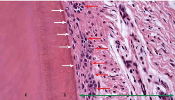
FIGURE 1 - On the root surface the cementum is covered by cementoblasts (white arrows). Collagen fibers—called Sharpey's fibers— penetrate amid these cells and attach themselves to the cementum (C). In the periodontal ligament (green arrow) epithelial cell islands and cords can be observed (red arrows) which form a three-dimensional network around the root, like a basketball hoop. This epithelial component of the periodontal ligament, called Epithelial Rests of Malassez (red arrows), constantly releases Epithelial (or Epidermal) Growth Factor (EGF), whose molecules diffuse through the cells in the extracellular matrix and stimulate osteoclasia on the periodontal bone surface, thereby promoting the maintenance of periodontal space (D = dentin; F = fibroblasts; V = blood vessels. HE; X25).
FIGURE 2 - Epithelial Rests of Malassez (red arrows) continuously release—for their maintenance and function—EGF molecules ( aste-risks) that diffuse throughout the extracellular matrix among the fibroblasts (F) and upon reaching the bone surface (B), stimulate osteoc-lasia to maintain the periodontal ligament (PL). A highlights the distance from the gingival (GE) and junctional (JE) epithelium to the alveolar bone crest (blue arrow), showing enough space for the EGF molecules to diffuse and be metabolized without causing underlying bone resorption. The green curly brace encompasses the connective attachment (white arrow = cementoblasts; purple arrow = osteoblasts;
C = cementum, D = dentin; V = blood vessel. Masson's Trichrome Stain, X10).
D C
V F
GE
JE
D C
B
PL
C
B
F
V
Epidermal Growth Factor (EGF) History and functions
Cells release EGF mediators to regulate and stimulate proliferation and differentiation, especially in epithelia.10,11,15 EGF's presence in
the body and in various body fluids is ubiq-uitous. It is found in urine (100 µg/ml), milk (80 µg/ml), saliva (12 µg/ml), plasma (2 µg/ ml) and amniotic fluid (1 µg/ml). The gene that controls EGF production in humans is on chromosome 4 and its molecule contains 53 amino acids with a molecular weight of 6,045 daltons. This molecule remains stable even in hot environments.
The specific receptors for this polypeptide (EGFr) consist of transmembrane proteins divided into three parts: extracellular,
trans-membrane and intracellular.15,16 When EGF
binds to the extracellular part of the recep-tor, the intracellular portion activates tyro-sine kinase and triggers cascading events that culminate in mitosis.10,11,12 EGFr is present in
epithelial cells of sites with high and low cell proliferation, high or low degree of differen-tiation.25 Other mediators also bind to EGFr
but induce different effects than those of EGF such as, for example, transforming growth fac-tor alpha or TGF-α. EGF receptors are part
FIGURE 3 - Epithelial Rests of Malassez (red arrows) stand out throughout the process of periodontal ligament (PL) reorganization during induced tooth movement and are usually associated with the repair of resorbed areas and with cementogenesis. This tooth appears as shown here after 7 days of experimental induced tooth movement in murines (white arrows = cementoblasts; purple arrow = osteoblasts; C = cementum; D = dentin; B = alveolar bone, P = pulp. HE; X10).
D
P
C B
FIGURE 4 - Epithelial Rests of Malassez (in red) in the periodontal liga-ment, redrawn from the original L. Malassez drawings (published in Arch Physiol Norm Pathol. 1885; 5: 309-340 6: 379-449) and republished by Racadot and Weill37 in 1966.
of a family of membrane receptors commonly referred to as EGFR1 or ERB B1. Receptor ERB B2, also known as HER-2/Neu, has at-tracted considerable attention because it is overexpressed in breast cancer and has been
considered a therapeutic target.41
EGF receptors are present in all oral tissue epithelia,58 including the junctional
epithe-lium.45 In other cells such as fibroblasts and
endothelial cells EGF also appears to act as a mitogen. However, EGFr has not been
detect-ed in pulp and periodontal tissues,58 but EGF
molecules have been detected in the interstice
of oral submucous connective tissue.42
Since it was first reported in 1962, EGF has played a role in regulating dental eruption and development.34,35,49,51 The first description of
EGF was provided by Stanley Cohen,15 who
identified it in the submandibular glands of rats, aiding in the acceleration of incisor erup-tion and the opening of newly-born rats' eyes. Cohen was awarded the Nobel Prize in
Medi-cine and Physiology in 1986.20 In 1989, Greg
Brown patented EGF for cosmetic use.8,53
The physiological importance of EGF in maintaining the integrity of oral tissues, both esophageal and gastric,19 is substantial. By
way of the saliva it helps to repair esopha-geal and gastric ulcers, inhibits gastric acid secretion and also stimulates DNA synthesis while protecting the mucosa against aggres-sive factors such as gastric acid, bile, pepsin and trypsin and against physical and bacterial
agents.54 EGF stimulates mitosis in a variety
of cell lineages such as the epithelium, fibro-blasts, chondrocytes, endothelium, smooth muscles and hepatocytes. Fibroblasts have 40,000 to 100,000 EGF receptors per cell. EGF stimulation requires the activity of at least 25% of those receptors.
EGF plays an essential role in tissue repair. In humans most of this substance is associated with platelets. It is synthesized by
megakaryo-cytes in the bone marrow3 and released in the
process of blood coagulation.29 A large amount
of EGF can be recovered from urine but it is almost entirely produced in the kidneys.
The presence of EGF in saliva and its prop-erties may explain some procedures adopted since 2,000 years ago in ancient Greece, when they applied snake saliva to open wounds to promote and accelerate tissue repair.2 When
EGF is produced in the salivary glands50 it is
excreted directly into the saliva.28,36 On
epi-thelial surfaces it stimulates the proliferation, differentiation, organization and keratiniza-tion of the superficial layers in the regenera-tive process of skin and mucosa ulcerations.8,31
A veritable EGF avalanche flows into the saliva after periodontal surgery and removal of impacted third molars.32,33 This EGF
in-crement is a response to the need to increase proliferation and differentiation, both phe-nomena typical of repair and regeneration. Ohshima et al31 also pointed out that salivary
dimensions are known as "biological distances": (a) Sulcus epithelium, (b) Junctional epithe-lium, and (c) The area of connective tissue at-tachment located in the root portion, positioned coronally to the alveolar bone crest21 (Fig 2).
The junctional epithelium and connective tissue attachment comprise the dentogingival complex.43 These structures have a constant
mean vertical dimension and were described by Gargiulo et al,21 who reported a microscopic
analysis of the dimensions and characteristics of the dentogingival junction in humans dur-ing the four phases of passive tooth eruption. 325 sound surfaces were obtained from human cadavers and analyzed, showing the following periodontal structure dimensions:
• Mean sulcus length: 0.69 mm;
• Mean junctional epithelium length:
0.97 mm;
• Mean supra-alveolar connective
attach-ment length: 1.07 mm (Mean connec-tive attachment length proved to be the most consistent measure).
When in an operative or restorative pro-cedure the connective attachment—which is the biological distance between the junctional epithelium and the alveolar bone crest—is "encroached upon", after a few days or weeks there will be noticeable resorption and loss of cervical bone level in the apical direction. This surgical encroachment of the biological space induces the junctional epithelium to proliferate and grow hyperplastically in or-der to keep the dentogingival junction cervi-cally at a more apical level. In other words, the junctional epithelium will move closer to the bone crest, and as it continuously produc-es EGF to maintain its structure under con-stant cell renewal, the concentration of this polypeptide increases to nearly bone level, stimulating bone resorption and lowering the alveolar bone crest. This mechanism also plays a key role in bone loss during periodontitis in EGF is linked to cancer etiology and
pathogenesis given its ability to boost DNA synthesis and stimulate cellular prolifera-tion.24 Thus, some medications are
primar-ily aimed at inhibiting EGF receptors in the oncological treatment of certain neoplasias. Monoclonal antibodies are substances used for this purpose.
In particular, EGF has shown a potent ac-tivity in inducing bone resorption,38,47
includ-ing in osteoclastogenesis.60 In mice deficient
in EGF receptors endochondral ossification proved to be severely altered by a defect in the recruitment of osteoclasts.57
EGF and biological distances
Nowhere in the human body is the epi-thelium a direct neighbor of bone tissue. Be-tween epithelium and bone tissue there is al-ways some connective tissue whose thickness and degree of fibrosis vary according to the different body parts (Figs 1 and 2).
The connective tissue interposed between epithelium and bone tissue may serve as a dilution and metabolism site for EGF, pre-venting it from reaching bone cell receptors in a high or average concentration, thereby stimulating osteoclastogenesis and the result-ing bone resorption38,47,60 (Fig 2). Every
mol-ecule has an average life and tends to be me-tabolized by enzymes and other products of cell and tissue metabolism. Molecules remain intact and capable of interacting with their receptors for a few seconds or minutes. Thus, we can explain why the spaces between epi-thelium and bone are usually constant or sta-ble in the human body such as between the junctional epithelium and the alveolar bone crest or between the gingival mucosa and the alveolar bone cortex (Fig 2).
conjunction with other cellular stress and in-flammation mediators.
ERM functions and EGF
EGF's ability to stimulate clast production, bone resorption and epithelial proliferation allows us to understand the function of the epithelial cords and islands that remain in the periodontal ligament (Figs 1 and 2) even after complete root formation. This component of the periodontal ligament is called Epithelial Rests of Malassez (ERM).
The three-dimensional configuration of ERM resembles a basketball hoop embracing the entire root portion of the tooth located inside (Fig 1). These are cords and islands 4 to 8 cells wide by 20 cells long, on average, which release EGF to enable their cells to self-stimulate, proliferate and maintain their structure.18,21,52 Additionally, ERM cells
re-lease prostaglandins.5,6,9,26,56
When cells release mediators that act on their similar neighbors of the same lineage, this is called autocrine effect. When released mediators act on neighboring cells of differ-ent lineage, this effect is called paracrine. EGF produces both autocrine and paracrine effects, i.e., it affects identical, neighboring cells and other nearby cells of different lineages.
In the periodontal ligament there is a con-stant release of EGF by ERM cells which, given their proximity, will induce resorption of the periodontal alveolar bone surface while en-suring that human periodontal space remains within a range of 0.20 and 0.40 mm thickness, i.e., 0.25 mm or 250 µm, on average.
ERM stem from Hertwig's Epithelial Root Sheath (HERS), arising from the enamel or-gan when its production ceases in the cervical region of a tooth germ (future tooth). As Her-twig's epithelial root sheath—a true epithelial skirt hanging out on the formed crown—is fragmented by apoptosis, the programmed
persistence of some cells occurs, whereby these cells remain in the form of epithelial is-lands and cords.
These epithelial islands and cords in the periodontal ligament were first described by Serres, in 1809, who believed they disappeared in adulthood,26,27,37,43,55 but in 1885 Malassez
insisted that they remained for life,26,27,37,43,55 as
was later shown to be true.
For many years it was believed that ERM cells were only involved in generating dis-ease mechanisms, such as periodontal pock-ets and periodontal cysts. EGF receptors
were also detected among ERM cells48
de-noting that these structures are active in the periodontal ligament.
For many decades ERM functions were unknown, but now it has been found that ERM cells:
1. Act in maintaining the periodontal space, avoiding alveolodental ankylosis26,27,56
through the continuous release of EGF (Fig 2). It is common for dental traumas to evolve into dental ankylosis due to the destruction of ERM cells. During orthodontic treatment al-veolodental ankylosis does not occur because ERM cells are not destroyed during induced tooth displacement.21,46,52
2. Participate in the process of periodon-tal ligament reorganization (Fig 3) by, among other benefits, protecting the areas where root resorption occurred and stimulating ce-mentogenesis.4,7,30,39,55
3. Participate in the induced tooth move-ment by increasing EGF production in peri-odontal tissues and helping to repair root resorption areas while stimulating cemento-genesis4,7,18,21,30,52,55 (Fig 3). On periodontal
4. Contain Merkel cells (Friedrich Sig-mund Merkel, 1845-1919, a Germanic anato-mist. The Germanic anatomist that gave the name to the Meckel's Cartilage was Johann Friedrich Meckel, 1781-1833) in their struc-ture and can release neuropeptides endowed with neurosensorial functions.44,59
ERM cells are not quiescent because when placed in cell cultures they secrete various types of proteins, peptides9 and
prostaglan-dins.5,9 The latter are important bone
resorp-tion mediators. Experiments show that when placed in cell cultures epithelial cells con-tinue to secrete mediators that induce bone resorption, even when indomethacine—an inhibitor of prostaglandin production—is introduced in the same environment. These results suggest that other factors account for
ERM's ability to induce bone resorption,5
es-pecially EGF, or Epidermal Growth Factor, as
demonstrated in vivo by Lindskog, Blomlöf
and Hammarström26 in 1988.
Isolated neuroendocrine cells known as Merkel cells are present in the basal layer of skin and mucous membrane epithelia. These cells can secrete specific mediators such as Calcitonin Gene Related Peptide (CGRP), Substance P (SP) and Vasoactive Intestinal Peptide (VIP). Immunocytochemical studies have revealed that ERM also contain Merkel cells that can secrete these mediators locally.44
ERM, EGF and Orthodontic Movement
During orthodontic movement intense bone resorption occurs, increasing the
amount of EGF and ERM cells.18
Through-out the orthodontic movement the oral mu-cosa secretes an increased amount of media-tors such as cytokines and growth facmedia-tors, and especially EGF, presumably to facilitate
tooth movement.23,52
Induced tooth movement stimulates ERM cell proliferation, enhancing their proliferative
capacity and size while facilitating periodon-tal ligament tissue renewal (Fig 3) and tooth displacement22,46 as a result of bone resorption
stimulation. ERM cells are present in orthodon-tic movement in humans and play a part in periodontal ligament reorganization, including in areas where root resorption has occurred.7,30
EGF involvement in induced tooth movement has been confirmed by some studies that in-creased the amount of EGF in periodontal tis-sues by drawing it from liposomes.1,40
Mature cementoblasts have been shown to not have EGF14 receptors. The evidence
suggests that progenitor cells in the peri-odontal ligament—when evolving to give rise to fibroblasts—maintain EGF receptors but when progressing to mature cemento-blasts they no longer keep such structures in
their membrane.13
During orthodontic movement ERM cells do not die or disappear but rather remain ac-tive and stimulated to proliferate and produce mediators that assist in tissue reorganization, cementogenesis and repair of any root surface that might have suffered resorption (Fig 3). There are no grounds to support the possibil-ity of alveolodental ankylosis happening as a result of induces orthodontic movement.
Final considerations
Cementoblasts "hide" root turnover be-cause they have receptors for mediators in-volved in bone turnover and ERM cells keep periodontal bone tissue away from the root by releasing osteoclasia inducing mediators— such as EGF. This mechanism for maintenance and functioning of human periodontium can be fractured in cases of trauma when large ce-mentoblasts and a significant part of the ERM network succumb to necrosis. If this happens, alveolodental ankylosis may ensue.
1. Alves JB, Ferreira CL, Martins AF, Silva GA, Alves GD, Paulino TP, et al. Local delivery of EGF-liposome mediated bone modeling in orthodontic tooth movement by increasing RANKL expression. Life Sci. 2009 Nov 4;85(19-20):693-9. 2. Angeletti LR, Agrimi U, Curia C, French D, Mariani-Costantini
R. Healing rituals and sacred serpents. Lancet. 1992 Jul 25;340(8813):223-5.
3. Ben-Ezra J, Sheibani K, Hwang DL, Lev-Ran A. Megakaryocyte synthesis is the source of epidermal growth factor in human platelets. Am J Pathol. 1990 Oct;137(4):755-9.
4. Bille ML, Nolting D, Kjær I. Immunohistochemical studies of the periodontal membrane in primary teeth. Acta Odontol Scand. 2009 Aug;21:1-6.
5. Birek C, Heersche JN, Jez D, Brunette DM. Secretion of a bone resorbing factor by epithelial cells cultured from porcine rests of Malassez, J Periodontal Res. 1983 Jan;18(1):75-81. 6. Birek C, Brunette DM, Heersche JN, Wang HM, Johnston
MG. A reverse hemolytic plaque assay for the detection of prostaglandin production by individual cells in vitro. Exp Cell Res. 1980 Sep;129(1):95-101.
7. Hasegawa N, Kawaguchi H, Ogawa T, Uchida T, Kurihara H. Immunohistochemical characteristics of epithelial cell rests of Malassez during cementum repair. J Periodontal Res. 2003 Feb;38(1):51-6.
8. Brown GL, Nanney LB, Griffen J, Cramer AB, Yancey JM, Curtsinger LJ 3rd, et al. Enhancement of wound healing by topical treatment with epidermal growth factor. N Engl J Med. 1989 Jul 13;321(2):76-9.
9. Brunette DM, Heersche JN, Purdon AD, Sodek J, Moe HK, Assuras JN. In vitro cultural parameters and protein and prostaglandin secretion of epithelial cells derived from porcine rests of Malassez. Arch Oral Biol. 1979;24(3):199-203. 10. Carpenter, G. Epidermal growth factor: biology and receptor
metabolism. J Cell Sci Suppl. 1985;3:1-9.
REFERENCES
11. Carpenter G. Receptors for epidermal growth factor and other polypeptide mitogens. Annu Rev Biochem. 1987;56:881-914.
12. Carpenter G, Cohen S. Epidermal growth factor. J Biol Chemistry. 1990 May;265 (14):7709-12.
13. Cho MI, Garant PR. Expression and role of epidermal growth factor receptors during differentiation of cementoblasts, osteoblasts, and periodontal ligament fibroblasts in the rat. Anat Rec. 1996 Jun;245(2):342-60.
14. Cho MI, Lin WL, Garant PR. Occurrence of epidermal growth factor-binding sites during differentiation of cementoblasts and periodontal ligament fibroblast of the young rat: a light and electron microscopic radioautographic study. Anat Rec. 1991 Sep;231(1):14-24.
15. Cohen S. Isolation of a mouse submaxillary gland protein accelerating incisor eruption and eyelid opening in the new-born animal. J Biol Chem. 1962 May;237:1555-62.
16. Cohen S. Epidermal growth factor. Bioscience Reports. 1986; 6:1017-28.
17. Cohen S, Ushiro H, Stoscheck C, Chinkers MA. A native 170000 epidermal growth factor receptor-kinase complex from shed plasma membrane residues. J Biol Chem. 1982 Feb;257(3):1523-31.
18. Dolce C, Anguita J, Brinkley L, Karnam P, Humphreys-Beher M, Nakagawa Y, et al. Effects of sialoadenectomy and exogenous EGF on molar drift and orthodontic tooth movement in rats. Am J Physiol. 1994 May;266(5 Pt 1): e731-8.
19. Eckley CA, Costa HO. Estudo da concentração salivar do fator de crescimento epidérmico em indivíduos com laringite crônica por reflexo laringofaríngeo. Rev Bras Otorrinolaringol. 2003 set-out;69(5)590-7.
20. The 1986 Nobel Prize for Physiology or Medicine. [Editorial]. Science. 1986 Oct;234(31):543-4
incomparably lower—in both extent and se-verity—than in dental trauma. Extensive loss of epithelial components has been reported in moderate and severe trauma, whereas in in-duced tooth movement studies show increased ERM proliferation and secretory capacity. The exuberant and rapid proliferation capacity of epithelial tissues and the spatial configuration of the periodontal epithelial network enable a speedy structural recovery and may explain ERM's major role in periodontal reorganiza-tion after minor trauma and, in particular,
during induced tooth movement.
Contact Address Alberto Consolaro
E-mail: [email protected]
21. Gargiulo AW, Wentz FM, Orban B. Dimensions and relations of the dentogingival junction in humans. J Periodontol. 1961;32(3):261-7.
22. Gilhuus-Moe O, Kvam E. Behavior of the epithelial remnants of Malassez following experimental movement of rat molars. Acta Odontol Scand. 1972 May;30(2):139-49.
23. Guajardo G, Okamoto Y, Gogen H, Shanfeld JL, Dobeck J, Herring AH, et al. Immunohistochemical localization of epidermal growth factor in cat paradental tissues during tooth movement. Am J Orthod Dentofacial Orthop. 2000 Aug;118(2):210-9.
24. Herbst RS. Review of epidermal growth factor receptor biology. Int J Radiat Oncol Biol Phys. 2004;59(2 Suppl):21-6. 25. Li TJ, Browne RM, Matthews JB. Expression of epidermal
growth factor receptors by odontogenic jaw cyst. Virchows Arch A Pathol Anat Histopathol. 1993;423(2):137-44. 26. Lindskog S, Blomlöf L, Hammarström L. Evidence for a role
of odontogenic epithelium in maintaining the periodontal space. J Clin Periodontol. 1988 Jul;15(6):371-3. 27. Loe H, Waerhaug J. Experimental replantation of teeth in
dogs and monkeys. Arch Oral Biol. 1961 Apr;3:176-84. 28. Mattila AL, Perheentupa J, Salmi J, Viinikka L. Human
epidermal growth factor concentrations in urine but not in saliva and serum depend on thyroid state. Life Sci. 1987 Dec 21;41(25):2739-47.
29. Mattila AL, Viinikka L, Saario I, Perheentupa J. Human epidermal growth: renal production and absence from plasma. Regul Pept. 1988 Oct;23(1):89-93.
30. Hasegawa N, Kawaguchi H, Ogawa T, Uchida T, Kurihara H. Immunohistochemical characteristics of epithelial cell rests of Malassez during cementum repair. J Periodontal Res. 2003 Feb;38(1):51-6.
31. Ohshima M, Sato M, Ishikawa M, Maeno M, Otsuka K. Physiologic levels of epidermal growth factor in saliva stimulate cell migration of an oral epithelial cell line, HO-1-N-1. Eur J Oral Sci. 2002 Apr;110(2):130-6.
32. Oxford GE, Nguyen KH, Alford CE, Tanaka Y, Humphreys-Beher MG. Elevated salivary EGF levels stimulated by periodontal surgery. J Periodontol. 1998 Apr;69(4):479-84. 33. Oxford GE, Jonsson R, Olofsson J, Zelles T,
Humphreys-Beher MG. Elevated levels of human salivary epidermal growth factor after oral and juxtaoral surgery. J Oral Maxillofac Surg. 1999 Feb;57(2):154-8.
34. Partanen AM, Thesleff I. Localization and quantization of I125-epidermal growth factor binding in mouse embryonic tooth and other embryonic tissues at different developmental stages. Dev Biol. 1987;120:186-97.
35. Partanen AM, Thesleff I. Growth factor and tooth development. Int J Dev Biol. 1989; 33:165-72. 36. Pesonen K, Viinikka L, Koskimies A, Banks AR, Nicolson
M, Perheentupa J. Size heterogeneity of epidermal growth factor in human body fluids. Life Sci. 1987 Jun 29;40(26):2489-94.
37. Racadot J, Weill R. Histologie dentaire: structure et développement de l’organe dentaire. Paris: Masson; 1966. 38. Raisz LG, Simmons HA, Sandberg AL, Canalis E. Direct
stimulation of bone resorption by epidermal growth factor. Endocrinology. 1980 Jul;107(1):270-3.
39. Rincon JC, Young WG, Bartold PM. The epithelial cell rests of Malassez: a role in periodontal regeneration? J Periodontal Res. 2006 Aug;41(4):245-52.
40. Saddi KR, Alves GD, Paulino TP, Ciancaglini P, Alves JB. Epidermal growth factor in lipossomes may enhance osteoclast recruitment during tooth movement in rats. Angle Orthod. 2008 Jul;78(4):604-9.
41. Schneider MR, Sibilia M, Erben RG. The EGFR network in bone biology and pathology. Trends Endocrinol Metab. 2009 Dec;20(10):517-24.
42. Shirasuna K, Hayashido Y, Sugiyama M, Yoshioka H, Matsuya T. Immunohistochemical localization of EGF and EGF receptor in human oral mucosa and its malignancy. Virchows Arch A Pathol Anat Histopathol. 1991;418(4):349-53.
43. Sicher, H. Changing concepts of the supporting dental structure. Oral Surg Oral Med Oral Pathol. 1959 Jan;12(1):31-5. 44. Tadokoro O, Maeda T, Heyeraas KJ, Vandevska-Radunovic
V, Kozawa Y, Hals Kvinnsland I. Merkel-like cells in Malassez epithelium in the periodontal ligament of cat: an immunohistochemical, confocal-laser scanning and immunoelectron-microscopic investigation. J Periodont Res. 2002 Dec;37(6):456.
45. Tajima Y, Yokose S, Kashimata M, Hiramatsu M, Minami N, Utsumi N. Epidermal growth factor expression in junctional epithelium of rat gingiva. J Periodontal Res. 1992 Jul;27(4 Pt 1):299-300.
46. Talic NF, Evans CA, Daniel JC, Zaki AEM. Proliferation of epithelial rest of Malassez during experimental tooth movement. Am J Orthod Dentofacial Orthop. 2003 May;123(5):527-33.
47. Tashjian AH Jr, Levine L. Epidermal growth factor stimulates prostaglandin production and bone resorption in cultured mouse calvaria. Biochem Biophys Res Commun. 1978 Dec 14;85(3):966-75.
48. Thesleff I. Epithelial cell rests of Malassez bind epidermal growth factor intensely. J Periodontal Res. 1987 Sep;22(5):419-21. 49. Thesleff I, Partanen AM, Rihtniemi L. Localization of epidermal
growth factor receptors in mouse incisors and human premolars during eruption. Eur J Orthod. 1987 Feb;9(1):24-32. 50. Thesleff I, Viinikka L, Saxén L, Lehtonen E, Perheentupa J. The parotid gland is the main source of human salivary epidermal growth factor. Life Sci. 1988;43(1):13-8.
51. Topham RT, Chiego DJ Jr, Smith AJ, Hinton DA, Gattone II VH, Klein RM. Effects of epidermal growth factor on tooth differentiation and eruption. In: Davidovitch A, editor. The biological mechanisms of tooth eruption and root resorption. Birmingham: Ebsco; 1988. p. 117-31.
52. Uematsu S, Mgi M, Deguchi T. Interleukin-1 beta, IL-6, tumor necrosis factor-alpha, epidermal growth factor, and beta 2-microglobulin levels are elevated in gingival crevicular fluid during human orthodontic tooth movement. J Dent Res. 1996;75(1):562-7.
53. Brown B, inventor. Dermatologics Inc. Method of decreasing cutaneous senescence. US patent 5618544: Method of decreasing cutaneous senescence.
54. Venturi S, Venturi M. Iodine in evolution of salivary glands and in oral health. Nutr Health. 2009;20(2):119-34.
55. Waerhaug, J. Effect of C-avitaminosis on the supporting structures of teeth. J Periodontol. 1958;29:87-97.
56. Wallace JA, Vergona K. Epithelial rest’s function in replantation: is splinting necessary in replantation? Oral Surg Oral Med Oral Pathol. 1990 Nov;70(5):644-9.
57. Wang K, Yamamoto H, Chin JR, Werb Z, Vu TH. Epidermal growth factor receptor-deficient mice have delayed primary endochondral ossification because of defective osteoclast recruitment. J Biol Chem. 2004 Dec 17;279(51):53848-56. 58. Whitcomb SS, Eversole LR, Lindemann RA.
Immunohistochemical mapping of epidermal growth-factor receptors in normal human oral soft tissue. Arch Oral Biol. 1993 Sep;38(9):823-6.
59. Yamashiro T, Fujiyama K, Fukunaga T, Wang Y, Takano-Yamamoto T. Epithelial Rests of Malassez express immunoreactivity of TrkA and its distribution is regulated by sensory nerve innervation. J Histochem Cytochem. 2000 Jul;48(7):979-84.