ON L I N E AR T I C L E*
Dental Press J. Orthod. 39 v. 15, no. 2, p. 39-41, Mar./Apr. 2010
Superimposition of 3D cone-beam CT models
in orthognathic surgery
Alexandre Trindade Simões da Motta**, Felipe de Assis Ribeiro Carvalho***, Ana Emília Figueiredo Oliveira****, Lúcia Helena Soares Cevidanes*****, Marco Antonio de Oliveira Almeida******
Introduction: Limitations of 2D quantitative and qualitative evaluation of surgical displace-ments can be overcome by CBCT and three-dimensional imaging tools. Objectives: The meth-od described in this study allows the assessment of changes in the condyles, rami, chin, max-illa and dentition by the comparison of CBCT scans before and after orthognathic surgery. Methods: 3D models are built and superimposed through a fully automated voxel-wise method using the pre-surgery cranial base as reference. It identifies and compares the grayscale of both three-dimensional structures, avoiding observer landmark identification. The distances between the anatomical surfaces pre and post-surgery are then computed for each pair of models in the same subject. The evaluation of displacement directions is visually done through color maps and semi-transparencies of the superimposed models. Conclusions: It can be concluded that this method, which uses free softwares and is mostly automated, shows advantages in the long-term evaluation of orthognathic patients when compared to conventional 2D methods. Accurate measurements can be acquired by images in real size and without anatomical superimpositions, and great 3D information is provided to clinicians and researchers.
Abstracts
Keywords: Cone Beam Computed Tomography. Three-dimensional image. Surgery, computer
assisted. Computer simulation. Orthodontics. Surgery, Oral.
** DDS, MSc, PhD. Professor, Department of Orthodontics, Fluminense Federal University, Niterói, Brazil. *** DDS, MSc. PhD student, Department of Orthodontics, State University of Rio de Janeiro, Brazil.
**** DDS, MSc, PhD. Professor, Department of Oral and Maxillofacial Radiology, Maranhão Federal University, São Luís, Brazil. ***** DDS, MSc, PhD. Assistant Professor, Department of Orthodontics, University of North Carolina at Chapel Hill.
Superimposition of 3D cone-beam CT models in orthognathic surgery
Dental Press J. Orthod. 40 v. 15, no. 2, p. 39-41, Mar./Apr. 2010
Editor’s summary
Novel orthodontic applications of advanced 3D imaging techniques include virtual models’ super-imposition for the assessment of growth, changes with treatment and stability, 3D soft-tissue analysis and computer simulation of surgical osteotomies. Quantitative and qualitative analysis of skeletal dis-placement, adaptive response and resorption that could not be attempted with 2D techniques can now be accomplished through 3D CBCT recon-structions and superimpositions.1,3,4 The complex movements during surgery for dentofacial deformi-ties clearly need to be assessed in three dimensions to improve outcome, stability and reduce symptoms of temporomandibular joint disorder after surgery.2
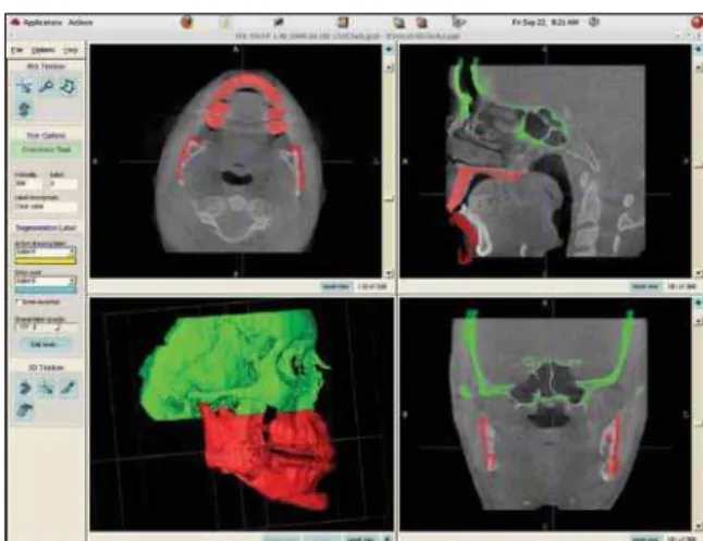
To evaluate within-subject changes, images of different phases were superimposed with the software Imagine (http://www.ia.unc.edu/dev/do-wnload/imagine/index.htm) in a fully automated method using voxel-wise registration to avoid ob-server-dependent location of points identified from overlap of anatomic landmarks. Since the cranial base is not altered by the surgery, its surfaces were used in the registration procedure, where the soft-ware compares the grey level intensity of each vox-el between two CT images. In this way, the cranial base of the pre-surgery CT is used as reference for the other time-points (Fig 1). Despite soft-tissue vi-sualization is better performed with magnetic reso-nance imaging and a better contrast between soft and hard-tissues is observed with spiral computed tomograhy, 3D models of the soft-tissue of the face can be precisely reconstructed with lower cost and radiation and still provide important information of facial esthetic response to surgical movements.4
The presented three-dimensional superimposi-tion method allows the assessment of important structural displacements following surgery, and its short and long-term stability. Despite all training, expertise, technical support, and time required, this methodology seems to have great validity for clinical, scientific and educational orthodontic and surgical application.
FIGURE 1 - After the registration procedure with the Imagine software, the superimposition between the post-surgery 3D model (color) and gray scale pre-surgery image can be observed, showing matching cra-nial bases and displaced mandibular structures (mandibular advance-ment and genioplasty). A correct superimposition between models of the two phases is then confirmed.
Questions
1) Which are the clinical applications of the 3D superimposition method described?
This method has been mostly used in orthosur-gery cases, assessing skeletal displacements follow-ing different osteotomies and verifyfollow-ing treatment outcomes, short and long-term stability. Complex cases, such as dentofacial deformities and severe asymmetries, for example hemifacial microsomia, can benefit from this method in the treatment planning and during the surgical procedure.
On the other hand, its application has already been tested and proved in growing patients, us-ing a superimposition on the anterior cranial base, which is early established. This possibility opens an extraordinary clinical field for a 3D follow-up of craniofacial growth and development of these pa-tients, providing comprehensive visual and quanti-tative analysis.
Motta ATS, Carvalho FAR, Oliveira AEF, Cevidanes LHS, Almeida MAO
Dental Press J. Orthod. 41 v. 15, no. 2, p. 39-41, Mar./Apr. 2010 Contact Address
Alexandre Trindade Simões da Motta Av. das Américas, 3500 - Bloco 7/sala 220
CEP: 22.640-102 – Barra da Tijuca - Rio de Janeiro/RJ, Brazil E-mail: alemotta@rjnet.com.br
that the use of 3D superimposition in case stud-ies at orthodontic graduate programs, allowing a thorough and detailed observation by students and professors, may be an important step on the intro-duction of this method in the clinical practice of the former residents.
2) Are there advantages on research purposes of the method described over the cephalomet-ric method?
Some advantages of the present method can be cited, such as the automated way of cranial base superimposition, avoiding errors associated to land-mark identification or structural contour determi-nation by the operator, representing a significant bias control in a scientific approach. Also, a 3D ob-servation of anatomic structures with real size and form instead of projected superimposed images is a clear differential, allowing the observation of bi-lateral structures in a more realistic way. Addition-ally, the comparison of three-dimensional surfaces instead of cephalometric points and lines can result in more reliable and detailed results. Otherwise, it is important to consider factors like simplicity and ease of working with 2D conventional images. When performing a quantitative analysis, the pres-ent method generates a great amount of informa-tion, leading sometimes to a difficult formulation of straight and concise conclusions of the observed phenomenon. Still, the determination of reliable directional tendencies is difficult because of vari-ous movement directions of the structures. This as-sessment may be improved by the development of vectorial analysis tools, defining in a clear way the displacement directions.
3) Could the method be used on the assessment of dentoalveolar changes following orthodontic treatment?
Yes, one of the possible applications would in-volve the visualization of dentoalveolar changes following orthopedic or orthodontic mechanics. Studies have tested the effects of dental expansion
mechanics, comparing 3D models before and after aligning and leveling, and showed that the expan-sion was mostly concentrated on the premolar re-gion. Otherwise, there are some drawbacks, since the segmentation of the teeth requires a good precision, but basic factors like the acquisition in centric occlusion or the presence of braces can rep-resent important image artifacts when building the 3D models. Another limitation lies on the simple fact that the superimposition requires stable ref-erence structures as the cranial base. For example, when assessing lower arch changes, a cranial base superimposition would show both skeletal and dental alterations, but for an accurate dentoalveolar visualization, an isolated superimposition should be done using the mandibular body, rami and other surface contours. This technology, known as shape correspondence, is still being developed.
1. Cevidanes LH, Bailey LJ, Tucker GR Jr, Styner MA, Mol A, Phil-lips CL, et al. Superimposition of 3D cone-beam CT models of orthognathic surgery patients. Dentomaxillofac Radiol. 2005 Nov;34(6):369-75.
2. Cevidanes LH, Bailey LJ, Tucker SF, Styner MA, Mol A, Phillips CL, et al. Three-dimensional cone-beam computed tomography for assessment of mandibular changes after orthognathic sur-gery. Am J Orthod Dentofacial Orthop. 2007 Jan;131(1):44-50. 3. Cevidanes L, Motta A, Styner M, Phillips C. 3D imaging for
early diagnosis and assessment of treatment response. In: McNnamara JA Jr, Kapila SD. Early orthodontic treatment: is the benefit worth the burden? 33rd Annual Moyers Sympo-sium. Ann Arbor; 2007. p. 305-21.
4. Motta AT. Avaliação da cirurgia de avanço mandibular através da superposição de modelos tridimensionais. [Tese]. Universi-dade do Estado do Rio de Janeiro (RJ); 2007.