CASE REPORT
Sclerosing mesenteritis as an unusual cause of fever
of unknown origin: a case report and review
Vivian Iida Avelino-Silva,I,IIFabio Eudes Leal,I,II,IIICaio Coelho-Netto,IIGuilherme Cutait de Castro Cotti,II Ricardo A. S. Souza,II Rodrigo Lautert Azambuja,IIManoel de Souza Rocha,II,IVEsper Georges KallasI,II,III IFaculdade de Medicina da Universidade de Sa˜o Paulo, Department of Infectious and Parasitic Diseases, Sa˜o Paulo/SP, Brazil.IIHospital Sı´rio-Libaneˆs, Sa˜o Paulo/SP, Brazil.IIIFaculdade de Medicina da Universidade de Sa˜o Paulo, Division of Clinic Immunology and Allergy, Sa˜o Paulo/SP, Brazil.IVFaculdade de Medicina da Universidade de Sa˜o Paulo, Department of Radiology, Sa˜o Paulo/SP, Brazil.
Email: [email protected] Tel.: 55 11 2661-6530
INTRODUCTION
Fever of unknown origin (FUO), defined as a temperature higher than 38.3
˚
C on several occasions and lasting longer than three weeks that is associated with a diagnosis that remains uncertain after one week of investigation (1), is a frequent condition in the infectious diseases specialty clinic. The condition is often associated with extensive diagnostic procedures and, not rarely, frustration for both the patient and the physician. An evidence-based approach has been used to group the causes of FUO (2) into four general categories: infectious, rheumatic/inflammatory, neoplastic, and miscellaneous disorders (3). Infectious diseases such as endocarditis, intra-abdominal abscesses, and tuberculosis, as well as inflammatory conditions such as temporal arteritis, adult’s Still disease, and late-onset rheumatoid arthritis, are commonly identified causes of FUO (3). However, unex-pected and rare causes are sometimes diagnosed, as reported below in a case of sclerosing mesenteritis.CASE DESCRIPTION
A 44-year-old, previously healthy man was referred to the infectious disease specialist for the investigation of fever that had occurred in the previous 10 days and fatigue that had been present for the previous 30 days. He reported being chronically constipated, which had worsened over the previous 30 days. The daily fever was the main complaint and arose at any time of the day without chills or notable sweats; no weight loss was reported. The physical examina-tion was unremarkable, and the initial laboratory findings revealed only a low positive protein levels as determined by urinalysis and slight increases in the levels of C-reactive protein and the erythrocyte sedimentation rate, with a normal complete blood count. Tests for mononucleosis-like agents were negative for active infections. The patient was hospitalized for additional exams and evaluation. Fever (38-38.5
˚
C) was confirmed, but it disappeared right after the introduction of a nonsteroidal anti-inflammatory medication(naproxen). Echocardiography, blood cultures, an eye fundo-scopic examination, and a colonoscopy were all unremark-able. The patient was then submitted to abdominal and pelvic computed tomography (CT) scans, which revealed a mural thickness in the sigmoid colon with nodular densities in adjacent adipose tissue (Figure 1A). Large numbers of lymph nodes up to 1.0 cm in diameter were observed near the inferior mesenteric vessels. An intestinal lymphoma was initially suspected, and the patient was submitted to abdominal laparoscopic surgery with rectosigmoidectomy and lymph node resection. The intraoperative aspect was of a chronic inflammatory lesion affecting the entire mural thickness and adjacent tissues in addition to moderate dilation of the proximal colon (Figure 1B). After hematox-ylin-eosin staining, the histopathologic exam revealed extensive lymphoplasmocytic inflammatory infiltrate with collagenic deposition involving the mesenteric adipocytes (Figures 1C and 1D). Staining for IgG4 was negative in the inflamed tissue.
After the surgical procedure, clinical recovery was adequate, and there was no rebound of fever or weakness after discontinuation of the nonsteroidal anti-inflammatory drug. At a follow-up visit eight months later, the patient remained asymptomatic and no longer complained of intestinal bowel symptoms or fever.
DISCUSSION
Sclerosing mesenteritis is a rare idiopathic condition characterized by a non-neoplastic inflammatory process in the mesenteric fat (4). Men are more commonly affected than women, and the incidence increases in middle-aged and older adults (5). The clinical presentation and radi-ological findings are nonspecific, which renders the condi-tion a diagnostic challenge for surgeons and internists.
Granulomatous and neoplastic diseases are part of the differential diagnosis for sclerosing mesenteritis on clinical and radiological grounds (6). Therefore, histopathology remains the main diagnostic tool (7). The histological findings are fibrosis, myofibroblasts, and inflammatory cell infiltration of the fatty tissue with degeneration or fat necrosis; aggregations of lipid-laden foamy macrophages are also present (4,8). The process can be predominantly inflammatory with fat tissue inflammation and necrosis (mesenteric panniculitis) or predominantly fibrotic, which leads to retractile mesenteritis (7-9).
Copyrightß2012CLINICS– This is an Open Access article distributed under the terms of the Creative Commons Attribution Non-Commercial License (http:// creativecommons.org/licenses/by-nc/3.0/) which permits unrestricted non-commercial use, distribution, and reproduction in any medium, provided the original work is properly cited.
No potential conflict of interest was reported.
CLINICS 2012;67(3):293-295 DOI:10.6061/clinics/2012(03)16
Sclerosing mesenteritis is sometimes observed in association with a multisystem clinical syndrome affecting the pancreas, biliary tract, liver, kidneys, and lungs; this condition has been described as ‘‘IgG4-related systemic sclerosing disease’’, and one of the most common histopathological features is the presence of IgG4+plasma cells within involved tissues (10).
However, cases of sclerosing mesenteritis have also been described in the absence of other sites of involvement.
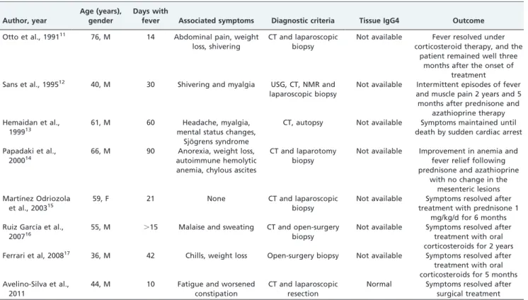
The likely causal relationship of the disease and FUO in this case is supported by the lack of any other diagnosis, the confirmatory histological findings of sclerosing mesenteritis, and the remission of fever with nonsteroidal anti-inflammatory drug use and the surgical removal of involved tissue. At a follow-up visit, the patient remained asymptomatic, suggesting that the local process was the only cause of FUO. Case reports of sclerosing mesenteritis as a cause of FUO are rare, and we
were able to identify only seven cases after a thorough review of the medical literature (Table 1).
The best treatment for sclerosing mesenteritis remains unclear. Asymptomatic or mild clinical forms may some-times be left untreated with spontaneous recovery (8). Surgical resection is required for patients with intestinal obstruction and perforation, and immunosuppressive ther-apy with corticosteroids, thalidomide, and other drugs has been recommended by some authors (7,8). In the present case, surgical removal was able to limit the process. The prognosis is mainly dependent on a correct diagnosis and on the extension of the fibrotic process (9).
Fever has not often been described as a symptom of sclerosing mesenteritis. In this case report, a long-lasting fever was the clinical hallmark of FUO. Although rare, physicians should be aware of this possibility.
Figure 1 - A: Abdominal and pelvic CT scan showing mural thickening of the sigmoid colon with densification of the adjacent mesenteric fat (white arrow) and an increased number of lymph nodes up to 1.0 cm in diameter near the inferior mesenteric vessels; B: Surgical photography showing the inflamed sigmoid; C and D: microscopic exam after hematoxylin-eosin staining showing an extensive lymphoplasmocytic inflammatory infiltrate with collagenic deposition involving the mesenteric adipocytes at 100x (C) and 400x (D) magnifications.
Table 1 -Case reports of sclerosing mesenteritis as a cause of FUO.
Author, year
Age (years), gender
Days with
fever Associated symptoms Diagnostic criteria Tissue IgG4 Outcome
Otto et al., 199111 76, M 14 Abdominal pain, weight
loss, shivering
CT and laparoscopic biopsy
Not available Fever resolved under corticosteroid therapy, and the
patient remained well three months after the onset of
treatment Sans et al., 199512 40, M 30 Shivering and myalgia USG, CT, NMR and
laparoscopic biopsy
Not available Intermittent episodes of fever and muscle pain 2 years and 5 months after prednisone and
azathioprine therapy Hemaidan et al.,
199913
61, M 60 Headache, myalgia,
mental status changes, Sjo¨grens syndrome
CT, autopsy Not available Symptoms maintained until death by sudden cardiac arrest
Papadaki et al., 200014
66, M 90 Anorexia, weight loss,
autoimmune hemolytic anemia, chylous ascites
CT and laparotomy biopsy
Not available Improvement in anemia and fever relief following prednisone and azathioprine
with no change in the mesenteric lesions Martı´nez Odriozola
et al., 200315
59, F 21 None CT and laparoscopic
biopsy
Not available Symptoms resolved after treatment with prednisone 1
mg/kg/d for 6 months Ruiz Garcı´a et al.,
200716
55, M .15 Malaise and sweating CT and open-surgery biopsy
Not available Symptoms resolved after treatment with oral corticosteroids for 2 years Ferrari et al, 200817 36, M 42 Chills, weight loss Open-surgery biopsy Not available Symptoms resolved after
treatment with oral corticosteroids for 5 months Avelino-Silva et al.,
2011
44, M 10 Fatigue and worsened
constipation
CT and laparoscopic resection
Normal Symptoms resolved after surgical treatment
M = male; F = female; CT = computed tomography; USG = ultrasonography; NMR = nuclear magnetic resonance. Sclerosing Mesenteritis and FUO
Avelino-Silva VI et al. CLINICS 2012;67(3):293-295
ACKNOWLEDGMENTS
This report is not a result of a specific funded grant. We acknowledge Professor Christopher D.M. Fletcher for his contribution to the histopathologic diagnosis.
AUTHOR CONTRIBUTIONS
Avelino-Silva VI conceived and coordinated the study and participated in the acquisition of data and manuscript writing. Leal FE participated in the design of the study, data acquisition and manuscript writing. Coelho-Netto C, Cotti GC, Souza RA, Azambuja RL, and Rocha MS participated in the acquisition of data and in manuscript writing. Kallas EG coordinated the study and participated in the acquisition of data and in manuscript writing.
REFERENCES
1. Petersdorf RG, Beeson PB. Fever of unexplained origin: report on 100 cases. Medicine (Baltimore). 1961;40:1-30, http://dx.doi.org/10.1097/ 00005792-196102000-00001.
2. Mourad O, Palda V, Detsky AS. A comprehensive evidence-based approach to fever of unknown origin. Arch Intern Med. 2003;163(5):545-51, http://dx.doi.org/10.1001/archinte.163.5.545.
3. Cunha BA. Fever of unknown origin: clinical overview of classic and current concepts. Infect Dis Clin North Am. 2007;21(4):867-915, vii, http://dx.doi.org/10.1016/j.idc.2007.09.002
4. Chawla S, Yalamarthi S, Shaikh IA, Tagore V, Skaife P. An unusual presentation of sclerosing mesenteritis as pneumoperitoneum: case report with a review of the literature. World J Gastroenterol. 2009;15(1):117-120, http://dx.doi.org/10.3748/wjg.15.117.
5. Akram S, Pardi DS, Schaffner JA, Smyrk TC. Sclerosing mesenteritis: clinical features, treatment, and outcome in ninety-two patients. Clin Gastroenterol Hepatol. 2007;5(5):589-596; quiz 523-584, http:// dx.doi.org/10.1016/j.cgh.2007.02.032.
6. Arora M, Dubin E. A clinical case study: sclerosing mesenteritis presenting as chylous ascites. Medscape J Med. 2008;10(2):30. 7. Gu GL, Wang SL, Wei XM, Ren L, Li DC, Zou FX. Sclerosing mesenteritis
as a rare cause of abdominal pain and intraabdominal mass: a cases report and review of the literature. Cases J. 2008;1(1):242, http:// dx.doi.org/10.1186/1757-1626-1-242.
8. Issa I, Baydoun H. Mesenteric panniculitis: various presentations and treatment regimens. World J Gastroenterol. 2009;15(30):3827-3830, http://dx.doi.org/10.3748/wjg.15.3827.
9. Nobili C, Degrate L, Caprotti R, et al. Extensive sclerosing mesenteritis of the rectosigmoid colon associated with erosive colitis. Gastroenterol Res Pract. 2009;2009:176793.
10. Bateman AC, Deheragoda MG. IgG4related systemic sclerosing disease -an emerging -and under-diagnosed condition. Histopathology. 2009;55(4):373-383, http://dx.doi.org/10.1111/j.1365-2559.2008.03217.x. 11. Otto F, Wedekind G. [Mesenteric panniculitis]. Z Gastroenterol.
1991;29(8):395-397.
12. Sans M, Varas M, Anglada A, Esperanza Bachs M, Navarro S, Brugues J. Mesenteric panniculitis presenting as fever of unknown origin. Am J Gastroenterol. 1995;90(7):1159-1161.
13. Hemaidan A, Vanegas F, Alvarez OA, Arroyo MA, Lee M. Mesenteric lipodystrophy with fever of unknown origin and mesenteric calcifica-tions. South Med J. 1999;92(5):513-516, http://dx.doi.org/10.1097/ 00007611-199905000-00013.
14. Papadaki HA, Kouroumalis EA, Stefanaki K, et al. Retractile mesenteritis presenting as fever of unknown origin and autoimmune haemolytic anaemia. Digestion. 2000;61(2):145-148, http://dx.doi.org/10.1159/ 000007748.
15. Martinez Odriozola P, Garcia Jimenez N, Cabeza Garcia S, Oceja Barrutieta E. [Sclerosing mesenteritis. Report of two cases with different clinical presentation]. An Med Interna. 2003;20(5):254-256.
16. Ruiz Garcia S, Yedra Alcaraz M, Ortega N, Villarinos Vivas MA. [Inusual presentation of mesenteric panniculitis]. An Med Interna. 2007;24(8):393-395.
17. Ferrari TC, Couto CM, Vilaca TS, Xavier MA, Faria LC. An unusual presentation of mesenteric panniculitis. Clinics (Sao Paulo). 2008;63(6): 843-844, http://dx.doi.org/10.1590/S1807-59322008000600023.
CLINICS 2012;67(3):293-295 Sclerosing Mesenteritis and FUO
Avelino-Silva VI et al.