363
CLINICS 2007;62(3):363-6
LETTER TO THE EDITORS
CORTICAL HYPEROSTOSIS SECONDARY TO
PROLONGED USE OF PROSTAGLANDIN E1
João Fernando Lourenço de Almeida, Helio Kimura, Luiz Henrique Hercowitz, Hélio Korkes, Eduardo Juan Troster.
INTRODUCTION
Prostaglandin E1 (Alprostadil), a drug used for ductal patency in cyanotic congenital heart disease, comprises an essential part of the treatment of infants waiting for car-diac surgery in hospitals with no specialized medical staff or in situations of inadequate weight for surgery. Prostag-landin E1 (PgE1) can also be used in elevation of presurgical oxygenation status in more severe cases.1,2
The duration of infusion is often short (from 6 hours to 20 days).3 However, in some cases, when there is low
birth weight, infection, or absence of a specialized hospi-tal to transfer the patient to (all of these being common situation in developing countries), the infusion time can extend for weeks or months.4,5
Common side effects of prostaglandin therapy include apnea, fever, convulsions, rash, skin flushing, vasodilata-tion with hypotension, diarrhea, and gastric outlet obstruc-tion.3 These effects are related to short-term use of PgE1,
and the Pediatric Intensive Care Unit staff has the exper-tise to easily recognize and treat them.
In rare situations of prolonged use of alprostadil, the most important side effects include soft tissue swelling and reversible cortical hyperostosis in the large bones.4,5 We
describe the case of an infant presenting this complication secondary to prolonged use of PgE1, with typical and ex-tensive radiological findings.
CASE REPORT
A 6-month-old male neonate, born at 38 weeks of ges-tation with APGAR scores of 7 and 9 at the first and fifth minute, respectively, weighed 2930 g at birth. His mother had no pregnancy or labor antecedent.
In the first hours of life, the neonate started to develop respiratory distress and cyanosis. On physical exam, a systolic murmur of a patent ductus arteriosus was found.
The child had no dysmorphic features.
The echocardiography revealed Ebstein’s anomaly, with a dysplastic tricuspid valve, nonrestrictive atrial septal de-fect (ostium secundum) of 7.5 mm, single aortic outlet from the left ventricle, and patent ductus arteriosus. The systolic function of the left heart was normal, with a left ventricu-lar ejection fraction (LVEF) of 65%.
Intravenous infusion of PgE1 was started on the first day of life, at a dosage of 0.05 µg/kg/minute.
The child progressed with mechanical ventilation depend-ency and had systemic and respiratory infections, requiring use of antibiotics. On 60th day, he underwent a tracheostomy
procedure. Despite the use of multiple and prolonged empiri-cal antibiotic treatments, the infant sustained a persistent fe-ver and had a C-reactive protein (CRP) level of 188 mg/L.
At this time, the clinical and laboratory diagnosis of per-sistent nosocomial sepsis was made, due to positive blood cultures that isolated an uncataloged, nonfermenting, Gram-negative bacillus. The patient had a number of positive blood and catheter cultures with the same bacillus. At the time, the antibiotic schedule included piperacillin and tazobactam associated with trimethoprim-sulfamethoxazole guided by the antibiogram. The cardiac surgical correction was delayed, and the time of utilization of PgE1 was increased.
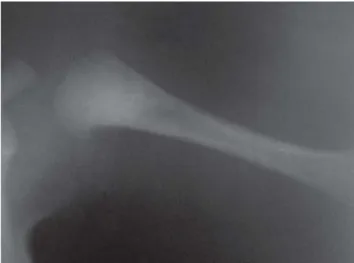
After 3 months, the child was transferred from another state to our hospital. On examination, he was noted to have painful bilateral swelling of ankles and fists. The pain with manipulation was intense in the 4 limbs and unresponsive to regular analgesics. The radiography of the large bones re-vealed an intense periosteal reaction with bilateral corticoperiosteal thickening of the diaphyses in clavicles, femur, tibia, humerus, radius, and ulna. The lesions were dis-tributed along the middle portion of the large bones de-scribed, with the exception of the humerus, where the af-fection was distributed more distally (Figures 1, 2, and 3).
Regarding laboratory findings, the maternal and child syphilis serology were negative. Alkaline phosphatase was 399 U/L (normal range, 145-320 U/L) with normal calcium and phosphate.
With these results and considering the dose and dura-Pediatric Intensive Care Unit Hospital Israelita Albert Einstein
364
CLINICS 2007;62(3):363-6 Cortical hyperostosis secondary to prolonged use of prostaglandin e1
Almeida JFL et al.
tion of PgE1 utilization, the diagnosis of cortical hyperos-tosis secondary to intravenous prostaglandin was made.
After this diagnosis, 2 attempts at reduction and inter-ruption of the intravenous PgE1 were made. The goal was to reduce the existing adverse events: hyperostosis, fever, and abdominal distention. The fever started within the first month of life, and the highest temperature was 38.6oC with
1 or 2 episodes per day. Even considering the infection to
be the main reason for the fever, the hypothesis of an ad-verse event secondary to PgE1 could not be ruled out. Ab-dominal distension was moderate, accompanied by vomit-ing or gastric residual content in the nasogastric tube.
The infant did not tolerate the reduction of the drug, experiencing rapid decrease in the arterial saturation (pulse oximetry saturation of 55% after 1 hour of reduction).
He was kept on PgE1 infusion for 162 days (5.4 months). The dose of PgE1 during the infusion period ranged from 0.025 to 0.1 µg/kg/minute. The total dose re-ceived was 45 mg, with mean dosage of 279 µg/day (0.043 µg/kg/minute).
After a period of 1 week during which the child re-ceived the drug by peripheral vascular access, the blood cultures become negative (3 samples). At this point, the cardiothoracic surgeons performed a modified Blalock-Taussig shunt. The patient had a good recovery from sur-gery, permitting the interruption of the drug in the operat-ing room.
After 2 weeks, the swelling of ankles and fists disap-peared, and the child had no longer pain after manipula-tion, with complete clinical regression. A radiograph of the left humerus after 47 days showed radiological regression of the bone hypertrophy (Figure 4).
DISCUSSION
The presence of cortical hyperostosis is frequent in in-fants with prolonged use of intravenous prostaglandin, but the final diagnosis is not always achieved. The investiga-tion performed by pediatricians and neonatologists is gen-erally focused on the more immediate and frequent adverse
Figure 2 - Radiograph of the right lower extremity showing cortical thickening of femur and tibia.
Figure 3 - Cortical hyperostosis of the left clavicle and humerus
365
CLINICS 2007;62(3):363-6 Cortical hyperostosis secondary to prolonged use of prostaglandin e1
Almeida JFL et al.
events such as apnea (19%), abdominal distension (16%), bradicardia (13%), enterocolitis (6.5%), hypotension (6.5%), fever (1.6%), and flushing (1.6%).3
In the case described above, our patient presented only fever and abdominal distension as acute adverse events. The fever, which had a clear infectious trigger, was probably related to the infusion of PgE1. This fact was corroborated by the maintenance of the presurgical fever even after the fall of CRP from 188 mg/L (highest value) to < 1 mg/L and the presence of negative blood cultures (sepsis treat-ment success). One day after interruption of the drug the fever disappeared.
Histological examination of bones with cortical hyper-ostosis shows rapid formation of primitive bone, extensive resorption of the outer cortical surface, and bone forma-tion in the inner surface. These changes are responsible for the increase in serum alkaline phosphatase concentrations that occurs in 53% of the patients.6,8
The cortical thickening seems to be related with the du-ration or dosage of continuous intravenous infusion of PgE1.5– 7 Forty-two percent of infants receiving PgE1 infusion for more
than 30 days develop hyperostosis; this increases to 100% in infants receiving the infusion for more than 60 days.8
It is notable that in many cases, the degree of hyperos-tosis is higher due to delay in diagnosis. There are reports of extremely early cortical changes, with initiation of ra-diographic findings in 9 days.5,8,9
The clinical history and physical exam are usually suf-ficient to elucidate the diagnosis. The most important dif-ferential diagnoses of this condition include congenital syphilis, infantile congenital hyperostosis, scurvy, and hy-pervitaminosis A. In contrast, when the cortical thicken-ing is restricted to 1 bone, it is usually secondary to trauma, tumor, or osteomyelitis.
In this case, the diagnosis of syphilis was unlikely due to absence of compatible clinical history and negative se-rology. In hypervitaminosis A, the cortical hyperostosis ap-pears after months or years of excessive ingestion. Regard-ing scurvy, the cortical thickenRegard-ing occurs durRegard-ing the heal-ing process of subperiosteal hematomas, which requires months of evolution and was unlikely in this patient.
Finally, infantile cortical hyperostosis is the diagnosis with the most difficult differentiation from prostaglandin-induced hyperostosis. Infantile cortical hyperostosis or Caffey disease is a genetic disorder, with autosomal domi-nant inheritance in its usual form, with incomplete penetrance. However, the severe and lethal form of the ease appears to be inherited as an autosomal recessive dis-order. Recently, a novel gene mutation on the alpha-1 chain of type I collagen has been described in patients with the autosomal dominant form of the disease.12 Clinically,
in-fantile cortical hyperostosis is characterized by hyperirri-tability, soft tissue swelling, and cortical hyperostosis. The median age of presentation is around 9 weeks of age. Fe-ver is present and may be due to the high prostaglandin levels or excessive metabolic activity in the bones.10,11In
the light of the fact that both diseases have the same clini-cal aspects and radiologiclini-cal findings, differentiation is pos-sible only due to the antecedent of prolonged therapy with prostaglandin. Because our patient had clinical, laboratory, and radiological aspects of the disease concomitant with the use of PgE1 of more than 5 months, we could deter-mine the diagnosis of prostaglandin-induced cortical hyper-ostosis. This fact was confirmed after the interruption of the drug and the subsequent reduction in the radiological and clinical findings.
Our patient had a similar evolution as for some other cases described in the literature, with an extremely pro-longed infusion period and a relatively rapid decrease in clinical and radiological findings after withdrawal of the drug as the most notable points. One unusual issue in this case was the presence of cortical hyperostosis involving the clavicles. The first description of clavicle involvement in PgE1 hyperostosis was made by Nadroo et al in 2000.5 Our
patient had clear bilateral clavicle involvement as seen in Figure 3, reinforcing the probability of bone hypertrophy in these bones.
CONCLUSION
Pediatric intensivists and neonatologists must be alert to the adverse effects of continuous intravenous infusion of prostaglandin. Attention must be given to the acute and more common adverse effects as well as to the ones related to pro-longed use. Early and frequent radiological investigation
366
CLINICS 2007;62(3):363-6 Cortical hyperostosis secondary to prolonged use of prostaglandin e1
Almeida JFL et al.
should be made in patients undergoing intravenous PgE1 treatment for more than 7 days. All efforts must be made to
attempt interruption of the drug as soon as possible, aiming at preventing the potential complications.
REFERENCES
1. Hoch M, Netz H. Heart failure in pediatric patients. Thorac Cardiovasc Surg. 2005;53:S129-S134.
2. Mattos SS, Rodrigues JV, Severi R, Nunes M, Cunha CEG, Melo VB, et al. Manuseio da atresia tricúspide em neonatos. Relato de três casos e revisão da literatura. J Pediatr (Rio J). 1994;70:33-8.
3. Lucron H, Chipaux M, Bosser G, Le Tacon S, Lethor JP, Feillet F, et al. Complications of prostaglandin E1 treatment of congenital heart disease in paediatric medical intensive care. Arch Mal Coeur Vaiss. 2005;98:524-30.
4. Caballero S, Torre I, Arias B, Blanco D, Zabala JI, Sanchez Luna M. Secondary effects of prostaglandin E1 on the management of hypoplastic left heart syndrome while waiting for heart transplantation. An Esp Pediatr. 1998;48:505-9.
5. Nadroo AM, Shringari S, Garg M, al-Sowailem AM. Prostaglandin induced cortical hyperostosis in neonates with cyanotic heart disease. J Perinatal Med. 2000;28:447-52.
6. Jorgensen HR, Svanholm H, Host A. Bone formation induced in an infant by systemic prostaglandin-E2 administration. Acta Orthop Scand. 1988;59:464-6.
7. Jureidini S, Chase NA, Alpert BS, Vanderzalm T, Sheneflet RE. Soft-tissue swelling in two neonates during prostaglandin E1 therapy.Pediatr Cardiol. 1986;7:157-60.
8. Woo K, Emery J, Peabody J. Cortical hyperostosis: a complication of prolonged prostaglandin infusion in infants awaiting cardiac transplantation. Pediatrics. 1994;93:417-9.
9. Kalloghlian AK, Frayha HH, de Moor MM. Cortical hyperostosis simulating osteomyelitis after short-term prostaglandin E1 infusion. Eur J Pediatr. 1996;155:173-4.
10. Velaphi S, Cilliers A, Beckh-Arnold E, Mokhachane M, Mphahlele R, Pettifor J. Cortical hyperostosis in an infant on prolonged prostaglandin infusion: case report and literature review. J Perinatol. 2004;24:263-5. 11. Delgado Azañero, Wilson Alejandro; Arrascue Dulanto, Víctor Manuel. Hiperostosis cortical infantil (enfermedad de Caffey) / Infantile cortical hyperostosis (Caffey’s disease). Rev Estomatol Hered. 2004;14:82-3. 12. Gensure RC, Mäkitie O, Barclay C, Chan C, DePalma SR, Bastepe M,