403
Atypical locations of cerebral hemorrhage in newborns
Radiol Bras. 2009 Nov/Dez;42(6):403–405
Case Report • Relato de Caso
Atypical locations of cerebral hemorrhage in newborns:
considerations about two cases*
Apresentações de localização atípica de hemorragia no cérebro de recém-nascidos: considerações acerca de dois casos
Alexandra Maria Vieira Monteiro1, Cláudio Márcio Amaral de Oliveira Lima2, Érica Barreiros Ribeiro2, Fernanda Camurati de Oliveira3, Silvia Miranda4, Luiz Eduardo Miranda5
Retrospective study of two cases of atypically localized cerebral hemorrhage among 777 newborns admitted to the neonatal intensive care unit at Casa de Saúde São José, Rio de Janeiro, RJ, Brazil. Clinical findings and imaging diagnoses were evaluated. The diagnostic effectiveness of ultrasonography was established by correlation with magnetic resonance imaging findings. Multifactorial etiology was observed, besides silent clinical presentation independently from localization. So far the neurological evaluation has satisfactorily progressed although the patients are still too young to allow a definite neurological evaluation.
Keywords: Brain; Newborn; Atypical hemorrhage; Posterior fossa hemorrhage; Ultrasonography; Magnetic resonance imaging.
Estudo retrospectivo de dois casos de hemorragia craniana de localização atípica em 777 recém-natos inter-nados na unidade de terapia intensiva neonatal da Casa de Saúde São José, Rio de Janeiro, RJ. Foram ava-liados os aspectos clínicos e o diagnóstico por métodos de imagem. Verificamos que a ultrassonografia foi diagnóstica nos dois casos quando comparada com a ressonância magnética. Em relação à etiologia, esta foi multifatorial, e a manifestação clínica silenciosa independente da localização. Até o presente momento, a avaliação neurológica tem tido curso satisfatório, embora os pacientes ainda tenham baixa idade para a ava-liação neurológica definitiva.
Unitermos: Cérebro; Recém-natos; Hemorragia atípica; Hemorragia na fossa posterior; Ultrassonografia; Imagem por ressonância magnética.
Abstract
Resumo
* Study developed at Casa de Saúde São José (CSSJ-RJ), Rio de Janeiro, RJ, Brazil.
1. PhD, MD, Radiologist at Casa de Saúde São José (CSSJ-RJ), Associate Professor of Radiology, Faculdade de Ciências Médicas da Universidade do Estado do Rio de Janeiro (FCM-UERJ), Rio de Janeiro, RJ, Brazil.
2. MDs, Radiologists, Unit of Magnetic Resonance Imaging – Hospital São José and Clínica Dr. Flávio Althoff, Crisciúma, SC, Brazil.
3. MD, Radiologist, Pontifícia Universidade Católica do Rio de Janeiro (PUC-Rio), Rio de Janeiro, RJ, Brazil.
4. MD, Pediatric Neurologist at Neonatal Intensive Care Unit, Casa de Saúde São José (CSSJ-RJ), Rio de Janeiro, RJ, Brazil. 5. MD, Neonatologist, Head for the Neonatal Intensive Care Unit, Casa de Saúde São José (CSSJ-RJ), Rio de Janeiro, RJ, Brazil.
Mailing address: Dr. Cláudio Márcio Amaral de Oliveira Lima. Rua São José, 322, Centro. Criciúma, SC, 88801-520, Brazil. E-mail: [email protected]; [email protected]
Received May 7, 2008. Accepted after revision June 10, 2008.
playing the most relevant role as compared with other imaging methods, besides being noninvasive, reproducible, portable and not requiring sedation(4). However, magnetic
resonance imaging (MRI) is the gold stan-dard for the diagnosis although the neces-sity of sedation and refrigerated environ-ment still represent a restriction to its fre-quent utilization in these patients(5,6).
The present study was aimed at corre-lating TFUS and MRI findings, determin-ing the hemorrhage location and clinical manifestations, describing possible etio-logic factors as well as the neuroetio-logical de-velopment up to the present moment by means of considerations about two cases of atypical presentation of hemorrhage in newborns of a population admitted to a neonatal intensive care unit.
CASES REPORT
In the period from March/2004 to April/ 2007, 777 neonates were admitted and sub-Monteiro AMV, Lima CMAO, Ribeiro EB, Oliveira FC, Miranda S, Miranda LE. Atypical locations of cerebral hemorrhage in newborns: considerations about two cases. Radiol Bras. 2009;42(6):403–405.
to be achieved. According to the literature, the location of hypoxic lesions in neonates tends to follow the physiological course of the blood circulation, and are predomi-nantly hemorrhagic in preterm newborns with location in the germinal matrix and peri- and/or intraventricular region through the ventriculopetal branches(2). In
term newborns, these lesions are predomi-nantly ischemic and located in the basal ganglia and cortical-subcortical regions of the brain through the ventriculofugal cir-culation(2). Generally, the physiopathology
of hypoxic lesions in the neonatal period occurs as a result from the loss of self-regulation with the consequential brain injury with different aspects and degrees, according to the level of development of the cerebrovascular system at the different gestational ages(3).
The utilization of imaging methods in patients of this age range has allowed an early and accurate diagnosis of lesions, with transfontanel ultrasonography (TFUS)
0100-3984 © Colégio Brasileiro de Radiologia e Diagnóstico por Imagem
INTRODUCTION
Cerebrovascular diseases represent a relevant cause of brain injury in neonates(1).
Both location and etiology of cerebral hem-orrhage in neonates have already been well known and described in the literature(2).
404
Monteiro AMV et al.
Radiol Bras. 2009 Nov/Dez;42(6):403–405 mitted to ultrasonography (US) at the
neo-natal intensive care unit (ICU) of Casa de Saúde São José, in the city of Rio de Janeiro, RJ, Brazil. Two of these patients underwent TFUS, being diagnosed with atypically localized brain hemorrhage later confirmed by MRI that is the method con-sidered as the gold standard for such diag-nosis.
Case 1 – Female, preterm, underweight (1470 g) newborn of a triplet gestation, with AB blood type, positive Rh factor, membranes rupture during labor, Apgar 3/6, requiring reanimation with positive pres-sure ventilation through orotracheal tube. Maximum total bilirubin level was 14.5. The patient remained under phototherapy for eight days. Hyaline membrane disease was found and the neonate received a sur-factant dose at the first hours of life. Neo-natal ICU discharge occurred 31 days af-ter admission, with a weight gain of 790 g
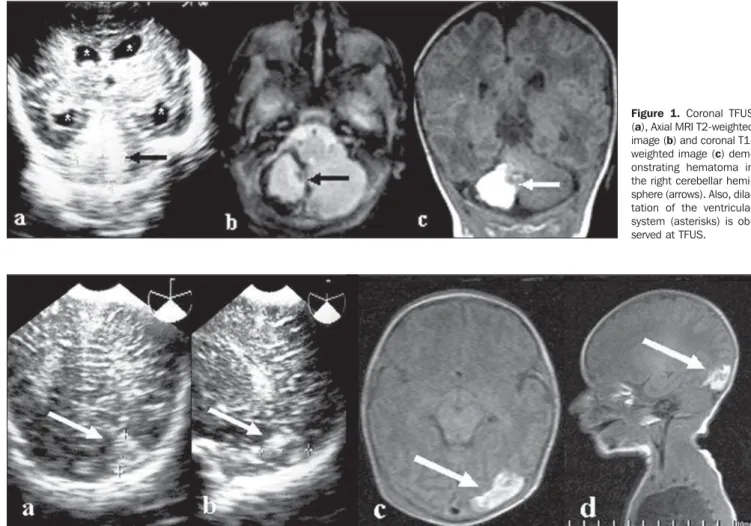
(total weight 2260 g). Echocardiogram was performed at the first day of life, demon-strating patent foramen ovale and persistent arterial canal with no significant clinical repercussion, besides subtle pulmonary artery hypertension. At the second day of life, the neonate was submitted to TFUS that demonstrated symmetrical, bilateral supratentorial ventricular dilatation and a hyperechogenic lesion in the right cerebel-lar hemisphere, measuring 2.7 × 2.1 cm. At 29 days of life, MRI identified the he-matoma in the right cerebellar hemisphere and in the postero-inferior region of the left cerebellar hemisphere, without dilatation of the ventricular system (Figure 1). Mag-netic resonance angiography (MRA) was normal.
Case 2 – Male, term newborn, with birth weight of 2145 g, B blood type, posi-tive Rh factor, normal labor and membrane rupture during the labor at the 37th
gesta-tional week, Apgar 6/8, requiring reanima-tion with positive pressure mask ventila-tion. Delayed intrauterine development and early, symptomatic polycythemia. The neonate was admitted to the neonatal ICU because of hypoglycemia. The patients progressed with concussion and hypogly-cemia (glyhypogly-cemia of 26 mg/dl). Concussion improved with the hypoglycemia correc-tion. A partial exogenous blood transfu-sion was performed through umbilical venous catheter. Echocardiogram per-formed one day after birth demonstrated small, persistent arterial canal with dias-tolic flow. The patient remained in the ICU for ten days. At the fifth day, TFUS dem-onstrated an echogenic image with hypoechoic center measuring about 2.0 cm in the left occipital lobe. At the eighth day, cranial MRI with MRA identified he-matoma with low fluid level in the left occipital lobe (Figure 2).
Figure 1. Coronal TFUS (a), Axial MRI T2-weighted image (b) and coronal T1-weighted image (c) dem-onstrating hematoma in the right cerebellar hemi-sphere (arrows). Also, dila-tation of the ventricular system (asterisks) is ob-served at TFUS.
405
Atypical locations of cerebral hemorrhage in newborns
Radiol Bras. 2009 Nov/Dez;42(6):403–405 DISCUSSION
According to the literature, atypical lo-cation of brain hemorrhage in newborns is found in exactly 0.25% of cases. Ghazi-Birry et al.(4) have proposed that such
lo-cation can be explained by the presence of germinal matrix not only in the caudo-thalamic region, but also in other regions such as occipital and temporal horn of the lateral ventricles, and in the infratentorial cerebellar region. In these locations, the germinal matrix rarely generates bleeding after 32 weeks of gestation since it has al-ready practically disappeared. In atypical cases, it is admitted that persistence of this matrix may occur, therefore increasing the incidence of bleedings(2).
Intracranial hemorrhages are frequent in preterm newborns, with highest prevalence in this age range(7), possibly affecting up to
40% newborns with gestational age < 32 weeks and birth-weight < 1500 g(5). On the
other hand, brain hemorrhages are less fre-quent in term newborns, with prevalence of 26% in asymptomatic newborns after vagi-nal delivery(8). Frequently, such
hemor-rhages are associated with apnea, bradycar-dia and convulsions, with several factors being related to increased risk, particularly peripartum complications such as pro-longed labor, use of forceps and fetal un-derweight(8).
In the present study, the authors could not isolate a single factor, as those de-scribed by other authors(1,3,7,8) identifying
factors such as tocotraumatism, hypoxia, hypoglycemia, hyaline membrane disease and preterm delivery, among others.
Moura-Ribeiro et al.(9) have reported
that, at this age range, the majority of cere-brovascular diseases present with some-times severe seizures about 24–48 postna-tal hours, even with a generally normal Apgar index; and within few days may progress to hemiparesis, hypotonia, tonus
asymmetry, drowsiness or irritability and also sucking difficulty(1), differently from
the present study where a neonate (case 2) initially presented concussion probably related to hypoglycemia. Campos-Castelló et al.(3) have also reported the presence of
hydrocephalus that may vary according to the extent and time of hemorrhage onset. In the present case, with presentation in the cerebellum (case 1), the authors identified an association with transitory hydroceph-alus with spontaneous resolution con-firmed by MRI.
As regards imaging diagnosis, several authors(2,6,9,10) agree that TFUS should be
the initial method of choice in the evalua-tion of a newborn brain. As far the locaevalua-tion in the cerebellum is concerned, the major-ity of findings was unilateral (71%) with predominance for the right hemisphere (64% at right versus 36% at left), in
agree-ment with the results of the present study. Other authors(4,10) also report a persistence
of lesions at US, on average, for four to six weeks.
It is a consensus that MRI is the gold standard for the diagnosis of cerebral le-sions(6,8), also with the assertion of the
fea-sibility of the study by functional MRI to stratify the newborns with highest risk for neurological damage, especially in the presence of an association with risk factors such as US demonstrating intraventricular hemorrhage at birth, among others(10).
Generally, cerebellar hemorrhages de-velop with hydrocephalus or ataxic syn-dromes, or even delayed cognitive devel-opment with cerebellar atrophy in most of patients who survive(3). In the present study,
the patients are still too young to allow a definite neurological evaluation.
CONCLUSION
Transfontanel ultrasonography was di-agnostic for atypical presentation of
cere-bral hemorrhage in newborns as compared with MRI. The clinical presentation was silent and, up to the present moment, the neurological development has been satis-factory, although the low age of the chil-dren precludes a definite evaluation.
REFERENCES
1. Moura-Ribeiro MVL, Pessoto MA, Marba STM. Cerebrovascular disease in neonates: evaluation of four cases. Arq Neuropsiquiatr. 1999;57:84–7. 2. Kaske TI, Rumack CM, Harlow CL. Imagens do cérebro de neonatos e crianças. In: Rumack CM, Wilson SR, Charboneau JW, editores. Tratado de ultra-sonografia diagnóstica. 2ª ed. Rio de Janei-ro: Guanabara Koogan; 1999. p. 1250–7. 3. Campos-Castelló J, Canelón de López MS,
San-tiago-Gómez R. Accidentes vasculares isquémi-cos y hemorrágiisquémi-cos cerebrales del recién nacido a término. Protocolo de estudio y orientaciones terapeuticas. Rev Neurol. 2000;31:632–44. 4. Ghazi-Birry HS, Brown WR, Moody DM, et al.
Human germinal matrix: venous origin of hem-orrhage and vascular characteristics. AJNR Am J Neuroradiol. 1997;18:219–29.
5. Antoniuk S, Silva RVC. Hemorragia periventri-cular e intraventriperiventri-cular de recién nacidos prema-turos. Rev Neurol. 2000;31:238–43.
6. Miller SP, Cozzio CC, Goldstein RB, et al. Com-paring the diagnosis of white matter injury in pre-mature newborns with serial MR imaging and transfontanel ultrasonography findings. AJNR Am J Neuroradiol. 2003;24:1661–9.
7. Farage L, Assis MC. Achados ultra-sonográficos da hemorragia intracraniana em recém-nascidos prematuros. Arq Neuropsiquiatr. 2005;63:814–6. 8. Looney CB, Smith JK, Merck LH, et al. Intracra-nial hemorrhage in asymptomatic neonates: prevalence on MR images and relationship to obstetric and neonatal risk factors. Radiology. 2007;242:535–41.
9. Moura-Ribeiro MVL, Ciasca SM, Vale-Caval-canti M, et al. Cerebrovascular disease in new-born infants: report of three cases with clinical follow-up and brain SPECT imaging. Arq Neuro-psiquiatr. 1999;57:1005–10.